This book is distributed under the terms of the Creative Commons Attribution-NonCommercial-NoDerivatives 4.0 International (CC BY-NC-ND 4.0) ( http://creativecommons.org/licenses/by-nc-nd/4.0/ ), which permits others to distribute the work, provided that the article is not altered or used commercially. You are not required to obtain permission to distribute this article, provided that you credit the author and journal.
NCBI Bookshelf. A service of the National Library of Medicine, National Institutes of Health.
StatPearls [Internet]. Treasure Island (FL): StatPearls Publishing; 2026 Jan-.
StatPearls [Internet].
Show detailsContinuing Education Activity
Brugada syndrome is a rare, potentially life-threatening inherited cardiac disorder that predisposes primarily young, otherwise healthy individuals to fatal ventricular arrhythmias and sudden cardiac death. The condition is most commonly caused by autosomal dominant mutations in the SCN5A gene, which encodes a cardiac sodium channel, although other genetic and environmental factors may modulate disease expression. These mutations impair sodium current, creating conduction abnormalities and predisposing to reentrant arrhythmias.
Clinical manifestations range from asymptomatic electrocardiographic abnormalities to syncope, nocturnal agonal respiration, or sudden cardiac arrest. Diagnosis relies on characteristic electrocardiographic findings, including right bundle branch block and ST-segment elevations in the right precordial leads (V1-V3), sometimes unmasked by fever or pharmacologic challenge. Management focuses on prevention of sudden death, primarily via implantable cardioverter-defibrillator placement, with adjunctive pharmacologic and lifestyle strategies in selected cases. Complications include recurrent ventricular arrhythmias and sudden cardiac death, and prognosis varies with risk stratification, early diagnosis, and adherence to recommended therapy.
This activity for healthcare professionals is designed to sharpen learners' skills in evaluating and managing Brugada syndrome. Participants will improve their understanding of the condition's etiology, risk factors, pathophysiology, clinical presentation, potential complications, and evidence-based diagnostic and therapeutic approaches. Greater proficiency will equip clinicians to collaborate successfully with interprofessional teams caring for individuals with Brugada syndrome.
Objectives:
- Identify the clinical and diagnostic features indicative of Brugada syndrome.
- Apply evidence-based, individualized strategies for managing Brugada syndrome and preventing its potential complications.
- Improve patient understanding of Brugada syndrome's etiology, trigger factors, symptoms, treatment options, and expected outcomes to promote adherence.
- Collaborate with the interprofessional team to educate, treat, and monitor patients with Brugada syndrome, guiding family-planning decisions and improving overall health outcomes.
Introduction
Brugada syndrome is an inherited cardiac channelopathy that increases the risk of sudden cardiac death (SCD) due to supraventricular and ventricular arrhythmias, often in young, otherwise healthy individuals. The characteristic electrocardiographic (ECG) changes were first reported in the 1950s, but the syndrome was formally described in 1992 by Josep and Pedro Brugada.[1][2]
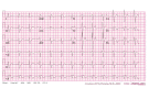
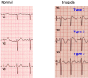
The syndrome is primarily identified by ECG findings of right bundle branch block and distinct ST-segment changes in the right precordial leads (V1-V3). These ECG patterns are classified into 3 morphologies. Type 1 is defined by coved ST-segment elevation of at least 2 mm in at least 1 right precordial lead, followed by T-wave inversions, in the absence of other causes of ST elevation (see Image. Brugada Syndrome Type 1 Morphology). Type 2 presents as saddleback-shaped ST-segment elevation of at least 2 mm, with a positive or biphasic T wave and J-point elevation of at least 1 mm. Type 3 is characterized by ST-segment elevation less than 1 mm with a positively deflecting T wave (see Image. Brugada Syndrome Electrocardiographic Patterns).
ECG patterns observed immediately after resuscitation or electrical cardioversion should not be considered diagnostic, as the ST segment can be dynamic. Multiple patterns may appear sequentially in patients who exhibit dynamic ECG changes or are exposed to certain medications.[3] Normal or nondiagnostic ECGs may also occur in individuals with a disease-associated genotype. Potent sodium channel-blocking drugs, including ajmaline, procainamide, and flecainide, can unmask ECG changes, although drug challenge reduces specificity.[4]
Diagnosis and management remain challenging due to an incomplete understanding of the syndrome’s genetic and molecular mechanisms. Clinical criteria continue to evolve as new research emerges.[5][6]
Etiology
Brugada syndrome is inherited in an autosomal dominant pattern. However, affected individuals may exhibit variable expressivity and reduced penetrance. The first genetic association identified was a loss-of-function mutation in the cardiac voltage-gated sodium channel gene SCN5A, present in an estimated 15% to 30% of cases.[7] Mutations in genes encoding calcium and potassium channels, associated regulatory proteins, and desmosomal components have also been implicated. Although approximately 19 genes have been linked to Brugada syndrome, SCN5A remains the most definitively associated.[8] Environmental and genetic factors, including elevated temperature, specific medications, electrolyte disturbances, and cocaine exposure, further modulate phenotypic expression.
Epidemiology
The true burden of Brugada syndrome is difficult to ascertain due to the unknown prevalence of asymptomatic individuals. Population studies report a prevalence of approximately 2 to 5 per 10,000 people. The syndrome is 8 to 10 times more common in men than in women. This sex difference is not observed in pediatric populations. The disparity has been attributed to higher testosterone levels after puberty and sex-specific discrepancies in ionic current proportions. Brugada syndrome is also more prevalent among individuals of Southeast Asian descent. The average age of affected individuals is 41 years. Notably, the syndrome accounts for 4% to 12% of all SCDs.[9]
Pathophysiology
The exact pathophysiological mechanism of Brugada syndrome remains unclear.[10] Two principal hypotheses have been proposed. The repolarization disorder model posits that a loss-of-function mutation in the cardiac sodium channel reduces sodium current, producing a more pronounced notch in the action potential of the right ventricular epicardium compared with the endocardium. This transmural current difference is thought to underlie the characteristic ECG patterns of Brugada syndrome and increase susceptibility to malignant arrhythmias.[11] In contrast, the depolarization disorder model attributes the ECG manifestations to delayed depolarization caused by slow conduction in the right ventricular outflow tract (RVOT), promoting arrhythmogenesis.[12]
No definitive evidence currently excludes the coexistence of both mechanisms in the same patient. Experimental data primarily support the repolarization model, but it has not been conclusively validated in clinical settings.[13] Further research is required to elucidate the precise pathophysiological mechanisms underlying Brugada syndrome.
Histopathology
When Brugada syndrome was first described, it was believed to occur exclusively in structurally normal hearts. However, more recent studies have identified RVOT abnormalities, including increased adipose tissue and fibrosis. These structural alterations support the depolarization disorder model as a potential contributor to slowed conduction in the RVOT. Whether these abnormalities are causal in arrhythmogenesis or secondary to the disease process and aging remains uncertain.[14]
History and Physical
Brugada syndrome exhibits a wide spectrum of clinical manifestations, ranging from asymptomatic presentation to SCD. The latter most frequently occurs during sleep, likely due to increased vagal tone, which can exacerbate arrhythmic risk in predisposed individuals. Among patients experiencing life-threatening arrhythmias such as ventricular tachycardia or ventricular fibrillation, approximately 80% report a preceding episode of syncope. Other common symptoms include palpitations, dizziness, and, less frequently, nocturnal agonal respirations.
Fever is an important clinical trigger, as elevated body temperature can unmask or exacerbate the Brugada ECG pattern and precipitate arrhythmias.[15] Type 1 Brugada pattern is pathognomonic for the syndrome, whereas type 2 and 3 patterns are not diagnostic unless they convert to type 1 on their own or upon provocation with sodium channel blockers (eg, ajmaline, flecainide, procainamide).
In addition to ventricular arrhythmias, up to 10% to 30% of patients may experience atrial arrhythmias, particularly supraventricular tachycardia, which is more prevalent in this population than in the general public. Despite potentially severe consequences, approximately 72% of individuals are asymptomatic at diagnosis. Moreover, 28% of cases occur without a known family history of SCD, highlighting the importance of maintaining a high index of suspicion even in the absence of apparent risk factors.[16]
Evaluation
A 12-lead ECG is essential for the diagnosis of Brugada syndrome and informs subsequent management strategies. Three distinct ECG patterns have been described. Type 1 is characterized by coved ST-segment elevation of greater than 2 mm in at least 1 right precordial lead (V1-V3), followed by a negatively deflecting T wave, and is the only pattern considered diagnostic. Type 2 presents as saddleback-shaped ST-segment elevation greater than 2 mm, with the terminal portion of the ST segment remaining elevated. This pattern is not diagnostic on its own. Type 3 exhibits saddleback-shaped ST-segment elevation of less than 2 mm and is nonspecific, also lacking independent diagnostic value.
Traditionally, a spontaneously occurring or pharmacologically induced type 1 pattern establishes the diagnosis of Brugada syndrome. However, a recently developed diagnostic scoring system highlights the limitations of ECG findings in asymptomatic individuals and recommends integrating clinical correlation to confirm the diagnosis.[17]
In patients with a normal baseline ECG but high clinical suspicion for Brugada syndrome, a drug challenge test may be indicated to unmask characteristic ST-segment elevations in the right precordial leads (V1-V3). High-risk features warranting provocative testing include a family history of Brugada syndrome or SCD and symptoms suggestive of the syndrome in the context of equivocal ECG findings.
The drug challenge typically employs sodium channel blockers, including Class IA (procainamide, ajmaline) or IC (flecainide, propafenone) agents, to elicit the diagnostic ECG pattern. Similar Brugada ECG manifestations may also be revealed following cocaine exposure or tricyclic antidepressant toxicity. Electrolyte disturbances, such as hyperkalemia and hypercalcemia, have likewise been reported to unmask ST-segment elevations in the right precordial leads.[18]
If the drug challenge yields a negative result in a pediatric patient, repetition after puberty may be required due to hormonal modulation of the Brugada phenotype. The full stomach test, in which ECGs are recorded before and after a large meal to exploit postprandial increases in vagal tone, has also been described as a diagnostic maneuver. Additional investigations that may aid risk stratification and management include genetic testing for SCN5A mutations and invasive electrophysiology studies.
Treatment / Management
Acute Management
Patients with Brugada syndrome typically present with polymorphic ventricular tachycardia or ventricular fibrillation precipitated by short-coupled premature ventricular complexes.[19] Individuals experiencing recurrent ventricular arrhythmias should be admitted to an intensive care unit and maintained under sedation. General anesthesia with endotracheal intubation may be considered if clinically indicated. Initial management focuses on eliminating potential triggers of an electrical storm. Common precipitating factors in Brugada syndrome include fever, administration of certain antiarrhythmic drugs, and myocardial ischemia.
Pharmacological management of electrical storm in Brugada syndrome includes isoproterenol, quinidine, and disopyramide. Isoproterenol normalizes the Brugada ECG pattern by enhancing inward calcium current (ICa-L) and is effective for the acute suppression of electrical storm. Ohgo et al reported that isoproterenol infusion at a dose of 0.003 (±0.003) μg/kg/min successfully suppressed electrical storm in all 5 patients studied.[20]
Quinidine, a class IA antiarrhythmic agent that blocks both the transient outward potassium current (Ito) and the rapid component of the delayed rectifier potassium current (IKr), has demonstrated efficacy in preventing phase II reentry and ventricular fibrillation in vitro. During electrophysiology studies, quinidine rendered ventricular arrhythmias noninducible in approximately 80% of patients with inducible ventricular fibrillation at baseline. This agent has proven effective in patients experiencing electrical storms or receiving multiple implantable cardioverter-defibrillator (ICD) shocks.[21]
Despite its effectiveness, quinidine may not be suitable for all patients due to potential side effects. In regions where this drug is unavailable, disopyramide can serve as an alternative for the acute management of ventricular tachycardia or ventricular fibrillation in Brugada syndrome.
Long Term Management
ICD implantation is the gold standard treatment for patients with Brugada syndrome, particularly those at high risk of SCD. Current guidelines recommend ICD placement in individuals who have a history of cardiac arrest, demonstrate spontaneous Type I Brugada ECG patterns accompanied by syncope, or exhibit diagnostic Brugada ECG changes during a drug challenge test. ICDs are highly effective in terminating life-threatening ventricular arrhythmias, providing a demonstrated survival benefit.
Pharmacologic therapy with quinidine, a class IA antiarrhythmic agent, is an adjunctive or alternative treatment option. Although evidence is mixed regarding its use as a primary substitute for ICD therapy, quinidine has been shown to reduce arrhythmic events. This agent is particularly valuable for patients who experience frequent ICD shocks or cannot undergo ICD implantation due to contraindications or refusal of the procedure. Quinidine exerts its effects by inhibiting the transient outward potassium current, thereby helping to normalize ST-segment elevations and suppress arrhythmias.[22]
Radiofrequency ablation is an emerging therapeutic option that has demonstrated encouraging results. This procedure targets arrhythmogenic substrates, particularly within the anterior epicardial region of the RVOT, and has shown potential in preventing recurrent ventricular arrhythmias, especially in patients experiencing frequent ICD discharges or drug-refractory symptoms.[23]
Management of asymptomatic Brugada ECG patterns is more nuanced and remains controversial. The risk of SCD in this population is lower but is not negligible. Personalized risk stratification, incorporating factors such as family history, genetic findings, inducibility of arrhythmias during electrophysiological study, and the presence of a spontaneous Type I pattern, is essential. An interprofessional approach involving cardiology, electrophysiology, and genetic counseling is recommended, along with close and regular follow-up to monitor clinical status and emerging risk.[24]
Differential Diagnosis
Several conditions that may mimic Brugada syndrome also commonly present with syncope. Standard evaluation of syncope includes obtaining a 12-lead ECG to assess for disorders that can resemble Brugada syndrome. Differential diagnoses include long QT syndrome, Wolff-Parkinson-White syndrome, pulmonary embolism, sick sinus syndrome, early repolarization syndrome, electrolyte disturbances, and atrial fibrillation.
Prognosis
The prognosis in Brugada syndrome depends on whether the patient is symptomatic or asymptomatic. Symptomatic individuals comprise a small proportion of the overall population. Although asymptomatic patients have a lower risk of SCD, monitoring the total burden of arrhythmic events is essential. Most patients with Brugada syndrome are relatively young and otherwise healthy. Consequently, these individuals often remain asymptomatic until a potentially fatal arrhythmic event occurs. Long-term survival is achievable with early detection and ICD placement. However, ICD implantation carries inherent risks of complications, which can influence outcomes in this patient population.[25]
Complications
After ICD implantation, device-related complications are the most frequent adverse events. Common complications include inappropriate shocks, lead fractures, and device pocket infections. The incidence of these complications is higher in patients with multilead devices compared with those receiving single-lead ICDs. Improved risk stratification in Brugada syndrome allows many patients to be treated safely without an ICD, using pharmacologic therapy with quinidine.[26][27]
Deterrence and Patient Education
The reported incidence of Brugada syndrome continues to rise, largely due to increased family screening. Although most identified individuals are asymptomatic, education regarding their elevated risk of cardiac arrest is essential. Counseling should also address potential triggers that may exacerbate the condition. Ongoing research seeks to validate risk stratification models that integrate clinical and genetic factors, comorbidities, age, sex, and environmental influences to more accurately predict the risk of arrhythmia and SCD in patients with Brugada syndrome. Currently, no strategies exist for primary prevention, reflecting the primarily genetic etiology of the disorder.
Enhancing Healthcare Team Outcomes
Brugada syndrome is relatively uncommon. Still, healthcare professionals must be familiar with the condition's ECG manifestations due to its association with sudden death. Management is best performed by an interprofessional team, including a cardiologist, electrophysiologist, and genetic counselor. Diagnosis relies on a comprehensive medical history, emphasizing syncopal episodes, chest discomfort, or dizziness.
Once the diagnosis is established, patients should be educated regarding their risk of cardiac arrest. Although ICD implantation is commonly indicated, it carries a risk of device-related complications and inappropriate shocks. The true incidence of death due to Brugada syndrome is unknown but may account for 3% to 20% of all sudden deaths in individuals with structurally normal hearts, with events typically occurring early after the 4th decade of life. Patients, families, and coworkers should receive instruction on the basics of cardiopulmonary resuscitation. Genetic counseling should also be offered to family members once a diagnosis of Brugada syndrome is confirmed.[28][29]
Review Questions
References
- 1.
- OSHER HL, WOLFF L. Electrocardiographic pattern simulating acute myocardial injury. Am J Med Sci. 1953 Nov;226(5):541-5. [PubMed: 13104407]
- 2.
- Brugada P, Brugada J. Right bundle branch block, persistent ST segment elevation and sudden cardiac death: a distinct clinical and electrocardiographic syndrome. A multicenter report. J Am Coll Cardiol. 1992 Nov 15;20(6):1391-6. [PubMed: 1309182]
- 3.
- Wilde AA, Antzelevitch C, Borggrefe M, Brugada J, Brugada R, Brugada P, Corrado D, Hauer RN, Kass RS, Nademanee K, Priori SG, Towbin JA., Study Group on the Molecular Basis of Arrhythmias of the European Society of Cardiology. Proposed diagnostic criteria for the Brugada syndrome: consensus report. Circulation. 2002 Nov 05;106(19):2514-9. [PubMed: 12417552]
- 4.
- Brugada R, Brugada J, Antzelevitch C, Kirsch GE, Potenza D, Towbin JA, Brugada P. Sodium channel blockers identify risk for sudden death in patients with ST-segment elevation and right bundle branch block but structurally normal hearts. Circulation. 2000 Feb 08;101(5):510-5. [PubMed: 10662748]
- 5.
- Marsman EMJ, Postema PG, Remme CA. Brugada syndrome: update and future perspectives. Heart. 2022 May;108(9):668-675. [PubMed: 34649929]
- 6.
- Krahn AD, Behr ER, Hamilton R, Probst V, Laksman Z, Han HC. Brugada Syndrome. JACC Clin Electrophysiol. 2022 Mar;8(3):386-405. [PubMed: 35331438]
- 7.
- Kapplinger JD, Tester DJ, Alders M, Benito B, Berthet M, Brugada J, Brugada P, Fressart V, Guerchicoff A, Harris-Kerr C, Kamakura S, Kyndt F, Koopmann TT, Miyamoto Y, Pfeiffer R, Pollevick GD, Probst V, Zumhagen S, Vatta M, Towbin JA, Shimizu W, Schulze-Bahr E, Antzelevitch C, Salisbury BA, Guicheney P, Wilde AA, Brugada R, Schott JJ, Ackerman MJ. An international compendium of mutations in the SCN5A-encoded cardiac sodium channel in patients referred for Brugada syndrome genetic testing. Heart Rhythm. 2010 Jan;7(1):33-46. [PMC free article: PMC2822446] [PubMed: 20129283]
- 8.
- Moras E, Gandhi K, Narasimhan B, Brugada R, Brugada J, Brugada P, Krittanawong C. Genetic and Molecular Mechanisms in Brugada Syndrome. Cells. 2023 Jul 05;12(13) [PMC free article: PMC10340412] [PubMed: 37443825]
- 9.
- Sarquella-Brugada G, Campuzano O, Arbelo E, Brugada J, Brugada R. Brugada syndrome: clinical and genetic findings. Genet Med. 2016 Jan;18(1):3-12. [PubMed: 25905440]
- 10.
- Behr ER, Ben-Haim Y, Ackerman MJ, Krahn AD, Wilde AAM. Brugada syndrome and reduced right ventricular outflow tract conduction reserve: a final common pathway? Eur Heart J. 2021 Mar 14;42(11):1073-1081. [PubMed: 33421051]
- 11.
- Yan GX, Antzelevitch C. Cellular basis for the Brugada syndrome and other mechanisms of arrhythmogenesis associated with ST-segment elevation. Circulation. 1999 Oct 12;100(15):1660-6. [PubMed: 10517739]
- 12.
- Nademanee K, Wilde AAM. Repolarization Versus Depolarization Defects in Brugada Syndrome: A Tale of 2 Different Electrophysiologic Settings? JACC Clin Electrophysiol. 2017 Apr;3(4):364-366. [PubMed: 29759448]
- 13.
- Blok M, Boukens BJ. Mechanisms of Arrhythmias in the Brugada Syndrome. Int J Mol Sci. 2020 Sep 25;21(19) [PMC free article: PMC7582368] [PubMed: 32992720]
- 14.
- Meregalli PG, Wilde AA, Tan HL. Pathophysiological mechanisms of Brugada syndrome: depolarization disorder, repolarization disorder, or more? Cardiovasc Res. 2005 Aug 15;67(3):367-78. [PubMed: 15913579]
- 15.
- Brugada J, Campuzano O, Arbelo E, Sarquella-Brugada G, Brugada R. Present Status of Brugada Syndrome: JACC State-of-the-Art Review. J Am Coll Cardiol. 2018 Aug 28;72(9):1046-1059. [PubMed: 30139433]
- 16.
- Casado-Arroyo R, Berne P, Rao JY, Rodriguez-Mañero M, Levinstein M, Conte G, Sieira J, Namdar M, Ricciardi D, Chierchia GB, de Asmundis C, Pappaert G, La Meir M, Wellens F, Brugada J, Brugada P. Long-Term Trends in Newly Diagnosed Brugada Syndrome: Implications for Risk Stratification. J Am Coll Cardiol. 2016 Aug 09;68(6):614-623. [PubMed: 27491905]
- 17.
- Antzelevitch C, Yan GX, Ackerman MJ, Borggrefe M, Corrado D, Guo J, Gussak I, Hasdemir C, Horie M, Huikuri H, Ma C, Morita H, Nam GB, Sacher F, Shimizu W, Viskin S, Wilde AA. J-Wave syndromes expert consensus conference report: Emerging concepts and gaps in knowledge. Heart Rhythm. 2016 Oct;13(10):e295-324. [PMC free article: PMC5035208] [PubMed: 27423412]
- 18.
- Littmann L, Monroe MH, Kerns WP, Svenson RH, Gallagher JJ. Brugada syndrome and "Brugada sign": clinical spectrum with a guide for the clinician. Am Heart J. 2003 May;145(5):768-78. [PubMed: 12766732]
- 19.
- Veerakul G, Nademanee K. Brugada syndrome: two decades of progress. Circ J. 2012;76(12):2713-22. [PubMed: 23149437]
- 20.
- Ohgo T, Okamura H, Noda T, Satomi K, Suyama K, Kurita T, Aihara N, Kamakura S, Ohe T, Shimizu W. Acute and chronic management in patients with Brugada syndrome associated with electrical storm of ventricular fibrillation. Heart Rhythm. 2007 Jun;4(6):695-700. [PubMed: 17556186]
- 21.
- Viskin S, Wilde AA, Guevara-Valdivia ME, Daoulah A, Krahn AD, Zipes DP, Halkin A, Shivkumar K, Boyle NG, Adler A, Belhassen B, Schapachnik E, Asrar F, Rosso R. Quinidine, a life-saving medication for Brugada syndrome, is inaccessible in many countries. J Am Coll Cardiol. 2013 Jun 11;61(23):2383-7. [PubMed: 23583244]
- 22.
- Cutler MJ, Eckhardt LL, Kaufman ES, Arbelo E, Behr ER, Brugada P, Cerrone M, Crotti L, deAsmundis C, Gollob MH, Horie M, Huang DT, Krahn AD, London B, Lubitz SA, Mackall JA, Nademanee K, Perez MV, Probst V, Roden DM, Sacher F, Sarquella-Brugada G, Scheinman MM, Shimizu W, Shoemaker B, Sy RW, Watanabe A, Wilde AAM. Clinical Management of Brugada Syndrome: Commentary From the Experts. Circ Arrhythm Electrophysiol. 2024 Jan;17(1):e012072. [PMC free article: PMC10824563] [PubMed: 38099441]
- 23.
- Doundoulakis I, Chiotis S, Pannone L, Della Rocca DG, Sorgente A, Kordalis A, Scacciavillani R, Zafeiropoulos S, Marcon L, Vetta G, Pagkalidou E, Bala G, Almorad A, Ströker E, Sieira J, La Meir M, Brugada P, Tsiachris D, Sarkozy A, Chierchia GB, de Asmundis C. Catheter ablation as an adjunctive therapy to ICD implantation in Brugada Syndrome. Eur Heart J Qual Care Clin Outcomes. 2024 Nov 05;10(7):590-601. [PubMed: 38777620]
- 24.
- Sieira J, Dendramis G, Brugada P. Pathogenesis and management of Brugada syndrome. Nat Rev Cardiol. 2016 Dec;13(12):744-756. [PubMed: 27629507]
- 25.
- Gaita F, Cerrato N, Giustetto C, Martino A, Bergamasco L, Millesimo M, Barbonaglia L, Carvalho P, Caponi D, Saglietto A, Bonacchi G, Bianchi F, Silvetti E, Crescenzi C, Canestrelli S, De Maio M, De Ferrari GM, Musumeci G, Rametta F, Scaglione M, Calò L. Asymptomatic Patients With Brugada ECG Pattern: Long-Term Prognosis From a Large Prospective Study. Circulation. 2023 Nov 14;148(20):1543-1555. [PMC free article: PMC10637308] [PubMed: 37830188]
- 26.
- Honarbakhsh S, Providencia R, Garcia-Hernandez J, Martin CA, Hunter RJ, Lim WY, Kirkby C, Graham AJ, Sharifzadehgan A, Waldmann V, Marijon E, Munoz-Esparza C, Lacunza J, Gimeno-Blanes JR, Ankou B, Chevalier P, Antonio N, Elvas L, Castelletti S, Crotti L, Schwartz P, Scanavacca M, Darrieux F, Sacilotto L, Mueller-Leisse J, Veltmann C, Vicentini A, Demarchi A, Cortez-Dias N, Antonio PS, de Sousa J, Adragao P, Cavaco D, Costa FM, Khoueiry Z, Boveda S, Sousa MJ, Jebberi Z, Heck P, Mehta S, Conte G, Ozkartal T, Auricchio A, Lowe MD, Schilling RJ, Prieto-Merino D, Lambiase PD., Brugada Syndrome Risk Investigators. A Primary Prevention Clinical Risk Score Model for Patients With Brugada Syndrome (BRUGADA-RISK). JACC Clin Electrophysiol. 2021 Feb;7(2):210-222. [PubMed: 33602402]
- 27.
- Siskin M, Cerrone M, Shokr M, Aizer A, Barbhaiya C, Dai M, Bernstein S, Holmes D, Knotts R, Park DS, Spinelli M, Chinitz LA, Jankelson L. ICD shocks and complications in patients with inherited arrhythmia syndromes. Int J Cardiol Heart Vasc. 2021 Dec;37:100908. [PMC free article: PMC8569698] [PubMed: 34765721]
- 28.
- Louis C, Calamaro E, Vinocur JM. Hereditary arrhythmias and cardiomyopathies: decision-making about genetic testing. Curr Opin Cardiol. 2018 Jan;33(1):78-86. [PubMed: 29059074]
- 29.
- Mizusawa Y. Recent advances in genetic testing and counseling for inherited arrhythmias. J Arrhythm. 2016 Oct;32(5):389-397. [PMC free article: PMC5063262] [PubMed: 27761163]
Disclosure: Krunal Shukla declares no relevant financial relationships with ineligible companies.
Disclosure: Eric Basile declares no relevant financial relationships with ineligible companies.
Disclosure: Jonathan Van Name declares no relevant financial relationships with ineligible companies.
Disclosure: Intisar Ahmed declares no relevant financial relationships with ineligible companies.
- [Doubts of the cardiologist regarding an electrocardiogram presenting QRS V1-V2 complexes with positive terminal wave and ST segment elevation. Consensus Conference promoted by the Italian Cardiology Society].[G Ital Cardiol (Rome). 2010][Doubts of the cardiologist regarding an electrocardiogram presenting QRS V1-V2 complexes with positive terminal wave and ST segment elevation. Consensus Conference promoted by the Italian Cardiology Society].Oreto G, Corrado D, Delise P, Fedele F, Gaita F, Gentile F, Giustetto C, Michelucci A, Padeletti L, Priori S. G Ital Cardiol (Rome). 2010 Nov; 11(11 Suppl 2):3S-22S.
- [Brugada ECG].[Herzschrittmacherther Elektrop...][Brugada ECG].Richter S. Herzschrittmacherther Elektrophysiol. 2015 Sep; 26(3):247-59. Epub 2015 Aug 7.
- ST segment elevation in the right precordial leads induced with class IC antiarrhythmic drugs: insight into the mechanism of Brugada syndrome.[J Cardiovasc Electrophysiol. 1...]ST segment elevation in the right precordial leads induced with class IC antiarrhythmic drugs: insight into the mechanism of Brugada syndrome.Fujiki A, Usui M, Nagasawa H, Mizumaki K, Hayashi H, Inoue H. J Cardiovasc Electrophysiol. 1999 Feb; 10(2):214-8.
- Prevalence of the Brugada electrocardiographic pattern at the Medical Center of Louisiana in New Orleans.[J La State Med Soc. 2003]Prevalence of the Brugada electrocardiographic pattern at the Medical Center of Louisiana in New Orleans.Greer RW, Glancy DL. J La State Med Soc. 2003 Sep-Oct; 155(5):242-6.
- Review The syndrome of right bundle branch block ST segment elevation in V1 to V3 and sudden death--the Brugada syndrome.[Europace. 1999]Review The syndrome of right bundle branch block ST segment elevation in V1 to V3 and sudden death--the Brugada syndrome.Brugada J, Brugada P, Brugada R. Europace. 1999 Jul; 1(3):156-66.
- Brugada Syndrome - StatPearlsBrugada Syndrome - StatPearls
Your browsing activity is empty.
Activity recording is turned off.
See more...