?
 
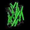
Lipid II flippase MurJ Peptidoglycan synthesis (PG) biosynthesis involves the formation of peptidoglycan precursor lipid II (undecaprenyl-pyrophosphate-linked N-acetyl glucosamine-N-acetyl muramic acid-pentapeptide) on the cytosolic face of the cell membrane. Lipid II is then translocated across the membrane and its glycopeptide moiety becomes incorporated into the growing cell wall mesh. MviN, renamed as MurJ, is a lipid II flippase essential for cell wall peptidoglycan synthesis. MurJ belongs to the MVF (mouse virulence factor) family of MOP superfamily transporters, which also includes the MATE (multidrug and toxic compound extrusion) transporter and eukaryotic OLF (oligosaccharidyl-lipid flippase) families. In addition to the canonical MOP transporter core consisting of 12 transmembrane helices (TMs), MurJ has two additional C-terminal TMs (13 and 14) of unknown function. Structural analysis indicates that the N lobe (TMs 1-6) and C lobe (TMs 7-14) are arranged in an inward-facing N-shape conformation, rather than the outward-facing V-shape conformation observed in all existing MATE transporter structures. Furthermore, a hydrophobic groove is formed by two C-terminal transmembrane helices, which leads into a large central cavity that is mostly cationic. Mutagenesis studies, revealed a solvent-exposed cavity that is essential for function. Mutation of conserved residues (Ser17, Arg18, Arg24, Arg52, and Arg255) at the proximal site failed to complement MurJ function, consistent with the idea that these residues are important for recognizing the diphosphate and/or sugar moieties of lipid II. It has also been suggested that the chloride ion in the central cavity and a zinc ion at the beginning of TM 7 might be functionally important. |
