Table
In vitro Rodents
Background
[PubMed]
Optical fluorescence imaging is increasingly used to study the biological functions of specific targets in vitro and in small animals (1, 2). However, the intrinsic fluorescence of biomolecules poses a problem when visible light-absorbing (350-700 nm) fluorophores are used because of high tissue absorption and scatter. Use of near-infrared (NIR) fluorescence (700-900 nm) for detection avoids the background fluorescence interference of natural biomolecules, providing a high contrast between target and background tissues. NIR fluorophores have wider dynamic ranges and minimal background as a result of reduced scattering compared with visible fluorescence detection. NIR fluorophores also have high sensitivity, resulting from low infrared background, and high extinction coefficients, which provide high quantum yields. The NIR region is also compatible with solid-state optical components such as diode lasers and silicon detectors. NIR fluorescence imaging is becoming a non-invasive alterative to radionuclide imaging in vitro and in small animals.
IRDye78 is a heptamethine indocyanine-type NIR fluorophore with peak absorption at 772 nm and peak emission at 790 nm (3). It provides a quantum yield of 9.0% and has a molecular mass of 1,083 Da. IRDye78 N-hydroxysuccinimide (NHS) ester can be conjugated to antibodies and low-molecular-weight ligands with one or more free primary amines.
Prostate-specific membrane antigen (PSMA) is a unique type II, transmembrane-bound glycoprotein that is overexpressed on prostate tumor cells and in the neovasculature of most solid prostate tumors but not in the vasculature of normal tissues (4, 5). This unique expression of PSMA makes it an important biomarker as well as a large extracellular target of imaging agents. PSMA was found to have N-acetyl α-linked acidic dipeptidase (NAALADase, or glutamate carboxypeptidase II) activity (6). PGI is a small molecule that is a potent inhibitor of NAALADase, with a Ki of 9.0 nM, whereas IRDye78 had a Ki of 500 nM (7). On the other hand, GPI-78 had a Ki of 0.4 nM. GPI-78 is being developed as an optical agent for imaging prostate cancer.
Synthesis
[PubMed]
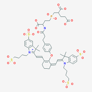
Humblet et al. (7) reported the synthesis of GPI-78 by reaction of IRDye78 monofunctional NHS ester with the single primary amino group of GPI. GPI-78 was purified by reverse-phase high-performance liquid chromatography, and its identity was confirmed by mass spectroscopy with >95% purity. GPI-78 had peak absorption at 773 nm and peak emission at 792 nm. The conjugation yields were 60%.
In Vitro Studies: Testing in Cells and Tissues
[PubMed]
Humblet et al. (7) determined that the dissociation constant (Kd) for GPI-78 binding to the active site of PSMA, as measured by fluorescence polarization, was 0.84 ± 0.15 nM. This value is very similar to the Ki (0.43 ± 0.04 nM) determined in enzymatic assays, suggesting that GPI-78 is binding to the active site.
GPI-78 bound to PSMA-negative PC-3 human prostate cancer cells overexpressing PSMA, but not to Erb-B2-overexpressing PC-3 cells (7). GPI-78 also bound to PSMA-positive LNCaP human cancer cells. There was 50% more fluorescent signal when the cells were incubated at 37 °C versus 4 °C because PSMA was being internalized at 37 °C. Experiments in which LNCaP cells were incubated with 2 μM GPI-78 at 4 °C for 20 min indicated that there were about 3.2 × 10⁵ receptor sites per cell.
Animal Studies
Rodents
[PubMed]
Nude mice bearing PSMA-positive LNCaP (NAALADase activity = 10 pmol/min/mg) cells on the right flank and PSMA-negative human bladder cancer TsuPRI cells (NAALADase activity = 0 pmol/min/mg) on the left flank received injections containing 5 nmol of GPI-78 or IRDye78 (7). Both IRDye78 and GPI-78 (injected intravenously) were cleared rapidly from the blood with similar half-lives: the early phase was 1.6 and 1.5 min and the late phase was 3.4 and 2.3 min, respectively. There was a rapid increase in signal intensities in the skin and kidney, reaching a plateau by 5 min with a gradual washout over a few hours. The peak GPI-78 tumor/background ratios were 1.80 ± 0.21 and 1.20 ± 0.05 (P = 0.024) for PSMA-positive and PSMA-negative tumors, respectively. The peak IRDye78 tumor/background ratios were 1.20 ± 0.11 and 1.30 ± 0.21 (P = 0.7) for PSMA-positive and PSMA-negative tumors, respectively. In vivo imaging of the tumors, performed with a small-animal NIR fluorescence imaging system, revealed that only PSMA-positive tumors exhibited any signal above background, with a brighter signal from GPI-78 than IRDye78.
NIH Support
CA88245
References
- 1.
- Achilefu S. Lighting up tumors with receptor-specific optical molecular probes. Technol Cancer Res Treat. 2004; 3 (4):393–409. [PubMed: 15270591]
- 2.
- Ntziachristos V. , Bremer C. , Weissleder R. Fluorescence imaging with near-infrared light: new technological advances that enable in vivo molecular imaging. Eur Radiol. 2003; 13 (1):195–208. [PubMed: 12541130]
- 3.
- Nakayama A. , del Monte F. , Hajjar R.J. , Frangioni J.V. Functional near-infrared fluorescence imaging for cardiac surgery and targeted gene therapy. Mol Imaging. 2002; 1 (4):365–77. [PubMed: 12940233]
- 4.
- Feneley M.R. , Jan H. , Granowska M. , Mather S.J. , Ellison D. , Glass J. , Coptcoat M. , Kirby R.S. , Ogden C. , Oliver R.T. , Badenoch D.F. , Chinegwundoh F.I. , Nargund V.H. , Paris A.M. , Britton K.E. Imaging with prostate-specific membrane antigen (PSMA) in prostate cancer. Prostate Cancer Prostatic Dis. 2000; 3 (1):47–52. [PubMed: 12497162]
- 5.
- Ghosh A. , Heston W.D. Tumor target prostate specific membrane antigen (PSMA) and its regulation in prostate cancer. J Cell Biochem. 2004; 91 (3):528–39. [PubMed: 14755683]
- 6.
- Luthi-Carter R. , Barczak A.K. , Speno H. , Coyle J.T. Molecular characterization of human brain N-acetylated alpha-linked acidic dipeptidase (NAALADase). J Pharmacol Exp Ther. 1998; 286 (2):1020–5. [PubMed: 9694964]
- 7.
- Humblet V. , Lapidus R. , Williams L.R. , Tsukamoto T. , Rojas C. , Majer P. , Hin B. , Ohnishi S. , De Grand A.M. , Zaheer A. , Renze J.T. , Nakayama A. , Slusher B.S. , Frangioni J.V. High-affinity near-infrared fluorescent small-molecule contrast agents for in vivo imaging of prostate-specific membrane antigen. Mol Imaging. 2005; 4 (4):448–62. [PubMed: 16285907]
Publication Details
Author Information and Affiliations
Publication History
Created: May 5, 2006; Last Update: August 12, 2008.
Copyright
Publisher
National Center for Biotechnology Information (US), Bethesda (MD)
NLM Citation
Leung K. GPI-78. 2006 May 5 [Updated 2008 Aug 12]. In: Molecular Imaging and Contrast Agent Database (MICAD) [Internet]. Bethesda (MD): National Center for Biotechnology Information (US); 2004-2013.
 In vitro