U.S. flag

An official website of the United States government

NCBI Bookshelf. A service of the National Library of Medicine, National Institutes of Health.

Adam MP, Feldman J, Mirzaa GM, et al., editors. GeneReviews® [Internet]. Seattle (WA): University of Washington, Seattle; 1993-2024.

Cover of GeneReviews®

GeneReviews® [Internet].

Show details

von Willebrand Disease

Synonym: von Willebrand Factor Deficiency

, PhD and , MD, FRCPC.

Author Information and Affiliations

Initial Posting: ; Last Update: October 5, 2017.

Estimated reading time: 44 minutes

Summary

Clinical characteristics.

Von Willebrand disease (VWD), a congenital bleeding disorder caused by deficient or defective plasma von Willebrand factor (VWF), may only become apparent on hemostatic challenge, and bleeding history may become more apparent with increasing age.

Recent guidelines on VWD have recommended taking a VWF level of 30 or 40 IU/dL as a cutoff for those diagnosed with the disorder. Individuals with VWF levels greater than 30 IU/dL and lower than 50 IU/dL can be described as having a risk factor for bleeding. This change in guidelines significantly alters the proportion of individuals with each disease type.

Type 1 VWD (~30% of VWD) typically manifests as mild mucocutaneous bleeding.

Type 2 VWD accounts for approximately 60% of VWD. Type 2 subtypes include:

  • Type 2A, which usually manifests as mild-to-moderate mucocutaneous bleeding;
  • Type 2B, which typically manifests as mild-to-moderate mucocutaneous bleeding that can include thrombocytopenia that worsens in certain circumstances;
  • Type 2M, which typically manifests as mild-moderate mucocutaneous bleeding;
  • Type 2N, which can manifest as excessive bleeding with surgery and mimics mild hemophilia A.

Type 3 VWD (<10% of VWD) manifests with severe mucocutaneous and musculoskeletal bleeding.

Diagnosis.

The diagnosis of VWD typically requires characteristic results of assays of hemostasis factors specific for VWD and/or identification of a heterozygous, homozygous, or compound heterozygous pathogenic variant(s) in VWF by molecular genetic testing. In addition, the diagnosis requires (in most cases) a positive family history. In those with a risk factor for bleeding (VWF levels >30 and <50 IU/dL), family history may not be positive because of incomplete penetrance and variable expressivity.

Management.

Treatment of manifestations: Affected individuals benefit from care in a comprehensive bleeding disorders program. The two main treatments are desmopressin (1-deamino-8-D-arginine vasopressin [DDAVP]) and clotting factor concentrates (recombinant and plasma-derived) containing both VWF and FVIII (VWF/FVIII concentrate). Indirect hemostatic treatments that can reduce symptoms include fibrinolytic inhibitors; hormones for menorrhagia are also beneficial. Individuals with VWD should receive prompt treatment for severe bleeding episodes. Pregnant women with VWD are at increased risk for bleeding complications at or following childbirth.

Prevention of primary manifestations: Prophylactic infusions of VWF/FVIII concentrates in individuals with type 3 VWD to prevent musculoskeletal bleeding and subsequent joint damage.

Prevention of secondary complications: Cautious use of desmopressin (particularly in those age <2 years because of the potential difficulty in restricting fluids in this age group). Vaccination for hepatitis A and B.

Surveillance: Follow up in centers experienced in the management of bleeding disorders. Periodic evaluation by a physiotherapist of those with type 3 VWD to monitor joint mobility.

Agents/circumstances to avoid: Activities involving a high risk of trauma, particularly head injury; medications with effects on platelet function (ASA, clopidogrel, or NSAIDS). Circumcision in infant males should only be considered following consultation with a hematologist.

Evaluation of relatives at risk: If the familial pathogenic variant(s) are known, molecular genetic testing for at-risk relatives to allow early diagnosis and treatment, if needed.

Pregnancy management: As VWF levels increase throughout pregnancy, women with baseline VWF and FVIII levels greater than 30 IU/dL are likely to achieve normal levels by the time of delivery. However, those with a basal level lower than 20 IU/dL and those with baseline VWF:RCo or other VWF activity measurement/VWF:Ag ratio <0.6 are likely to require replacement therapy. Desmopressin has been successfully used to cover delivery in women with type 1 VWD and a proportion of pregnant women with type 2 VWD; delayed, secondary postpartum bleeding may be a problem.

Genetic counseling.

VWD types 2B and 2M are inherited in an autosomal dominant manner. VWD types 1 and 2A are typically inherited in an autosomal dominant manner but may also be inherited in an autosomal recessive manner. VWD types 2N and 3 are inherited in an autosomal recessive manner.

  • AD inheritance. Most affected individuals have an affected parent. The proportion of cases caused by de novo pathogenic variants is unknown. Each child of an individual with AD VWD has a 50% chance of inheriting the pathogenic variant.
  • AR inheritance. At conception, each sib of an individual with AR VWD has a 25% chance of being affected, a 50% chance of being an asymptomatic carrier, and a 25% chance of being unaffected and not a carrier. Carrier testing for family members at risk for AR VWD is possible once the pathogenic variants have been identified in the family.

Prenatal and preimplantation genetic testing are possible if the pathogenic variant(s) in the family are known.

GeneReview Scope

von Willebrand Disease: Included Phenotypes
  • Type 1 von Willebrand disease
  • Type 2A von Willebrand disease
  • Type 2B von Willebrand disease
  • Type 2M von Willebrand disease
  • Type 2N von Willebrand disease
  • Type 3 von Willebrand disease

For synonyms and outdated names see Nomenclature.

Diagnosis

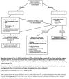
Several guidelines and testing algorithms have been published [Keeney et al 2008, Nichols et al 2008, Lassila et al 2011, Laffan et al 2014]. See Figures 1 and 2.

Figure 1.

Figure 1.

Initial testing algorithm for von Willebrand disease From Nichols et al [2008]. Reprinted with permission of John Wiley and Sons.

Figure 2.

Figure 2.

Algorithm for additional testing for von Willebrand disease subtype From Laffan et al [2014]. Reprinted with permission of John Wiley and Sons.

Von Willebrand disease (VWD) is caused by deficient or defective plasma von Willebrand factor (VWF), a large multimeric glycoprotein that plays a pivotal role in primary hemostasis by mediating platelet hemostatic function and stabilizing blood coagulation factor VIII (FVIII).

There are three types of VWD [Sadler et al 2006]:

  • Type 1. Partial quantitative deficiency of essentially normal VWF
  • Type 2. Qualitative deficiency of defective VWF; divided into four subtypes depending on VWF function perturbed: 2A, 2B, 2M, 2N
  • Type 3. Complete quantitative deficiency of (virtually absent) VWF

Suggestive Findings

Von Willebrand disease (VWD) should be suspected in individuals with excessive mucocutaneous bleeding including the following:

  • Bruising without recognized trauma
  • Prolonged, recurrent nosebleeds
  • Bleeding from the gums after brushing or flossing teeth or prolonged bleeding following dental cleaning or dental extractions
  • Menorrhagia, particularly if occurring since menarche
  • Prolonged bleeding following surgery, trauma, or childbirth
  • Gastrointestinal bleeding

The utility of standard clinical assessment tools to score occurrence of symptoms and their severity as part of VWD diagnosis is increasingly recognized [Tosetto et al 2006, Rodeghiero et al 2010, Elbatarny et al 2014, Mittal et al 2015]. These tools can: determine if there is more bleeding than in the general population; justify the diagnosis of a bleeding disorder; quantify the extent of symptoms; indicate situations requiring clinical intervention; and be used to indicate that a bleeding disorder is unlikely [Tosetto et al 2011]. Additionally, bleeding severity assessment correlates with the long-term probability of bleeding [Tosetto 2016].

Establishing the Diagnosis

The diagnosis of VWD is established in a proband with excessive mucocutaneous bleeding and characteristic results of assays of hemostasis factors specific for VWD (see Clinical Laboratory Testing and Table 1) and/or a heterozygous, homozygous, or compound heterozygous pathogenic (or likely pathogenic) variant(s) in VWF identified by molecular genetic testing (see Table 2).

Note: (1) Per ACMG/AMP variant interpretation guidelines, the terms "pathogenic variants" and "likely pathogenic variants" are synonymous in a clinical setting, meaning that both are considered diagnostic and both can be used for clinical decision making [Richards et al 2015]. Reference to "pathogenic variants" in this section is understood to include any likely pathogenic variants. (2) Identification of a variant(s) of uncertain significance cannot be used to confirm or rule out the diagnosis.

In addition, the diagnosis requires (in most cases) a positive family history. Note: In those with a risk factor for bleeding (VWF levels >30 and <50 IU/dL), family history may not be positive because of incomplete penetrance and variable expressivity.

Clinical Laboratory Testing

Screening tests

  • Complete blood count (CBC) may be normal, but could also show a microcytic anemia (if the individual is iron deficient) or a low platelet count (thrombocytopenia), specifically in type 2B VWD.
  • Activated partial thromboplastin time (aPTT) is often normal, but may be prolonged when the factor VIII (FVIII:C) level is reduced to below 30-40 IU/dL, as can be seen in severe type 1 VWD, type 2N VWD, or type 3 VWD. The normal range for FVIII:C clotting activity is approximately 50-150 IU/dL.
  • Prothrombin time is normal in VWD.
  • Other. Although some laboratories may also include a skin bleeding time and platelet function analysis (PFA closure time) in their evaluation of an individual with suspected VWD, these tests lack sensitivity in persons with mild bleeding disorders.

Hemostasis factor assays. The following specific hemostasis factor assays (see Table 1) should be performed even if the screening tests are normal in an individual in whom VWD is suspected [Budde et al 2006]. Note: Normal ranges are determined by the individual laboratory and thus are indicative only.

The International Society on Thrombosis and Haemostasis has recently published new guidance on assays that measure von Willebrand factor activity (VWF:Act) [Bodó et al 2015]. These tests include:

  • VWF:RCo. Ristocetin cofactor activity: all assays that use platelets and ristocetin. Ability of VWF to agglutinate platelets, initiated by the antibiotic ristocetin (normal range ~50-200 IU/dL)
  • VWF:GPIbR. All assays that are based on the ristocetin-induced binding of VWF to a recombinant WT GPIb fragment
  • VWF:GPIbM. All assays that are based on the spontaneous binding of VWF to a gain-of-function variant GPIb fragment
  • VWF:Ab. All assays that are based on the binding of a monoclonal antibody (mAb) to a VWF A1 domain epitope
  • VWF:Ag. Quantity of VWF protein (antigen) in the plasma, measured antigenically using enzyme-linked immunosorbant assay (ELISA) or by latex immunoassay (LIA) [Castaman et al 2010a] (normal range ~50-200 IU/dL). A reduced ratio (<0.6) of VWF:Act to VWF:Ag can indicate loss of high-molecular-weight (HMW) multimers.
  • Factor VIII:C level. Functional FVIII assay (i.e., activity of FVIII in the coagulation cascade) (normal range ~50-150 IU/dL)

If abnormalities in the tests above are identified, specialized coagulation laboratories may also perform the following assays to determine the subtype of VWD:

  • VWF multimer analysis. SDS-agarose electrophoresis used to determine the complement of VWF oligomers in the plasma. Normal plasma contains VWF ranging from dimers to multimers comprising more than 40 dimers and molecular weight into gigadaltons. Multimers are classified as low (1-5-dimer), intermediate (6-10-dimer), and high (≥10-dimer) molecular weight. HMW multimers are decreased or missing in type 2A VWD and often in 2B VWD; intermediate MW may also be lost in type 2A VWD. Abnormalities in satellite ("triplet") band patterns can give clues as to pathogenesis and help to classify subtypes of type 2 VWD [Budde et al 2008].
  • Ristocetin-induced platelet agglutination (RIPA). Ability of VWF to agglutinate platelets at two to three concentrations of ristocetin. Agglutination at a low concentration (~0.5-0.7 mg/mL) is abnormal and may indicate type 2B or platelet-type pseudo VWD (PT-VWD) caused by pathogenic variants in GP1BA (see Differential Diagnosis), in which enhanced VWF-platelet binding is present.
  • Binding of FVIII by VWF (VWF:FVIIIB). Ability of VWF to bind FVIII. Useful, but not widely used to identify type 2N VWD.
  • Collagen binding assay (VWF:CB). Ability of VWF to bind to collagen (a sub-endothelial matrix component). Used to help define functional VWF discordance (i.e., to help distinguish types 1 and 2 VWD) [Flood et al 2013]. Collagen I/III mixture is often used, but isolated deficient binding to collagen types IV and VI has recently been recognized [Flood et al 2012]. Normal range is approximately 50-200 IU/dL. A reduced ratio of VWF:CB/VWF:Ag can indicate loss of HMW multimers.
  • VWF:GP1BA. Functional tests are used to determine how well VWF binds to GpIbα. Previously, this was assessed using the VWF:RCo assay. Currently, this analysis is undertaken as part of the newer activity assays.

Table 1.

Classification of VWD Based on Specific VWF Tests

VWD TypeVWF:Act 1VWF:Ag 1Act/AgFVIII:C IU/dL 1Multimer Pattern 2Other
1LowLowEquivalent~1.5x VWF:AgEssentially normal
2ALowLowVWF:Act < VWF:AgLow or normalAbnormal ↓ HMW↓ VWF:GP1BA binding
2BLowLowVWF:Act < VWF:AgLow or normalOften abnormal ↓ HMW↑ RIPA 3 (↓ platelet count)
2MLowLowVWF:Act << VWF:AgLow or normalNo loss of HMW↓ VWF:GP1BA binding
↓VWF:collagen binding 4
2NNormal/lowNormal/lowEquivalent<40Normal in most cases↓ VWF:FVIIIB 5
3AbsentAbsentNA<10Absent
1.

Relative to the reference range (approximate values); VWF:Act (50-200 IU/dL), VWF:Ag (50-200 IU/dL), FVIII:C (50-150 IU/dL). VWF activity (VWF:Act) includes VWF:RCo and the newer VWF activity assays in this instance.

2.

HMW multimers

3.

Increased agglutination at low concentrations of ristocetin

4.

Reduction in the ability of VWF to bind to collagen. Types I/III are bound by the A3 domain while types IV and VI are bound by the A1 domain. The latter types are not commonly analyzed.

5.

Ability of VWF to bind and protect FVIII is reduced. VWF and FVIII levels can look exactly like those in males with mild hemophilia A or in symptomatic hemophilia A carrier females.

Molecular Genetic Testing

Molecular genetic testing approaches can include single-gene testing, use of a multigene panel, and more comprehensive genomic testing:

  • Single-gene testing. Sequence analysis of VWF is performed first and followed by gene-targeted deletion/duplication analysis if no pathogenic variant is found.
  • A multigene panel that includes VWF and other genes of interest (see Differential Diagnosis) may be considered. Note: (1) The genes included in the panel and the diagnostic sensitivity of the testing used for each gene vary by laboratory and are likely to change over time. (2) Some multigene panels may include genes not associated with the condition discussed in this GeneReview; thus, clinicians need to determine which multigene panel is most likely to identify the genetic cause of the condition while limiting identification of variants of uncertain significance and pathogenic variants in genes that do not explain the underlying phenotype. (3) In some laboratories, panel options may include a custom laboratory-designed panel and/or custom phenotype-focused exome analysis that includes genes specified by the clinician. (4) Methods used in a panel may include sequence analysis, deletion/duplication analysis, and/or other non-sequencing-based tests.
    For an introduction to multigene panels click here. More detailed information for clinicians ordering genetic tests can be found here.
    Note: Analysis of exons 23 to 34 of VWF is complicated by the presence of a partial pseudogene, VWFP1.
  • More comprehensive genomic testing (when available) including exome sequencing and genome sequencing may be considered. Such testing may provide or suggest a diagnosis not previously considered (e.g., mutation of a different gene or genes that results in a similar clinical presentation).
    For an introduction to comprehensive genomic testing click here. More detailed information for clinicians ordering genomic testing can be found here.

Table 2.

Molecular Genetic Testing Used in von Willebrand Disease (VWD)

Gene 1VWD Type(s) 2Proportion of VWD Attributed to This TypeMethodProportion of Probands with a Pathogenic Variant 3 Detectable by Method
VWF 1~30%Sequence analysis 480% 5
Gene-targeted deletion/duplication analysis 66% 7
All type 2 forms~60%Sequence analysis 4~90% 7
Gene-targeted deletion/duplication analysis 60.2% 7
3<10% 8Sequence analysis 4~90% 7
Gene-targeted deletion/duplication analysis 63.7% 7
1.
2.

Recent changes in the von Willebrand factor (VFW) level used for diagnosis have significantly altered the proportion of patients classified with each disease type [Lassila et al 2011, Castaman et al 2013, Laffan et al 2014].

3.

See Molecular Genetics for information on allelic variants detected in this gene.

4.

Sequence analysis detects variants that are benign, likely benign, of uncertain significance, likely pathogenic, or pathogenic. Variants may include small intragenic deletions/insertions and missense, nonsense, and splice site variants; typically, exon or whole-gene deletions/duplications are not detected. For issues to consider in interpretation of sequence analysis results, click here.

5.
6.

Gene-targeted deletion/duplication analysis detects intragenic deletions or duplications. Methods used may include a range of techniques such as quantitative PCR, long-range PCR, multiplex ligation-dependent probe amplification (MLPA), and a gene-targeted microarray designed to detect single-exon deletions or duplications.

7.
8.

In populations with frequent consanguineous partnerships, the rate of recessive forms of VWD may be elevated and type 3 VWD comprises a larger proportion of affected individuals.

Clinical Characteristics

Clinical Description

Von Willebrand disease (VWD) is a congenital bleeding disorder; however, symptoms may only become apparent on hemostatic challenge and bleeding history may become more apparent with increasing age. Thus, it may take some time before a bleeding history becomes apparent.

Recent guidelines on VWD have recommended taking von Willebrand factor (VWF) levels of 30 or 40 IU/dL as a cutoff for those diagnosed with the disorder. Individuals with VWF levels greater than 30 IU/dL and lower than 50 IU/dL can be described as having a risk factor for bleeding. This change in guidelines significantly alters the proportion of individuals with each disease type [Lassila et al 2011, Castaman et al 2013, Laffan et al 2014].

Bleeding history also depends on disease severity; type 3 VWD is often apparent early in life, whereas mild type 1 VWD may not be diagnosed until midlife, despite a history of bleeding episodes.

Individuals with VWD primarily manifest excessive mucocutaneous bleeding (e.g., bruising, epistaxis, menorrhagia) and do not tend to experience musculoskeletal bleeding unless the FVIII:C level is lower than 10 IU/dL, as can be seen in type 2N or type 3 VWD.

Bleeding score. In general, there is an inverse relationship between the VWF level and the severity of bleeding [Tosetto et al 2006]. Bleeding scores (BS) have been documented in several cohort studies and give an indication of the range of bleeding severity associated with different VWD types:

Table 3.

Bleeding Scores (BS) Reported in VWD by Type

Patient GroupStudy# of PatientsBS MedianBS Range
Type 1 Goodeve et al [2007] 1509-1-24
Type 2A Castaman et al [2012] 46116-16
Type 2B Federici et al [2009] 4054-24
Type 2M Castaman et al [2012] 6174-28
Type 3 Solimando et al [2012] 9156-26
Type 3 Bowman et al [2013] 42133-30

The higher the bleeding score, the greater the bleeding severity

Note: While the studies reported have all used similar bleeding assessment tools, slight variations in the tools and their application may have contributed to differences in bleeding scores.

Recently established cutoffs for an abnormal BS (≥4 for adult males, ≥6 for adult females, ≥3 for children) can be utilized to objectively assess the affected status of individuals tested using the ISTH-bleeding assessment tool (BAT) in a standard fashion [Elbatarny et al 2014].

BS in adults has also been shown to be a predictor of future bleeding [Federici et al 2014].

Type 1 VWD accounts for approximately 30% of all VWD in populations with infrequent consanguineous partnerships [Batlle et al 2016, Veyradier et al 2016]. It typically manifests as mild mucocutaneous bleeding; however, symptoms can be more severe when VWF levels are lower than 15 IU/dL. Epistaxis and bruising are common symptoms among children. Menorrhagia is the most common finding in women of reproductive age [Ragni et al 2016].

Type 2 VWD accounts for approximately 60% of all VWD. The relative frequency of the subtypes is 2A>2M>2N>2B in European populations [Batlle et al 2016, Veyradier et al 2016].

  • Type 2A VWD. Individuals with type 2A VWD usually present with mild to moderate mucocutaneous bleeding [Veyradier et al 2016].
  • Type 2B VWD. Individuals typically present with mild-moderate mucocutaneous bleeding. Thrombocytopenia may be present. A hallmark of type 2B VWD is a worsening of thrombocytopenia during stressful situations, such as severe infection or during surgery or pregnancy, or if treated with desmopressin [Federici et al 2009].
  • Type 2M VWD. Individuals typically present with mild-moderate mucocutaneous bleeding symptoms, but bleeding episodes can be severe, particularly in the presence of very low or absent VWF:RCo [Castaman et al 2012, Larsen et al 2013].
  • Type 2N VWD. Symptoms are essentially the same as those seen in mild hemophilia A and include excessive bleeding at the time of surgery or procedures as both disorders result from reduced FVIII:C [van Meegeren et al 2015].

Type 3 VWD accounts for up to 10% of VWD (except in areas where consanguineous partnerships are common, where a higher proportion may be found). It manifests with severe bleeding including both excessive mucocutaneous bleeding and musculoskeletal bleeding [Metjian et al 2009, Ahmad et al 2013, Kasatkar et al 2014].

Associated complications

  • Gastrointestinal angiodysplasia occurs most commonly in middle-aged/elderly individuals with types 2A and 3 VWD and affects the colon, small intestine, and stomach [Franchini & Mannucci 2014]. Lack of VWF in Weibel-Palade bodies promotes angiogenesis in endothelial cells [Starke et al 2011]. The disorder has also been reported in types 1 and 2B VWD [Hertzberg et al 1999, Siragusa et al 2008].
  • Menorrhagia is experienced by a large proportion of women with VWD.
  • The development of alloantibodies against VWF is an uncommon but serious complication of VWD treatment. An estimated 5%-10% of individuals with type 3 VWD may experience this complication. Affected individuals present with reduced or absent response to infused VWF concentrates or, in rare cases, with anaphylactic reaction. Individuals who have had multiple transfusions are at highest risk for this complication.

Genotype-Phenotype Correlations

The three phenotypes reflect a partial (type 1 VWD) or complete (type 3 VWD) quantitative deficiency of VWF or qualitative deficits (type 2 VWD) of VWF. See Molecular Genetics, Pathogenic variants for details regarding the genotypes associated with each subtype of VWD.

Individuals with large deletions of VWF are at highest risk for alloantibody development, although some with other null alleles have also been reported to develop this complication [James et al 2013].

Penetrance

Type 1 VWD (AD)

  • VWF level. Pathogenic variants resulting in plasma VWF levels lower than 25 IU/dL are mostly fully penetrant. Those resulting in higher VWF levels are often incompletely penetrant.
  • ABO blood group appears to be an important contributor to penetrance and reduced VWF level in type 1 VWD [Goodeve et al 2007, James et al 2007a]. Blood group contributes approximately 25% of the variance in plasma VWF level; ABO glycosylation of VWF influences its rate of clearance [Jenkins & O'Donnell 2006]. Individuals with non-O blood groups have higher VWF levels than those with O blood group; those with group AB have the highest levels.

Other AD types (2A, 2B, and 2M). Pathogenic variants are often fully penetrant.

Nomenclature

Changes in nomenclature:

  • von Willebrand's disease has been replaced by von Willebrand disease.
  • vWF has been replaced by VWF.
  • vWD has been replaced by VWD.
  • RiCof (ristocetin cofactor activity) has been replaced by VWF:RCo [Mazurier & Rodeghiero 2001].
  • FVIII RAg (FVIII related antigen) has been replaced by VWF:Ag.
  • Platelet-type pseudo von Willebrand (PT-VWD), also called pseudo-VWD, is caused by pathogenic variants in GP1BA and, thus, is not a form of VWD (see Differential Diagnosis).
  • Acquired von Willebrand syndrome (AVWS), previously known as acquired VWD, is the preferred terminology for defects in VWF concentration, structure, or function that are neither inherited nor reflective of pathogenic variants in VWF, but arise as consequences of other medical conditions (see brief discussion of AVWS under Differential Diagnosis).

See also Mazurier & Rodeghiero [2001] and Bodó et al [2015].

Prevalence

VWD affects 0.1% to 1% of the population; 1:10,000 seek tertiary care referral.

VWD type 3 affects 0.5:1,000,000-6:1,000,000 population, increasing with the rate of consanguinity.

Differential Diagnosis

Two disorders can be difficult to distinguish phenotypically from von Willebrand disease (VWD):

  • Mild hemophilia A, caused by pathogenic variants in F8, resembles type 2N VWD in that reduced levels of FVIII:C (~5-40 IU/dL) and normal-to-borderline low levels of VWF can be seen in both disorders. In families with reduced FVIII:C, an X-linked pattern of inheritance can help identify those with mild hemophilia A.
    The VWF:FVIIIB test, which determines the ability of VWF to bind FVIII, can be used to discriminate between the two disorders [Casonato et al 2007] and a commercial assay is now available [Veyradier et al 2011], although on a limited basis. Alternatively, molecular genetic testing can be used to distinguish the two disorders. Both molecular and phenotypic testing have some fallibilities in interpretation.
  • PT-VWD (pseudo VWD) (OMIM 177820), caused by pathogenic variants in GP1BA, may be difficult to distinguish from type 2B VWD; one study identified pathogenic variants in GP1BA in up to 15% of persons diagnosed with 2B VWD [Hamilton et al 2011]. The two disorders can be distinguished by mixing patient/control plasma and platelets to determine which component is defective [Othman et al 2016] or by molecular testing.
    PT-VWD has been shown to be less severe than type 2B VWD using a bleeding assessment tool [Kaur et al 2014].
    The two disorders require different treatment. In PT-VWD, VWF concentrate is needed to correct the reduced VWF level, but platelet transfusion may also be required if there is significant thrombocytopenia. The half-life of replaced VWF is reduced as a result of binding to the abnormal GpIbα, necessitating more frequent administration of VWF concentrate than in VWD.

Acquired von Willebrand syndrome (AVWS) is a mild-moderate bleeding disorder that can occur in a variety of conditions [Sucker et al 2009, Federici et al 2013, Mital 2016] but is not caused by pathogenic variants in VWF. It is most often seen in persons older than age 40 years with no prior bleeding history. AVWS has diverse pathology and a number of possible causes:

  • Lymphoproliferative or plasma cell proliferative disorders, paraproteinemias (monoclonal gammopathy of unknown significance [MGUS]), multiple myeloma, and Waldenstrom macroglobulinemia. Antibodies against VWF have been detected in some of these cases.
  • Autoimmune disorders including systemic lupus erythrematosus (SLE), scleroderma, and antiphospholipid antibody syndrome
  • Shear-induced VWF conformational changes leading to increased VWF proteolysis (e.g., aortic valve stenosis, ventricular septal defect)
  • Markedly increased blood platelet count (e.g., essential thrombocythemia or other myeloproliferative disorders)
  • Removal of VWF from circulation by aberrant binding to tumor cells (e.g., Wilm's tumor or certain lymphoproliferative disorders)
  • Decreased VWF synthesis (e.g., hypothyroidism)
  • Certain drugs (e.g., valproic acid, ciprofloxacin, griseofulvin, hydroxyethyl starch)

Management

Evaluations Following Initial Diagnosis

To establish the extent of disease and needs in an individual diagnosed with von Willebrand disease (VWD), the following evaluations are recommended:

  • A personal and family history of bleeding to help predict severity and tailor treatment. Use of a bleeding assessment tool can facilitate standardized assessment [Kaur et al 2016, Tosetto 2016, ISTH-BAT].
  • A joint and muscle evaluation for those with type 3 VWD (Musculoskeletal bleeding is rare in types 1 and 2 VWD.)
  • Screening for hepatitis B and C as well as HIV if the diagnosis is type 3 VWD or if the individual received blood products or plasma-derived clotting factor concentrates before 1985
  • Baseline serum concentration of ferritin to assess iron stores, as many individuals with VWD (particularly women with menorrhagia) are iron deficient
  • Gynecologic evaluation for women with menorrhagia [Rodeghiero 2008]
  • Consultation with a clinical geneticist and/or genetic counselor

Treatment of Manifestations

See Nichols et al [2008] (full text), Castaman et al [2013] (full text), and Laffan et al [2014] (full text) for treatment guidelines.

Individuals with VWD benefit from referral to a comprehensive bleeding disorders program for education, treatment, and genetic counseling.

Medical Treatments

The two main treatments are desmopressin (1-deamino-8-D-arginine vasopressin [DDAVP]) and clotting factor concentrates (recombinant and plasma-derived) containing both VWF and FVIII (VWF/FVIII concentrate). Indirect hemostatic treatments are also beneficial. Individuals with VWD should receive prompt treatment for severe bleeding episodes.

Desmopressin

  • Most individuals with type 1 VWD and some with type 2 VWD respond to intravenous or subcutaneous treatment with desmopressin [Castaman et al 2008, Federici 2008, Leissinger et al 2014], which promotes release of stored VWF and raises levels three- to fourfold. Intranasal preparations are also available.
  • Following VWD diagnosis, a desmopressin challenge is advisable to assess VWF response.
  • Desmopressin is the treatment of choice for acute bleeding episodes or to cover surgery.
  • It has been used successfully to cover delivery in women with type 1 VWD and also for a proportion of pregnant women with type 2 VWD (where a desmopressin trial has previously proved efficacious) [Castaman et al 2010b] (see Pregnancy Management).
  • Desmopressin is contraindicated in individuals with arteriovascular disease and in those older than age 70 years, for whom VWF/FVIII concentrate is required.
  • In persons who do not tolerate desmopressin or who have a poor VWF response, clotting factor concentrate is required.

Note: Because desmopressin can cause hyponatremia (which can lead to seizures and coma), fluid intake should be restricted for 24 hours following its administration to minimize this risk.

Intravenous infusion of VWF/FVIII clotting factor concentrates

  • In those who are non-responsive to desmopressin (i.e., VWF deficiency is not sufficiently corrected) and for those in whom desmopressin is contraindicated (see Treatment by VWD Type), bleeding episodes can be prevented or controlled with intravenous infusion of recombinant VWF or virally inactivated plasma-derived clotting factor concentrates containing both VWF and FVIII [Castaman & Linari 2016, Windyga et al 2016a, Windyga et al 2016b].
  • Plasma-derived concentrates are prepared from pooled blood donations from many donors. Virus inactivation procedures eliminate potential pathogens.
  • Recombinant VWF Vonvendi® was licensed for use in adults (18+ years) by the US FDA in December 2015 [Franchini & Mannucci 2016].

Indirect treatments. In addition to treatments that directly increase VWF levels, individuals with VWD often benefit from indirect hemostatic treatments, including:

  • Fibrinolytic inhibitors (i.e., tranexamic acid for treatment or prevention of bleeding episodes);
  • Hormonal treatments (i.e., the combined oral contraceptive pill for the treatment of menorrhagia).

Combined treatments for menorrhagia. Current treatments and a proposed future treatment trial are described by Ragni et al [2016]. 1,321 women with VWD were assessed between 2011 and 2014, and of these 816 (61.8%) had menorrhagia.

  • Treatments used most commonly were combined oral contraceptives, tranexamic acid, and desmopressin as first- and second-line therapies, whereas VWF concentrate was the most common third-line therapy used by 13 women (1.6%).
  • Review of information on 88 women from six published studies showed that a VWF dose of 33-100 IU kg-1 reduced menorrhagia on days one to six of the menstrual cycle in 101 women.

Treatment by VWD Type

Type 1 VWD. Treatments that directly increase VWF levels (e.g., desmopressin or VWF/FVIII clotting factor concentrates) are usually only needed for the treatment or prevention of severe bleeding, as with major trauma or surgery.

Indirect treatment with fibrinolytic inhibitors or hormones is often effective.

Type 2A VWD. Treatment with clotting factor concentrates is usually only required for the treatment or prevention of severe bleeding episodes such as during surgery.

Responsiveness to desmopressin is variable and should be confirmed prior to therapeutic use.

Indirect treatments can be beneficial.

Type 2B VWD. Clotting factor concentrates are usually required to treat severe bleeding or at the time of surgery.

Treatment with desmopressin should be undertaken cautiously as it can precipitate a worsening of any thrombocytopenia. Individuals with mild or atypical type 2B VWD (caused by p.Pro1266Leu, p.Pro1266Glu, and p.Arg1308Leu variants), however, do not appear to develop thrombocytopenia when exposed to desmopressin [Federici et al 2009].

Indirect treatments (i.e., fibrinolytic inhibitors) can be useful.

Type 2M VWD. Because desmopressin response is generally poor, VWF/FVIII concentrate is the treatment of choice.

Type 2N VWD. Desmopressin can be used for minor bleeding, but because the FVIII level will drop rapidly (as FVIII is not protected by VWF), concentrate containing VWF as well as FVIII is required to cover surgical procedures.

Type 3 VWD. Treatment often requires the repeated infusion of VWF/FVIII clotting factor concentrates [Franchini & Mannucci 2016, Lavin & O'Donnell 2016, Lissitchkov et al 2017].

Desmopressin is not effective in type 3 VWD.

Indirect treatments may also be beneficial.

Pediatric Issues

Special considerations for the care of infants and children with VWD include the following:

  • Infant males should be circumcised only after consultation with a pediatric hemostasis specialist.
  • Desmopressin should be used with caution, particularly in those younger than age two years, because of the potential difficulty in restricting fluids in this age group.
  • VWF levels are higher in the neonatal period [Klarmann et al 2010]; thus, phenotypic testing for milder forms of VWD should be delayed until later in childhood.

Prevention of Primary Manifestations

Individuals with type 3 VWD are often given prophylactic infusions of VWF/FVIII concentrates to prevent musculoskeletal bleeding and subsequent joint damage.

Prevention of Secondary Complications

Desmopressin should be used with caution, particularly in those younger than age two years, because of the potential difficulty in restricting fluids in this age group.

Individuals with VWD should be vaccinated for hepatitis A and B [Nichols et al 2008, Castaman et al 2013].

Prevention of chronic joint disease is a concern for individuals with type 3 VWD. However, controversy exists regarding the specific schedule and dosing of prophylactic regimens. An international trial that investigated prophylactic treatment for symptoms including joint bleeding, nosebleeds, and menorrhagia concluded that rates of bleeding within individuals during prophylaxis were significantly lower than levels prior to prophylaxis [Berntorp et al 2010, Abshire et al 2013]. Additionally, rates of bleeding were also significantly reduced in a trial of the recombinant VWF product Vonvendi®, which was licensed for use in adults in 2015 [Franchini & Mannucci 2016].

Surveillance

Individuals with milder forms of VWD can benefit from being followed by treatment centers with experience in the management of bleeding disorders.

Individuals with type 3 VWD should be followed in experienced centers and should have periodic evaluations by a physiotherapist to monitor joint mobility.

Agents/Circumstances to Avoid

Activities with a high risk of trauma, particularly head injury, should be avoided.

Medications that affect platelet function (ASA, clopidogrel, or NSAIDs) should be avoided as they can worsen bleeding symptoms.

Infant males should be circumcised only after consultation with a pediatric hemostasis specialist.

Evaluation of Relatives at Risk

It is appropriate to evaluate apparently asymptomatic at-risk relatives of an affected individual to allow early diagnosis and treatment as needed [Goodeve 2016].

Evaluations can include:

See Genetic Counseling for issues related to testing of at-risk relatives for genetic counseling purposes.

Pregnancy Management

VWF levels increase throughout pregnancy with the peak occurring four hours after delivery [James et al 2015]. Nonetheless, pregnant women with VWD are at increased risk for bleeding complications and care should be provided in centers with experience in perinatal management of bleeding disorders [Pacheco et al 2010, Castaman 2013, Biguzzi et la 2015, Reynen & James 2016, Roth & Syed 2016].

Women with baseline VWF and FVIII levels higher than 30 IU/dL are likely to achieve normal levels by the time of delivery, whereas those with a basal level lower than 20 IU/dL and those with baseline VWF:Act/VWF:Ag ratio <0.6 are likely to require replacement therapy [Castaman et al 2010b, James et al 2015, Hawke et al 2016].

Although deliveries should occur based on obstetric indications, instrumentation should be minimized [Demers et al 2005].

Delayed, secondary postpartum bleeding may be a problem. VWF level rapidly returns to pre-pregnancy level following delivery [Castaman et al 2013].

Therapies Under Investigation

Search ClinicalTrials.gov in the US and EU Clinical Trials Register in Europe for access to information on clinical studies for a wide range of diseases and conditions. Note: There may not be clinical trials for this disorder.

Genetic Counseling

Genetic counseling is the process of providing individuals and families with information on the nature, mode(s) of inheritance, and implications of genetic disorders to help them make informed medical and personal decisions. The following section deals with genetic risk assessment and the use of family history and genetic testing to clarify genetic status for family members; it is not meant to address all personal, cultural, or ethical issues that may arise or to substitute for consultation with a genetics professional. —ED.

Mode of Inheritance

Von Willebrand disease (VWD) types 2B and 2M are inherited in an autosomal dominant manner.

VWD types 1 and 2A are typically inherited in an autosomal dominant manner but may also be inherited in an autosomal recessive manner.

VWD types 2N and 3 are inherited in an autosomal recessive manner.

A single case of uniparental disomy has been reported in type 3 VWD. The two copies of the pathogenic variant resulted from maternal uniparental isodisomy [Boisseau et al 2011].

Risk to Family Members – Autosomal Dominant Inheritance

Parents of a proband

  • Most individuals diagnosed with one of the autosomal dominant types of VWD have an affected parent.
  • A proband with autosomal dominant VWD may have the disorder as the result of a de novo VWF pathogenic variant. The proportion of cases caused by a de novo VWF variant is unknown.
  • If the VWF pathogenic variant causing autosomal dominant VWD found in the proband cannot be detected in the leukocyte DNA of either parent, two possible explanations are germline mosaicism in a parent or de novo pathogenic variant in the proband. Neither possibility has been sufficiently investigated to comment on relative likelihood of occurrence. Unpublished data on type 1 VWD indicated that approximately 4% of individuals had a de novo pathogenic variant [Goodeve et al, unpublished data].
  • The family history of some individuals diagnosed with AD VWD may appear to be negative because of failure to recognize the disorder in family members, early death of the parent before the recognition of symptoms, or delayed onset of significant hemostatic challenges in the affected parent. Therefore, an apparently negative family history cannot be confirmed unless appropriate evaluations (e.g., VWD hemostasis factor assays and/or molecular genetic testing if the proband's pathogenic variant is known) have been performed on the parents of the proband.

Sibs of a proband. The risk to the sibs of the proband depends on the genetic status of the proband's parents:

  • If a parent of the proband is affected, the risk to the sibs is 50%.
  • The sibs of a proband with clinically unaffected parents are still at increased risk for the disorder because of the possibility of reduced penetrance in a parent.
  • If the VWF variant found in the proband cannot be detected in the leukocyte DNA of either parent, the empiric recurrence risk to sibs is approximately 1% because of the theoretic possibility of parental germline mosaicism.

Offspring of a proband. Each child of an individual with autosomal dominant VWD has a 50% chance of inheriting the VWF pathogenic variant.

Other family members. The risk to other family members depends on the status of the proband's parents: if a parent is affected, his or her family members may be at risk.

Risk to Family Members – Autosomal Recessive Inheritance

Parents of a proband

  • The parents of an individual with autosomal recessive VWD are usually heterozygotes (i.e., carriers of one VWF pathogenic variant).
  • Heterozygotes (carriers) of type 3 VWD are often asymptomatic. However, between 15% and 50% may show some mild bleeding symptoms and may be diagnosed with type 1 VWD [Nichols et al 2008, Bowman et al 2013].
  • Heterozygotes (carriers) of type 2N VWD are often asymptomatic. However, a small proportion may show some mild bleeding symptoms and may be diagnosed with type 1 VWD.

Sibs of a proband

  • When both parents are heterozygous for a VWF pathogenic variant, each sib of an individual with autosomal recessive VWD has at conception a 25% chance of being affected, a 50% chance of being heterozygous, and a 25% chance of being unaffected and not a carrier.
    • Heterozygotes (carriers) of type 3 VWD are often asymptomatic. However, 15%-50% may show some mild bleeding symptoms and may be diagnosed with type 1 VWD [Nichols et al 2008, Bowman et al 2013].
    • Heterozygotes (carriers) of type 2N VWD are often asymptomatic. However, a small proportion may show some mild bleeding symptoms and may be diagnosed with type 1 VWD.
  • Note: When only one parent is a carrier (in the case of uniparental isodisomy [Boisseau et al 2011]), the risk to sibs of a proband to be affected is unknown, but likely very low (<1%). However, each sib has a 50% chance of being heterozygous and a 50% chance of being unaffected and not a carrier.

Offspring of a proband. The offspring of an individual with autosomal recessive VWD are obligate heterozygotes (carriers) for a pathogenic variant in VWF.

Other family members. Each sib of the proband's parents is at 50% risk of being a carrier of a VWF pathogenic variant.

Carrier (Heterozygote) Detection

Carrier testing for at-risk relatives requires prior identification of the VWF pathogenic variant(s) in the family.

Related Genetic Counseling Issues

See Management, Evaluation of Relatives at Risk for information on evaluating at-risk relatives for the purpose of early diagnosis and treatment.

Considerations in families with an apparent de novo pathogenic variant. When neither parent of a proband with an autosomal dominant condition has the pathogenic variant identified in the proband or clinical evidence of the disorder, the pathogenic variant is likely de novo. However, non-medical explanations including alternate paternity or maternity (e.g., with assisted reproduction) and undisclosed adoption could also be explored.

Family planning

  • The optimal time for determination of genetic risk, clarification of carrier status, and discussion of the availability of prenatal/preimplantation genetic testing is before pregnancy.
  • It is appropriate to offer genetic counseling (including discussion of potential risks to offspring and reproductive options) to young adults who are affected, are carriers, or are at risk of being carriers.

DNA banking. Because it is likely that testing methodology and our understanding of genes, pathogenic mechanisms, and diseases will improve in the future, consideration should be given to banking DNA from probands in whom a molecular diagnosis has not been confirmed (i.e., the causative pathogenic mechanism is unknown). For more information, see Huang et al [2022].

Prenatal Testing and Preimplantation Genetic Testing

Once the VWF pathogenic variant(s) have been identified in an affected family member, prenatal and preimplantation genetic testing for von Willebrand disease are possible.

Differences in perspective may exist among medical professionals and within families regarding the use of prenatal testing. While most centers would consider use of prenatal testing to be a personal decision, discussion of these issues may be helpful.

Resources

GeneReviews staff has selected the following disease-specific and/or umbrella support organizations and/or registries for the benefit of individuals with this disorder and their families. GeneReviews is not responsible for the information provided by other organizations. For information on selection criteria, click here.

Molecular Genetics

Information in the Molecular Genetics and OMIM tables may differ from that elsewhere in the GeneReview: tables may contain more recent information. —ED.

Table A.

von Willebrand Disease: Genes and Databases

Data are compiled from the following standard references: gene from HGNC; chromosome locus from OMIM; protein from UniProt. For a description of databases (Locus Specific, HGMD, ClinVar) to which links are provided, click here.

Table B.

OMIM Entries for von Willebrand Disease (View All in OMIM)

193400VON WILLEBRAND DISEASE, TYPE 1; VWD1
277480VON WILLEBRAND DISEASE, TYPE 3; VWD3
613160VON WILLEBRAND FACTOR; VWF
613554VON WILLEBRAND DISEASE, TYPE 2; VWD2

Gene structure. VWF spans 178 kb of genomic DNA in 52 exons that encode an 8.8-kb mRNA and a 2,813-amino acid protein [Sadler 1998]. For a detailed summary of gene and protein information, see Table A, Gene.

VWF has a partial pseudogene, VWFP1 (exons 23-34), which complicates analysis of these exons. Domain structure and exons encoding each VWF domain are shown in Figure 3.

Figure 3. . VWF protein structure [adapted from Zhou et al 2012] and location of VWF pathogenic variants by VWD type.

Figure 3.

VWF protein structure [adapted from Zhou et al 2012] and location of VWF pathogenic variants by VWD type. Bold horizontal lines indicate the approximate position of exons where pathogenic variants are most prevalent; thinner lines indicate exons with (more...)

Pathogenic variants. Most cases of VWD result from single-nucleotide variants; however, large deletions and gene conversion events have been described (Table 4, Figure 3) [James & Lillicrap 2006]. Pathogenic variants are catalogued in the EAHAD VWF Database (see Table 5). The types of variants most commonly associated with VWD subtypes are described here.

Type 1 VWD. Partial quantitative deficiency in type 1 VWD is mostly associated with missense variants. Pathogenic variants have been identified in an estimated 60%-65% of individuals with type 1 VWD [Cumming et al 2006, Goodeve et al 2007, James et al 2007a, Yadegari et al 2012]. However, with recent guidelines recommending a VWF level of 30 or 40 IU/dL as a cutoff for VWD, the detection rate is much higher. In a recent Spanish study of individuals with VWD, 133 (27.7%) had type 1 VWD [Batlle et al 2016]; in a French study, 25% had type 1 [Veyradier et al 2016]. Variants associated with type 1 include:

  • Fully penetrant, dominantly inherited missense variants that are often identified when VWF:Ag and VWF:RCo levels are lower than 30 IU/dL;
  • Incompletely penetrant, dominantly inherited missense variants (e.g., p.Tyr1584Cys), identified in approximately 50% of individuals whose VWF:Ag levels are 20-80 IU/dL [O'Brien et al 2003, Goodeve et al 2007].

Of note, approximately 50% of pathogenic variants in type 1 VWD are located between exons 18 and 28.

Missense variants, predominantly in the D3 and A1 domains [Haberichter et al 2008, Millar et al 2008, Eikenboom et al 2013], reduce the residence time of VWF in plasma by many fold. The so-called "Vicenza" variant, p.Arg1205His, is the best characterized and the most common of these pathogenic variants. Such pathogenic variants have been referred to as type 1 clearance (1C) [Haberichter et al 2006].

Type 2 VWD. Qualitative deficiency (type 2 VWD) results from pathogenic missense variants in functionally important areas of VWF. Most pathogenic variants seen in types 2A and 2M and all pathogenic missense variants in type 2B are located in exon 28.

Type 3 VWD. Severe quantitative deficiency in type 3 VWD typically results from homozygosity or compound heterozygosity for null alleles, but also a small proportion of missense variants. Pathogenic variants associated with type 3 VWD are found throughout the entire coding region of VWF (Figure 3). Sequence analysis of the entire coding region plus deletion/duplication analysis identifies two pathogenic variants in approximately 90% of individuals with type 3 VWD and a single heterozygous variant in the remaining approximately 10% [Kasatkar et al 2014, EAHAD VWF Database].

Deletion/duplication analysis of VWF detects approximately 2%-5% of pathogenic variants reported in individuals with VWD [EAHAD VWF Database]. Out-of-frame deletions predominate in type 3 VWD, while in-frame deletions of up to nine exons have also been reported in types 1 and 2 VWD [Sutherland et al 2009, Casari et al 2010]. Duplications of one or two exons have also been reported [Boisseau et al 2013, Obser et al 2016, Veyradier et al 2016].

Gene conversion events with VWFP1 occur from the 3' end of intron 27 into the 5' end of exon 28 [Goodeve 2010]. There are sufficient differences between VWF and VWFP1 to recognize a conversion event through two or more sequential sequence variants from the pseudogene sequence (VWFP1). Conversions of 6 bp to 335 bp are most commonly seen and have been reported in VWD types 1, 2B, 2M, and 3. They have been reported in both multiethnic and Indian populations in higher proportions than often found in other studies [Gupta et al 2005, Kasatkar et al 2014].

Table 4.

Selected VWF Pathogenic Variants

VWD Type 1DNA Nucleotide ChangePredicted Protein ChangeVWF ExonReference Sequences
1c.3614G>Ap.Arg1205His27 NM_000552​.3
NP_000543​.2
1c.4751A>Gp.Tyr1584Cys28
2Ac.4517C>Tp.Ser1506Leu28
2Ac.4789C>Tp.Arg1597Trp28
2Bc.3797C>Tp.Pro1266Leu28
2Bc.3797C>Ap.Pro1266Glu28
2Bc.3916C>Tp.Arg1306Trp28
2Bc.3923G>Tp.Arg1308Leu28
2Bc.3946G>Ap.Val1316Met28
2Bc.4022G>Ap.Arg1341Gln28
2Bc.4135C>Tp.Arg1379Cys28
2Mc.3835G>Ap.Val1279Ile28
2Mc.4273A>Tp.Ile1425Phe28
2Nc.2372C>Tp.Thr791Met18
2Nc.2446C>Tp.Arg816Trp19
2Nc.2561G>Ap.Arg854Gln20
3c.2435delCp.Pro812ArgfsTer3118
3c.4975C>Tp.Arg1659Ter28
3c.7603C>Tp.Arg2535Ter45

Variants listed in the table have been provided by the authors. GeneReviews staff have not independently verified the classification of variants.

GeneReviews follows the standard naming conventions of the Human Genome Variation Society (varnomen​.hgvs.org). See Quick Reference for an explanation of nomenclature.

1.

Examples of the most frequent variants identified in each VWD type are shown. See EAHAD VWF Database for further information on allelic variants and frequencies.

Normal gene product. The 2,813-amino acid VWF protein comprises:

VWF has two key functions:

  • Binding collagen in the sub-endothelium at sites of vascular damage, which initiates repair through platelet recruitment and clot formation plus binding; and
  • Protecting FVIII from premature proteolytic degradation and transporting it to sites where fibrin generation is required.

VWF has two sites of synthesis: endothelial cells and megakaryocytes, the precursors of platelets. During synthesis, tail-to-tail disulfide-linked dimers are formed through the CK domains, followed by head-to-head VWF oligomers of up to 40 dimers in length.

During multimer production, the propeptide (VWFpp) is cleaved by furin and is secreted into the plasma along with VWF. The ratio of VWFpp to mature VWF (von Willebrand factor antigen or VWF:Ag) can be used to estimate relative half-life of mature VWF [Haberichter et al 2008] and provide information about the mechanism of pathogenicity [Eikenboom et al 2013].

To render HMW VWF less thrombogenic, it is cleaved by ADAMTS13 (a disintegrin and metalloprotease with thrombospondin type 1 motif) between amino acids 1605 and 1606 following secretion. The pattern of multimer proteolysis products by agarose-SDS gel analysis can be suggestive of VWD subtype [Schneppenheim & Budde 2011].

Abnormal gene product. Abnormalities in VWF depend on the type of pathogenic variant. The protein and nucleotide abnormalities both play a role in determining VWD type:

  • Type 1. Missense variants predominate but may affect VWF through different mechanisms. Intracellular retention is a common mechanism for type 1 VWD pathogenicity [Eikenboom et al 2009, Eikenboom et al 2013]. Haploinsufficiency resulting from a heterozygous null allele results in reduced VWF expression in a small proportion of cases.
  • Type 2A. Missense variants result in a loss of high- and sometimes intermediate-molecular-weight multimers through a number of mechanisms that may act together: (1) impaired dimer assembly, (2) impaired multimer assembly, (3) enhanced susceptibility to VWF cleaving protease encoded by ADAMTS13 [Hassenpflug et al 2006], and (4) intracellular retention [Schneppenheim et al 2010]. All result in the loss of HMW forms of VWF with fewer GpIbα binding sites and less effective platelet clot formation.
  • Type 2B. Missense variants (see Pathogenic variants for specific examples) that allow spontaneous binding of VWF to platelet glycoprotein GpIbα, without the conformational change in VWF precipitated by its binding to collagen following subendothelial damage, are associated with type 2B. The platelet-VWF complex is removed from the circulation and can result in mild-moderate thrombocytopenia in up to 50% of individuals [Castaman & Federici 2016]. Higher-molecular-weight multimers bind platelets preferentially, favoring their loss. VWF platelet binding can also enhance susceptibility to the VWF cleaving protease (encoded by ADAMTS13), contributing to the loss of HMW multimers.
  • Type 2M. Missense variants (often in the A1 domain) that result in poor binding of VWF to GpIbα (see Figure 3) alter protein confirmation and render the domain incapable of binding GpIbα, but without the loss of HMW multimers associated with type 2A [James et al 2007b]. Missense variants in the A1 domain, and less commonly the A3 domain, may also reduce affinity for subendothelial collagen types I/III (A3 domain) and IV/VI (A1 domain) [Flood et al 2012, Flood et al 2015].
  • Type 2N. Affinity of VWF for FVIII is reduced as a result of alteration of key amino acids in the FVIII binding site or of conformational change having an indirect effect on VWF-FVIII binding (through lack of cleavage of the propeptide from mature VWF).
  • Type 3. Both alleles are affected by pathogenic variants (null or missense) that result in lack of VWF secretion from the cell. Most individuals with type 3 VWD have two null alleles and therefore produce no significant quantity of VWF. Approximately 20% of alleles have missense variants (see Figure 3). Some may impair VWF multimerization, resulting in intracellular retention and lack of secretion into plasma.

EAHAD VWF Variant Database summary. The von Willebrand factor LOVD database currently lists more than 700 unique variants representing more than 1,400 affected individuals. Table 5 summarizes the numbers of affected individuals with propeptide and mature VWF pathogenic variants and the ratio between the two.

Table 5.

EAHAD VWF Mutation Database Summary (January 2017)

VWD TypeProtein LocationAll
PropeptideMature protein
124144168
2A17104121
2B04040
2M05050
2N44650
3121148269
Total166532698
% by VWF location24%76%100%

Summary of separate patient entries per VWD type. Only a single entry for each patient is included; multiple entries are omitted.

Chapter Notes

Acknowledgments

The authors would like to thank Professor David Lillicrap, Kingston, Canada, for critical reading of the review and Dr Mackenzie Bowman, Kingston, Canada, for preparation of the Figure 3.

Revision History

  • 5 October 2017 (ha) Comprehensive update posted live
  • 24 July 2014 (me) Comprehensive update posted live
  • 13 October 2011 (me) Comprehensive update posted live
  • 26 October 2010 (cd) Revision: deletion/duplication analysis available clinically
  • 4 June 2009 (et) Review posted live
  • 4 December 2008 (ag) Original submission

References

Published Guidelines / Consensus Statements

  • Castaman G, Goodeve A, Eikenboom J; European Group on von Willebrand Disease. Principles of care for the diagnosis and treatment of von Willebrand disease. Available online. 2013. Accessed 9-27-22.
  • James AH, Kouides PA, Abdul-Kadir R, Edlund M, Federici AB, Halimeh S, Kamphuisen PW, Konkle BA, Martínez-Perez O, McLintock C, Peyvandi F, Winikoff R. Von Willebrand disease and other bleeding disorders in women: consensus on diagnosis and management from an international expert panel. Available online. 2009. Accessed 9-27-22.
  • Keeney S, Bowen D, Cumming A, Enayat S, Goodeve A, Hill M. The molecular analysis of von Willebrand disease: a guideline from the UK Haemophilia Centre Doctors' Organisation Haemophilia Genetics Laboratory Network. Available online. 2008. Accessed 9-27-22.
  • Laffan MA, Lester W, O'Donnell JS, Will A, Tait RC, Goodeve A, Millar CM, Keeling DM. The diagnosis and management of von Willebrand disease: a United Kingdom Haemophilia Centre Doctors Organization guideline approved by the British Committee for Standards in Haematology. Available online. 2014. Accessed 9-27-22.
  • NHLBI. The diagnosis, evaluation, and management of von Willebrand disease (includes a more detailed version and synopsis of the Nichols et al 2008 guidelines and patient education information). Available online. Accessed 9-27-22.
  • Nichols WL, Hultin MB, James AH, Manco-Johnson MJ, Montgomery RR, Ortel TL, Rick ME, Sadler JE, Weinstein M, Yawn BP. Von Willebrand disease (VWD): evidence-based diagnosis and management guidelines, the National Heart, Lung, and Blood Institute (NHLBI) Expert Panel report (USA). Available online. 2008. Accessed 9-27-20.
  • Richards S, Aziz N, Bale S, Bick D, Das S, Gastier-Foster J, Grody WW, Hegde M, Lyon E, Spector E, Voelkerding K, Rehm HL, et al. Standards and guidelines for the interpretation of sequence variants: a joint consensus recommendation of the American College of Medical Genetics and Genomics and the Association for Molecular Pathology. Available online. 2015. Accessed 9-27-20.
  • Sadler JE. Von Willebrand disease. In: Valle D, Beaudet al Vogelstein B, Kinzler KW, Antonarakis SE, Ballabio A, Gibson K, Mitchell G, eds. The Online Metabolic and Molecular Bases of Inherited Disease (OMMBID). New York, NY: McGraw-Hill. Chap 174.

Literature Cited

  • Abshire TC, Federici AB, Alvarez MT, Bowen J, Carcao MD, Cox Gill J, Key NS, Kouides PA, Kurnik K, Lail AE, Leebeek FW, Makris M, Mannucci PM, Winikoff R, Berntorp E, et al. Prophylaxis in severe forms of von Willebrand's disease: results from the von Willebrand Disease Prophylaxis Network (VWD PN). Haemophilia. 2013;19:76–81. [PMC free article: PMC4514514] [PubMed: 22823000]
  • Ahmad F, Budde U, Jan R, Oyen F, Kannan M, Saxena R, Schneppenheim R. Phenotypic and molecular characterisation of type 3 von Willebrand disease in a cohort of Indian patients. Thromb Haemost. 2013;109:652–60. [PubMed: 23407766]
  • Batlle J, Pérez-Rodríguez A, Corrales I, López-Fernández MF, Rodríguez-Trillo Á, Lourés E, Cid AR, Bonanad S, Cabrera N, Moret A, Parra R, Mingot-Castellano ME, Balda I, Altisent C, Pérez-Montes R, Fisac RM, Iruín G, Herrero S, Soto I, de Rueda B, Jiménez-Yuste V, Alonso N, Vilariño D, Arija O, Campos R, Paloma MJ, Bermejo N, Toll T, Mateo J, Arribalzaga K, Marco P, Palomo Á, Sarmiento L, Iñigo B, Nieto Mdel M, Vidal R, Martínez MP, Aguinaco R, César JM, Ferreiro M, García-Frade J, Rodríguez-Huerta AM, Cuesta J, Rodríguez-González R, García-Candel F, Cornudella R, Aguilar C, Borràs N, Vidal F. Molecular and clinical profile of von Willebrand disease in Spain (PCM-EVW-ES): Proposal for a new diagnostic paradigm. Thromb Haemost. 2016;115:40–50. [PubMed: 26245874]
  • Berntorp E, de Moerloose P, Ljung RC. The role of prophylaxis in bleeding disorders. Haemophilia. 2010;16:189–93. [PubMed: 20590880]
  • Biguzzi E, Siboni SM, Ossola MW, Zaina B, Migliorini AC, Peyvandi F. Management of pregnancy in type 2B von Willebrand disease: case report and literature review. Haemophilia. 2015;21:e98–103. [PubMed: 25431025]
  • Bodó I, Eikenboom J, Montgomery R, Patzke J, Schneppenheim R, Di Paola J, et al. Platelet-dependent von Willebrand factor activity. Nomenclature and methodology: communication from the SSC of the ISTH. J Thromb Haemost. 2015;13:1345–50. [PMC free article: PMC5576173] [PubMed: 25858564]
  • Boisseau P, Giraud M, Ternisien C, Veyradier A, Fressinaud E, Lefrancois A, Bezieau S, Fouassier M. An unexpected transmission of von Willebrand disease type 3: the first case of maternal uniparental disomy 12. Haematologica. 2011;96:1567–8. [PMC free article: PMC3186323] [PubMed: 21750090]
  • Boisseau P, Ternisien C, Caron C, Giraud M, Talarmain P, Zawadzki C, Beziau S, Fressinaud E, Goudemand J, Veyradier A. Incidence of large VWF gene deletions and duplications in the French cohort of 1182 patients with von Willebrand disease (VWD). Amsterdam, Netherlands: XXIV Congress of the International Society on Thrombosis and Haemostasis. 2013.
  • Bowman M, Tuttle A, Notley C, Brown C, Tinlin S, Deforest M, Leggo J, Blanchette VS, Lillicrap D, James P, et al. The genetics of Canadian type 3 von Willebrand disease: further evidence for co-dominant inheritance of mutant alleles. J Thromb Haemost. 2013;11:512–20. [PMC free article: PMC3904644] [PubMed: 23311757]
  • Budde U, Pieconka A, Will K, Schneppenheim R. Laboratory testing for von Willebrand disease: contribution of multimer analysis to diagnosis and classification. Semin Thromb Hemost. 2006;32:514–21. [PubMed: 16862525]
  • Budde U, Schneppenheim R, Eikenboom J, Goodeve A, Will K, Drewke E, Castaman G, Rodeghiero F, Federici A, Batlle J, Perez A, Meyer D, Mazurier C, Goudemand J, Ingerslev J, Habart D, Vorlova Z, Holmberg L, Lethagen S, Pasi J, Hill F, Peake I. Detailed von Willebrand factor multimer analysis in patients with von Willebrand disease in the European study, molecular and clinical markers for the diagnosis and management of type 1 von Willebrand disease (MCMDM-1VWD). J Thromb Haemost. 2008;6:762–71. [PubMed: 18315556]
  • Casari C, Pinotti M, Lancellotti S, Adinolfi E, Casonato A, De Cristofaro R, Bernardi F. The dominant-negative von Willebrand factor gene deletion p.P1105_C1926delinsR: molecular mechanism and modulation. Blood. 2010;116:5371–6. [PubMed: 20570857]
  • Casonato A, Gallinaro L, Cattini MG, Pontara E, Padrini R, Bertomoro A, Daidone V, Pagnan A. Reduced survival of type 2B von Willebrand factor, irrespective of large multimer representation or thrombocytopenia. Haematologica. 2010;95:1366–72. [PMC free article: PMC2913086] [PubMed: 20305138]
  • Casonato A, Pontara E, Sartorello F, Cattini MG, Perutelli P, Bertomoro A, Gallinaro L, Pagnan A. Identifying carriers of type 2N von Willebrand disease: procedures and significance. Clin Appl Thromb Hemost. 2007;13:194–200. [PubMed: 17456630]
  • Castaman G. Changes of von Willebrand factor during pregnancy in women with and without von Willebrand disease. Mediterr J Hematol Infect Dis. 2013;5:e2013052. [PMC free article: PMC3736880] [PubMed: 23936623]
  • Castaman G, Federici AB. Type 2B von Willebrand disease: a matter of plasma plus platelet abnormality. Semin Thromb Hemost. 2016;42:478–82. [PubMed: 27148840]
  • Castaman G, Federici AB, Tosetto A, La Marca S, Stufano F, Mannucci PM, Rodeghiero F. Different bleeding risk in type 2A and 2M von Willebrand disease: a 2-year prospective study in 107 patients. J Thromb Haemost. 2012;10:632–8. [PubMed: 22329792]
  • Castaman G, Goodeve A, Eikenboom J, et al. Principles of care for the diagnosis and treatment of von Willebrand disease. Haematologica. 2013;98:667–74. [PMC free article: PMC3640108] [PubMed: 23633542]
  • Castaman G, Lethagen S, Federici AB, Tosetto A, Goodeve A, Budde U, Batlle J, Meyer D, Mazurier C, Fressinaud E, Goudemand J, Eikenboom J, Schneppenheim R, Ingerslev J, Vorlova Z, Habart D, Holmberg L, Pasi J, Hill F, Peake I, Rodeghiero F. Response to desmopressin is influenced by the genotype and phenotype in type 1 Von Willebrand disease (VWD), results from the European study MCMDM-1VWD. Blood. 2008;111:3531–9. [PubMed: 18230755]
  • Castaman G, Linari S. Human von Willebrand factor/factor VIII concentrates in the management of pediatric patients with von Willebrand disease/hemophilia A. Ther Clin Risk Manag. 2016;12:1029–37. [PMC free article: PMC4936816] [PubMed: 27445481]
  • Castaman G, Tosetto A, Cappelletti A, Goodeve A, Federici AB, Batlle J, Meyer D, Goudemand J, Eikenboom JC, Schneppenheim R, Budde U, Ingerslev J, Lethagen S, Hill F, Peake IR, Rodeghiero F. Validation of a rapid test (VWF-LIA) for the quantitative determination of von Willebrand factor antigen in type 1 von Willebrand disease diagnosis within the European multicenter study MCMDM-1VWD. Thromb Res. 2010a;126:227–31. [PubMed: 20650506]
  • Castaman G, Tosetto A, Rodeghiero F. Pregnancy and delivery in women with von Willebrand's disease and different von Willebrand factor mutations. Haematologica. 2010b;95:963–9. [PMC free article: PMC2878795] [PubMed: 19951969]
  • Cumming A, Grundy P, Keeney S, Lester W, Enayat S, Guilliatt A, Bowen D, Pasi J, Keeling D, Hill F, Bolton-Maggs PH, Hay C, Collins P. An investigation of the von Willebrand factor genotype in UK patients diagnosed to have type 1 von Willebrand disease. Thromb Haemost. 2006;96:630–41. [PubMed: 17080221]
  • Demers C, Derzko C, David M, Douglas J. Gynaecological and obstetric management of women with inherited bleeding disorders. J Obstet Gynaecol Can. 2005;27:707–32. [PubMed: 16100628]
  • Eikenboom J, Federici AB, Dirven RJ, Castaman G, Rodeghiero F, Budde U, Schneppenheim R, Batlle J, Canciani MT, Goudemand J, Peake I, Goodeve A, et al. VWF propeptide and ratios between VWF, VWF propeptide, and FVIII in the characterization of type 1 von Willebrand disease. Blood. 2013;121:2336–9. [PMC free article: PMC4191434] [PubMed: 23349392]
  • Eikenboom J, Hilbert L, Ribba AS, Hommais A, Habart D, Messenger S, Al-Buhairan A, Guilliatt A, Lester W, Mazurier C, Meyer D, Fressinaud E, Budde U, Will K, Schneppenheim R, Obser T, Marggraf O, Eckert E, Castaman G, Rodeghiero F, Federici AB, Batlle J, Goudemand J, Ingerslev J, Lethagen S, Hill F, Peake I, Goodeve A. Expression of 14 von Willebrand factor mutations identified in patients with type 1 von Willebrand disease from the MCMDM-1VWD study. J Thromb Haemost. 2009;7:1304–12. [PubMed: 19566550]
  • Elbatarny M, Mollah S, Grabell J, Bae S, Deforest M, Tuttle A, Hopman W, Clark DS, Mauer AC, Bowman M, Riddel J, Christopherson PA, Montgomery RR., Zimmerman Program Investigators. Rand ML, Coller B, James PD. Normal range of bleeding scores for the ISTH-BAT: adult and pediatric data from the merging project. Haemophilia. 2014;20:831–5. [PMC free article: PMC4251588] [PubMed: 25196510]
  • Federici AB. The use of desmopressin in von Willebrand disease: the experience of the first 30 years (1977-2007). Haemophilia. 2008;14 Suppl 1:5–14.
  • Federici AB, Bucciarelli P, Castaman G, Mazzucconi MG, Morfini M, Rocino A, Schiavoni M, Peyvandi F, Rodeghiero F, Mannucci PM. The bleeding score predicts clinical outcomes and replacement therapy in adults with von Willebrand disease. Blood. 2014;123:4037–44. [PubMed: 24786773]
  • Federici AB, Budde U, Castaman G, Rand JH, Tiede A. Current diagnostic and therapeutic approaches to patients with acquired von Willebrand syndrome: a 2013 update. Semin Thromb Hemost. 2013;39:191–201. [PubMed: 23397553]
  • Federici AB, Mannucci PM, Castaman G, Baronciani L, Bucciarelli P, Canciani MT, Pecci A, Lenting PJ, De Groot PG. Clinical and molecular predictors of thrombocytopenia and risk of bleeding in patients with von Willebrand disease type 2B: a cohort study of 67 patients. Blood. 2009;113:526–34. [PubMed: 18805962]
  • Flood VH, Gill JC, Christopherson PA, Bellissimo DB, Friedman KD, Haberichter SL, Lentz SR, Montgomery RR. Critical von Willebrand factor A1 domain residues influence type VI collagen binding. J Thromb Haemost. 2012;10:1417–24. [PMC free article: PMC3809952] [PubMed: 22507569]
  • Flood VH, Gill JC, Friedman KD, Christopherson PA, Jacobi PM, Hoffmann RG, Montgomery RR, Haberichter SL, et al. Collagen binding provides a sensitive screen for variant von Willebrand disease. Clin Chem. 2013;59:684–91. [PMC free article: PMC3852672] [PubMed: 23340442]
  • Flood VH, Schlauderaff AC, Haberichter SL, Slobodianuk TL, Jacobi PM, Bellissimo DB, Christopherson PA, Friedman KD, Gill JC, Hoffmann RG, Montgomery RR, et al. Crucial role for the VWF A1 domain in binding to type IV collagen. Blood. 2015;125:2297–304. [PMC free article: PMC4383803] [PubMed: 25662333]
  • Franchini M, Mannucci PM. Gastrointestinal angiodysplasia and bleeding in von Willebrand disease. Thromb Haemost. 2014;112:427–31. [PubMed: 24898873]
  • Franchini M, Mannucci PM. Von Willebrand factor (Vonvendi(R)): the first recombinant product licensed for the treatment of von Willebrand disease. Expert Rev Hematol. 2016;9:825–30. [PubMed: 27427955]
  • Goodeve A. Diagnosing von Willebrand disease: genetic analysis. Hematology Am Soc Hematol Educ Program. 2016;2016:678–82. [PMC free article: PMC6065508] [PubMed: 27913546]
  • Goodeve A, Eikenboom J, Castaman G, Rodeghiero F, Federici AB, Batlle J, Meyer D, Mazurier C, Goudemand J, Schneppenheim R, Budde U, Ingerslev J, Habart D, Vorlova Z, Holmberg L, Lethagen S, Pasi J, Hill F, Hashemi Soteh M, Baronciani L, Hallden C, Guilliatt A, Lester W, Peake I. Phenotype and genotype of a cohort of families historically diagnosed with type 1 von Willebrand disease in the European study, Molecular and Clinical Markers for the Diagnosis and Management of Type 1 von Willebrand Disease (MCMDM-1VWD). Blood. 2007;109:112–21. [PubMed: 16985174]
  • Goodeve AC. The genetic basis of von Willebrand disease. Blood Rev. 2010;24:123–34. [PubMed: 20409624]
  • Gupta PK, Adamtziki E, Budde U, Jaiprakash M, Kumar H, Harbeck-Seu A, Kannan M, Oyen F, Obser T, Wedekind I, Saxena R, Schneppenheim R. Gene conversions are a common cause of von Willebrand disease. Br J Haematol. 2005;130:752–8. [PubMed: 16115133]
  • Haberichter SL, Balistreri M, Christopherson P, Morateck P, Gavazova S, Bellissimo DB, Manco-Johnson MJ, Gill JC, Montgomery RR. Assay of the von Willebrand factor (VWF) propeptide to identify patients with type 1 von Willebrand disease with decreased VWF survival. Blood. 2006;108:3344–51. [PMC free article: PMC1895439] [PubMed: 16835381]
  • Haberichter SL, Castaman G, Budde U, Peake I, Goodeve A, Rodeghiero F, Federici AB, Batlle J, Meyer D, Mazurier C, Goudemand J, Eikenboom J, Schneppenheim R, Ingerslev J, Vorlova Z, Habart D, Holmberg L, Lethagen S, Pasi J, Hill FG, Montgomery RR. Identification of type 1 von Willebrand disease patients with reduced von Willebrand factor survival by assay of the VWF propeptide in the European study: molecular and clinical markers for the diagnosis and management of type 1 VWD (MCMDM-1VWD). Blood. 2008;111:4979–85. [PMC free article: PMC2384129] [PubMed: 18344424]
  • Hamilton A, Ozelo M, Leggo J, Notley C, Brown H, Frontroth JP, Angelillo-Scherrer A, Baghaei F, Enayat SM, Favaloro E, Lillicrap D, Othman M. Frequency of platelet type versus type 2B von Willebrand disease. An international registry-based study. Thromb Haemost. 2011;105:501–8. [PubMed: 21301777]
  • Hassenpflug WA, Budde U, Obser T, Angerhaus D, Drewke E, Schneppenheim S, Schneppenheim R. Impact of mutations in the von Willebrand factor A2 domain on ADAMTS13-dependent proteolysis. Blood. 2006;107:2339–45. [PubMed: 16322474]
  • Hawke L, Grabell J, Sim W, Thibeault L, Muir E, Hopman W, Smith G, James P. Obstetric bleeding among women with inherited bleeding disorders: a retrospective study. Haemophilia. 2016;22:906–11. [PubMed: 27704714]
  • Hertzberg MS, Facey SA, Favaloro EJ, Koutts J. Type 2B von Willebrand's disease and angiodysplasia. Thromb Haemost. 1999;82:1557–8. [PubMed: 10595657]
  • Huang SJ, Amendola LM, Sternen DL. Variation among DNA banking consent forms: points for clinicians to bank on. J Community Genet. 2022;13:389–97. [PMC free article: PMC9314484] [PubMed: 35834113]
  • James AH, Konkle BA, Kouides P, Ragni MV, Thames B, Gupta S, Sood S, Fletcher SK, Philipp CS. Postpartum von Willebrand factor levels in women with and without von Willebrand disease and implications for prophylaxis. Haemophilia. 2015;21:81–7. [PubMed: 25333737]
  • James P, Lillicrap D. Genetic testing for von Willebrand disease: the Canadian experience. Semin Thromb Hemost. 2006;32:546–52. [PubMed: 16862529]
  • James PD, Lillicrap D, Mannucci PM. Alloantibodies in von Willebrand disease. Blood. 2013;122:636–40. [PMC free article: PMC3731926] [PubMed: 23297130]
  • James PD, Notley C, Hegadorn C, Leggo J, Tuttle A, Tinlin S, Brown C, Andrews C, Labelle A, Chirinian Y, O'Brien L, Othman M, Rivard G, Rapson D, Hough C, Lillicrap D. The mutational spectrum of type 1 von Willebrand disease: Results from a Canadian cohort study. Blood. 2007a;109:145–54. [PubMed: 17190853]
  • James PD, Notley C, Hegadorn C, Poon MC, Walker I, Rapson D, Lillicrap D. Challenges in defining type 2M von Willebrand disease: results from a Canadian cohort study. J Thromb Haemost. 2007b;5:1914–22. [PubMed: 17596142]
  • Jenkins PV, O'Donnell JS. ABO blood group determines plasma von Willebrand factor levels: a biologic function after all? Transfusion. 2006;46:1836–44. [PubMed: 17002642]
  • Kasatkar P, Shetty S, Ghosh K. Genetic heterogeneity in a large cohort of Indian type 3 von Willebrand disease patients. PLoS One. 2014;9:e92575. [PMC free article: PMC3967998] [PubMed: 24675615]
  • Kaur H, Borhany M, Azzam H, Costa-Lima C, Ozelo M, Othman M. The utility of International Society on Thrombosis and Haemostasis-Bleeding Assessment Tool and other bleeding questionnaires in assessing the bleeding phenotype in two platelet function defects. Blood Coagul Fibrinolysis. 2016;27:589–93. [PubMed: 27100304]
  • Kaur H, Ozelo M, Scovil S, James PD, Othman M. Systematic analysis of bleeding phenotype in PT-VWD compared to type 2B VWD using an electronic bleeding questionnaire. Clin Appl Thromb Hemost. 2014;20:765–71. [PubMed: 25063765]
  • Keeling D, Beavis J, Marr R, Sukhu J, Bignell P. A family with type 2M VWD with normal VWF:RCo but reduced VWF:CB and a M1761K mutation in the A3 domain. Haemophilia. 2012;18:e33. [PubMed: 22004444]
  • Keeney S, Bowen D, Cumming A, Enayat S, Goodeve A, Hill M. The molecular analysis of von Willebrand disease: a guideline from the UK Haemophilia Centre Doctors' Organisation Haemophilia Genetics Laboratory Network. Haemophilia. 2008;14:1099–111. [PubMed: 18637846]
  • Klarmann D, Eggert C, Geisen C, Becker S, Seifried E, Klingebiel T, Kreuz W. Association of ABO(H) and I blood group system development with von Willebrand factor and Factor VIII plasma levels in children and adolescents. Transfusion. 2010;50:1571–80. [PubMed: 20210927]
  • Laffan MA, Lester W, O'Donnell JS, Will A, Tait RC, Goodeve A, Millar CM, Keeling DM. The diagnosis and management of von Willebrand disease: a United Kingdom Haemophilia Centre Doctors Organization guideline approved by the British Committee for Standards in Haematology. Br J Haematol. 2014;167:453–65. [PMC free article: PMC4283483] [PubMed: 25113304]
  • Larsen DM, Haberichter SL, Gill JC, Shapiro AD, Flood VH. Variability in platelet- and collagen-binding defects in type 2M von Willebrand disease. Haemophilia. 2013;19:590–4. [PMC free article: PMC3688679] [PubMed: 23496210]
  • Lassila R, Holme PA, Landorph A, Petrini P, Onundarson PT, Hillarp A. Nordic Haemophilia Council's practical guidelines on diagnosis and management of von Willebrand disease. Semin Thromb Hemost. 2011;37:495–502. [PubMed: 22102192]
  • Lavin M, O'Donnell JS. New treatment approaches to von Willebrand disease. Hematology Am Soc Hematol Educ Program. 2016;2016:683–9. [PMC free article: PMC6142427] [PubMed: 27913547]
  • Leissinger C, Carcao M, Gill JC, Journeycake J, Singleton T, Valentino L. Desmopressin (DDAVP) in the management of patients with congenital bleeding disorders. Haemophilia. 2014;20:158–67. [PubMed: 23937614]
  • Lissitchkov TJ, Buevich E, Kuliczkowski K, Stasyshyn O, Cerqueira MH, Klukowska A, Joch C, Seifert W. Pharmacokinetics, efficacy, and safety of a plasma-derived VWF/FVIII concentrate (VONCENTO) for on-demand and prophylactic treatment in patients with von Willebrand disease (SWIFT-VWD study). Blood Coagul Fibrinolysis. 2017;28:152–62. [PMC free article: PMC5312726] [PubMed: 27203734]
  • Mazurier C, Hilbert L. Type 2N von Willebrand disease. Curr Hematol Rep. 2005;4:350–8. [PubMed: 16131435]
  • Mazurier C, Rodeghiero F. Recommended abbreviations for von Willebrand factor and its activities. Thromb Haemost. 2001;86:712. [PubMed: 11522027]
  • Metjian AD, Wang C, Sood SL, Cuker A, Peterson SM, Soucie JM, Konkle BA. Bleeding symptoms and laboratory correlation in patients with severe von Willebrand disease. Haemophilia. 2009;15:918–25. [PubMed: 19473418]
  • Millar CM, Riddell AF, Brown SA, Starke R, Mackie I, Bowen DJ, Jenkins PV, van Mourik JA. Survival of von Willebrand factor released following DDAVP in a type 1 von Willebrand disease cohort: influence of glycosylation, proteolysis and gene mutations. Thromb Haemost. 2008;99:916–24. [PubMed: 18449422]
  • Mital A. Acquired von Willebrand Syndrome. Adv Clin Exp Med. 2016;25:1337–44. [PubMed: 28028990]
  • Mittal N, Naridze R, James P, Shott S, Valentino LA. Utility of a Paediatric Bleeding Questionnaire as a screening tool for von Willebrand disease in apparently healthy children. Haemophilia. 2015;21:806–11. [PMC free article: PMC4673659] [PubMed: 25982122]
  • Nichols WL, Hultin MB, James AH, Manco-Johnson MJ, Montgomery RR, Ortel TL, Rick ME, Sadler JE, Weinstein M, Yawn BP. Von Willebrand disease (VWD): evidence-based diagnosis and management guidelines, the National Heart, Lung, and Blood Institute (NHLBI) Expert Panel report (USA). Haemophilia. 2008;14:171–232. [PubMed: 18315614]
  • O'Brien LA, James PD, Othman M, Berber E, Cameron C, Notley CR, Hegadorn CA, Sutherland JJ, Hough C, Rivard GE, O'Shaunessey D, Lillicrap D. Founder von Willebrand factor haplotype associated with type 1 von Willebrand disease. Blood. 2003;102:549–57. [PubMed: 12649144]
  • Obser T, Ledford-Kraemer M, Oyen F, Brehm MA, Denis CV, Marschalek R, Montgomery RR, Sadler JE, Schneppenheim S, Budde U, Schneppenheim R. Identification and characterization of the elusive mutation causing the historical von Willebrand disease type IIC Miami. J Thromb Haemost. 2016;14:1725–35. [PMC free article: PMC5035592] [PubMed: 27344059]
  • Othman M, Kaur H, Favaloro EJ, Lillicrap D, Di Paola J, Harrison P, Gresele P, Willebrand D, Platelet P, et al. Platelet type von Willebrand disease and registry report: communication from the SSC of the ISTH. J Thromb Haemost. 2016;14:411–4. [PubMed: 26882161]
  • Pacheco LD, Costantine MM, Saade GR, Mucowski S, Hankins GD, Sciscione AC. von Willebrand disease and pregnancy: a practical approach for the diagnosis and treatment. Am J Obstet Gynecol. 2010;203:194–200. [PubMed: 20417473]
  • Ragni MV, Machin N, Malec LM, James AH, Kessler CM, Konkle BA, Kouides PA, Neff AT, Philipp CS, Brambilla DJ. Von Willebrand factor for menorrhagia: a survey and literature review. Haemophilia. 2016;22:397–402. [PMC free article: PMC4874860] [PubMed: 26843404]
  • Reynen E, James P. Von Willebrand disease and pregnancy: a review of evidence and expert opinion. Semin Thromb Hemost. 2016;42:717–23. [PubMed: 27648611]
  • Richards S, Aziz N, Bale S, Bick D, Das S, Gastier-Foster J, Grody WW, Hegde M, Lyon E, Spector E, Voelkerding K, Rehm HL, et al. Standards and guidelines for the interpretation of sequence variants: a joint consensus recommendation of the American College of Medical Genetics and Genomics and the Association for Molecular Pathology. Genet Med. 2015;17:405–24. [PMC free article: PMC4544753] [PubMed: 25741868]
  • Rodeghiero F. Management of menorrhagia in women with inherited bleeding disorders: general principles and use of desmopressin. Haemophilia. 2008;14 Suppl 1:21–30.
  • Rodeghiero F, Tosetto A, Abshire T, Arnold DM, Coller B, James P, Neunert C, Lillicrap D. ISTH/SSC bleeding assessment tool: a standardized questionnaire and a proposal for a new bleeding score for inherited bleeding disorders. J Thromb Haemost. 2010;8:2063–5. [PubMed: 20626619]
  • Roth CK, Syed LJ. von Willebrand disease in pregnancy. Nurs Womens Health. 2016;20:501–5. [PubMed: 27719779]
  • Sadler JE. Biochemistry and genetics of von Willebrand factor. Ann Rev Biochem. 1998;67:395–424. [PubMed: 9759493]
  • Sadler JE, Budde U, Eikenboom JC, Favaloro EJ, Hill FG, Holmberg L, Ingerslev J, Lee CA, Lillicrap D, Mannucci PM, Mazurier C, Meyer D, Nichols WL, Nishino M, Peake IR, Rodeghiero F, Schneppenheim R, Ruggeri ZM, Srivastava A, Montgomery RR, Federici AB. Update on the pathophysiology and classification of von Willebrand disease: a report of the Subcommittee on von Willebrand Factor. J Thromb Haemost. 2006;4:2103–14. [PubMed: 16889557]
  • Schneppenheim R, Budde U. Von Willebrand factor: the complex molecular genetics of a multidomain and multifunctional protein. J Thromb Haemost. 2011;9 Suppl 1:209–15.
  • Schneppenheim R, Michiels JJ, Obser T, Oyen F, Pieconka A, Schneppenheim S, Will K, Zieger B, Budde U. A cluster of mutations in the D3 domain of von Willebrand factor correlates with a distinct subgroup of von Willebrand disease: type 2A/IIE. Blood. 2010;115:4894–901. [PubMed: 20351307]
  • Siragusa S, Malato A, Lo Coco L, Cigna V, Saccullo G, Abbene I, Anastasio R, Caramazza D, Patti R, Arcara M, Di Vita G. Gastrointestinal bleeding due to angiodysplasia in patients with type 1 von Willebrand disease: report on association and management. Haemophilia. 2008;14:150–2. [PubMed: 18070064]
  • Solimando M, Baronciani L, La Marca S, Cozzi G, Asselta R, Canciani MT, Federici AB, Peyvandi F. Molecular characterization, recombinant protein expression, and mRNA analysis of type 3 von Willebrand disease: Studies of an Italian cohort of 10 patients. Am J Hematol. 2012;87:870–4. [PubMed: 22674667]
  • Starke RD, Ferraro F, Paschalaki KE, Dryden NH, McKinnon TA, Sutton RE, Payne EM, Haskard DO, Hughes AD, Cutler DF, Laffan MA, Randi AM. Endothelial von Willebrand factor regulates angiogenesis. Blood. 2011;117:1071–80. [PMC free article: PMC3035068] [PubMed: 21048155]
  • Sucker C, Michiels JJ, Zotz RB. Causes, etiology and diagnosis of acquired von Willebrand disease: a prospective diagnostic workup to establish the most effective therapeutic strategies. Acta Haematol. 2009;121:177–82. [PubMed: 19506364]
  • Sutherland MS, Cumming AM, Bowman M, Bolton-Maggs PH, Bowen DJ, Collins PW, Hay CR, Will AM, Keeney S. A novel deletion mutation is recurrent in von Willebrand disease types 1 and 3. Blood. 2009;114:1091–8. [PubMed: 19372260]
  • Tosetto A. Bleeding assessment tools: limits and advantages for the diagnosis and prognosis of inherited bleeding disorders. Semin Thromb Hemost. 2016;42:463–70. [PubMed: 27096763]
  • Tosetto A, Castaman G, Plug I, Rodeghiero F, Eikenboom J. Prospective evaluation of the clinical utility of quantitative bleeding severity assessment in patients referred for hemostatic evaluation. J Thromb Haemost. 2011;9:1143–48. [PubMed: 21435168]
  • Tosetto A, Rodeghiero F, Castaman G, Goodeve A, Federici AB, Batlle J, Meyer D, Fressinaud E, Mazurier C, Goudemand J, Eikenboom J, Schneppenheim R, Budde U, Ingerslev J, Vorlova Z, Habart D, Holmberg L, Lethagen S, Pasi J, Hill F, Peake I. A quantitative analysis of bleeding symptoms in type 1 von Willebrand disease: results from a multicenter European study (MCMDM-1 VWD). J Thromb Haemost. 2006;4:766–73. [PubMed: 16634745]
  • Valentijn KM, Eikenboom J. Weibel-Palade bodies: a window to von Willebrand disease. J Thromb Haemost. 2013;11:581–92. [PubMed: 23398618]
  • van Meegeren ME, Mancini TL, Schoormans SC, van Haren BJ, van Duren C, Diekstra A, Laros-van Gorkom BA, Brons PP, Simons A, Hoefsloot L, van Heerde WL. Clinical phenotype in genetically confirmed von Willebrand disease type 2N patients reflects a haemophilia A phenotype. Haemophilia. 2015;21:e375–83. [PubMed: 26207643]
  • Veyradier A, Boisseau P, Fressinaud E, Caron C, Ternisien C, Giraud M, Zawadzki C, Trossaert M, Itzhar-Baikian N, Dreyfus M, d'Oiron R, Borel-Derlon A, Susen S, Bezieau S, Denis CV, Goudemand J., et al. A laboratory phenotype/genotype correlation of 1167 French patients from 670 families with von Willebrand disease: a new epidemiologic picture. Medicine (Baltimore). 2016;95:e3038. [PMC free article: PMC4839904] [PubMed: 26986123]
  • Veyradier A, Caron C, Ternisien C, Wolf M, Trossaert M, Fressinaud E, Goudemand J. Validation of the first commercial ELISA for type 2N von Willebrand's disease diagnosis. Haemophilia. 2011;17:944–51. [PubMed: 21371195]
  • Windyga J, Dolan G, Altisent C, Katsarou O, Lopez Fernandez MF, Zulfikar B, et al. Practical aspects of DDAVP use in patients with von Willebrand Disease undergoing invasive procedures: a European survey. Haemophilia. 2016a;22:110–120. [PubMed: 26207933]
  • Windyga J, Dolan G, Altisent C, Katsarou O, Lopez Fernandez MF, Zulfikar B, et al. Practical aspects of factor concentrate use in patients with von Willebrand disease undergoing invasive procedures: a European survey. Haemophilia. 2016b;22:739–751. [PubMed: 27292438]
  • Yadegari H, Driesen J, Pavlova A, Biswas A, Hertfelder HJ, Oldenburg J. Mutation distribution in the von Willebrand factor gene related to the different von Willebrand disease (VWD) types in a cohort of VWD patients. Thromb Haemost. 2012;108:662–71. [PubMed: 22871923]
  • Zhou YF, Eng ET, Zhu J, Lu C, Walz T, Springer TA. Sequence and structure relationships within von Willebrand factor. Blood. 2012;120:449–58. [PMC free article: PMC3398765] [PubMed: 22490677]
Copyright © 1993-2024, University of Washington, Seattle. GeneReviews is a registered trademark of the University of Washington, Seattle. All rights reserved.

GeneReviews® chapters are owned by the University of Washington. Permission is hereby granted to reproduce, distribute, and translate copies of content materials for noncommercial research purposes only, provided that (i) credit for source (http://www.genereviews.org/) and copyright (© 1993-2024 University of Washington) are included with each copy; (ii) a link to the original material is provided whenever the material is published elsewhere on the Web; and (iii) reproducers, distributors, and/or translators comply with the GeneReviews® Copyright Notice and Usage Disclaimer. No further modifications are allowed. For clarity, excerpts of GeneReviews chapters for use in lab reports and clinic notes are a permitted use.

For more information, see the GeneReviews® Copyright Notice and Usage Disclaimer.

For questions regarding permissions or whether a specified use is allowed, contact: ude.wu@tssamda.

Bookshelf ID: NBK7014PMID: 20301765

Views

Tests in GTR by Gene

Related information

  • MedGen
    Related information in MedGen
  • OMIM
    Related OMIM records
  • PMC
    PubMed Central citations
  • PubMed
    Links to PubMed
  • Gene
    Locus Links

Similar articles in PubMed

See reviews...See all...

Recent Activity

Your browsing activity is empty.

Activity recording is turned off.

Turn recording back on

See more...