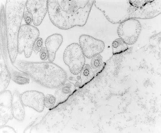
Figure 82-5. Transmission electron micrograph showing flagellates of T cruzi attached by hemidesmosomes to the epithelium of the rectal gland of Triatoma dimidiata.

Figure 82-5Transmission electron micrograph showing flagellates of T cruzi attached by hemidesmosomes to the epithelium of the rectal gland of Triatoma dimidiata

[From Zeledón R: Life cycle of Trypanosoma cruzi in the insect vector. In Brenner RR, Stoka AM (eds): Chagas’ Disease Vectors. Vol. 2 Anatomic and Physiological Aspects. CRC Press, Boca Raton, FL, 1987, with permission.]

From: Chapter 82, Hemoflagellates
Cover of Medical Microbiology
Medical Microbiology. 4th edition.
Baron S, editor.
Copyright © 1996, The University of Texas Medical Branch at Galveston.

NCBI Bookshelf. A service of the National Library of Medicine, National Institutes of Health.