This publication is provided for historical reference only and the information may be out of date.
Summary
NOTE: THIS PUBLICATION HAS BEEN RETIRED. THIS ARCHIVAL VERSION IS FOR HISTORICAL REFERENCE ONLY, AND THE INFORMATION MAY BE OUT OF DATE.
Clinical characteristics.
9q22.3 microdeletion, which includes deletion of PTCH1, the gene that is mutated in Gorlin syndrome (nevoid basal cell carcinoma syndrome), is characterized by the clinical findings of this well-described disorder as well as developmental delay and/or intellectual disability, metopic craniosynostosis, obstructive hydrocephalus, pre- and postnatal macrosomia, and seizures. Affected individuals are also at increased risk for Wilms tumor. Common findings in Gorlin syndrome include: calcification of the falx cerebri prior to age 20 years; basal cell carcinomas (BCCs) of the skin; jaw keratocysts; palmar/plantar skin pits; and increased risk for childhood medulloblastomas as well as cardiac and ovarian fibromas. The clinical spectrum of the 9q22.3 microdeletion is variable and the clinical findings depend somewhat on the size of the microdeletion.
Diagnosis/testing.
The diagnosis of the 9q22.3 microdeletion is confirmed by demonstration of a heterozygous microdeletion at chromosome 9q22.3. The minimal critical region that is deleted recurrently in affected individuals (but not in controls) is 352 kb, and includes PTCH1 and FANCC. The 9q22.3 microdeletion cannot be identified by routine analysis of G-banded chromosomes or other conventional cytogenetic banding techniques, except with extremely large deletions.
Management.
Treatment of manifestations: Routine treatment and management by appropriate specialists for cardiac, neurologic, and dermatologic findings. Comprehensive physical, occupational, and speech therapy services as needed. Surgical intervention as needed for excision or treatment of mandibular keratocysts, basal cell carcinomas, or other tumors that develop, or for management or correction of physical anomalies.
Prevention of primary manifestations: Limiting exposure to ionizing radiation, such as by computed tomography and x-rays.
Surveillance: Routine monitoring of head circumference and neurologic status throughout childhood with prompt evaluation by a neurologist and/or neurosurgeon for increasing head size, behavioral changes, or change in consciousness for evidence of obstructive hydrocephalus, medulloblastoma, and/or other cerebral tumors. Regular abdominal ultrasound for Wilms tumor, similar to surveillance for Beckwith-Wiedemann syndrome, is recommended. In those over age eight years, orthopantogram every 12-18 months to identify jaw keratocysts and skin examination at least annually.
Agents/circumstances to avoid: Excessive sun exposure; use of radiotherapy because of risk of developing multiple BCCs in the treated area.
Pregnancy management: Cesarean section delivery may be required for affected fetuses with macrocephaly.
Genetic counseling.
The 9q22.3 microdeletion is inherited in an autosomal dominant manner. In the majority of individuals, the microdeletion appears to result from either a de novo event or inheritance of an unbalanced chromosome rearrangement from a parent with a balanced rearrangement; however, inheritance of a deletion from a symptomatic parent mosaic for the deletion has been reported. When neither parent has a balanced chromosome rearrangement or deletion, recurrence risk for future pregnancies is low (probably <5%), but greater than that of the general population because parents may have germline mosaicism or low-level somatic mosaicism that also includes the germline. Prenatal testing is possible for pregnancies at increased risk based on identification of a balanced chromosome rearrangement or a deletion in a parent, and for parents concerned about the possibility of germline mosaicism.
Diagnosis
Clinical Diagnosis
The clinical spectrum of the 9q22.3 microdeletion is variable and the clinical findings depend somewhat on the size of the microdeletion.
All reported 9q22.3 microdeletions include PTCH1, the gene that is mutated in Gorlin syndrome (nevoid basal cell carcinoma syndrome); therefore, all individuals with 9q22.3 microdeletion have the clinical findings of this well-described disorder [Kimonis et al 2004].
Major features of Gorlin syndrome include:
- Lamellar calcification of the falx cerebri prior to age 20 years
- Five or more basal cell carcinomas in a lifetime or one prior to age 30 years
- Jaw keratocysts
- Palmar/plantar pits
- First-degree relative with Gorlin syndrome
Minor features of Gorlin syndrome include [Kimonis et al 2004]:
- Cleft lip and/or cleft palate
- Pre- or postaxial polydactyly
- Macrocephaly (occipital-frontal circumference of >97th centile)
- Ocular anomalies (including microphthalmia, cataracts, retinal anomalies, developmental defects)
- Rib and/or vertebral anomalies
- Cardiac and ovarian fibromas
- Childhood medulloblastoma (also called primitive neuroectodermal tumor [PNET])
- Lymphomesenteric or pleural cysts
Additional findings common in 9q22.3 microdeletion include [Muller et al 2012]:
- Developmental delay and/or intellectual disability
- Short nose and long and tented philtrum
- Metopic craniosynostosis
- Obstructive hydrocephalus
- Pre- and postnatal height and weight of >95th centile
- Seizures
Occasional abnormalities in 9q22.3 microdeletion may include renal anomalies, Wilms tumor, Chiari malformation, and dysgenesis of the corpus callosum.
Testing
Cytogenetic testing. The 9q22.3 microdeletion cannot be identified by routine analysis of G-banded chromosomes or other conventional cytogenetic banding techniques, except with extremely large deletions.
Molecular Genetic Testing
Critical region. The diagnosis of the 9q22.3 microdeletion is confirmed by demonstration of a heterozygous microdeletion at chromosome 9q22.3. The minimal critical region that is deleted recurrently in affected individuals, but not in controls, is 352 kb, and includes the genes PTCH1 (human homolog 1 of Drosophila Patched) and FANCC (Fanconi anemia complementation group C) [Muller et al 2012].
Gene. PTCH1 is the only gene for which deletion is known to account for the majority of features in 9q22.3 microdeletion; however, deletion of this gene does not appear to be sufficient to cause the features that are distinct from those usually seen in Gorlin syndrome. Among the two to 273 genes included within the interval of this contiguous gene deletion are genes that encode microRNAs, transcription factors, uncharacterized open reading frames, and proteins of unknown function [Muller et al 2012]. Many of these genes remain uncharacterized as to their individual deletion or pathogenic variant phenotypes.
Note: The genes that are deleted vary with the size and breakpoints of the microdeletion.
Table 1.
Molecular Genetic Testing Used in 9q22.3 Microdeletion
Interpretation of Test Results
Deletion analysis. Depending on the initial test that identifies the deletion, confirmation of the deletion by an independent method may be warranted. If high-density genomic microarray platforms have been used for the identification of the deletion, confirmation of the deletion may not be necessary, as it is unlikely that many adjacent targets would show an abnormal copy number by chance.
Testing Strategy
To confirm/establish the diagnosis in a proband requires detection of the 352-kb minimal critical deletion common in 9q22.3 microdeletion or any larger overlapping deletion.
- If the 9q22.3 microdeletion is suspected based on the clinical features, a targeted technique (e.g., FISH, MLPA) can be employed.
- Deletions may also be detected by genomic chromosomal microarray (CMA) analysis performed as part of the evaluation of developmental delay or intellectual disability, and/or after detection of a whole PTCH1 deletion.
Note: The deletion cannot be identified by routine chromosome analysis.
Clinical Characteristics
Clinical Description
Many individuals with 9q22.3 microdeletion exhibit hypotonia in infancy and all exhibit gross motor delay. Hypotonia may persist even into late childhood and adolescence in individuals with larger deletions [Shimojima et al 2009, Yamamoto et al 2009, Muller et al 2012]. Individuals with the smallest deletions may have resolution of their motor delay and have no other impairments or delays.
Individuals with deletions of approximately 2 Mb or larger exhibit persistent delays in attaining motor, speech, and behavioral/social milestones [Shimojima et al 2009, Muller et al 2012]. Individuals with these larger-sized deletions typically have intellectual impairment that becomes apparent at school age, requiring special education. More severe or profound disability is expected with increasing deletion size; IQ or developmental quotient (DQ) scores in the 30s to 40s or lower have been reported [Kroes et al 1994, Redon et al 2006, Fujii et al 2007, Nowakowska et al 2007, Yamamoto et al 2009, Muller et al 2012].
A fraction of all individuals with 9q22.3 microdeletion develop seizures [Shimojima et al 2009, Yamamoto et al 2009, Muller et al 2012].
Many (16/37) individuals reported with 9q22.3 microdeletion have cerebral ventricular dilation that ranges from severe to mild and asymmetric, and can be associated with cerebral atrophy or a space-occupying lesion (e.g., medulloblastoma) [Muller et al 2012]. Of those, a fraction (7/16) will have severe obstructive hydrocephalus of unknown etiology that requires ventricular shunting [Muller et al 2012].
Approximately 20% of individuals with 9q22.3 microdeletion have prenatal onset of macrosomia characterized by birth length and weight above the 95th centile, which continues postnatally [Muller et al 2012].
A few case reports describe either macrosomia or hemihyperplasia in individuals with 9q22.3 microdeletion [Cajaiba et al 2006, Chen et al 2006, Redon et al 2006, Shimojima et al 2009, Yamamoto et al 2009, Muller et al 2012, Isidor et al 2013].
Eight of the 37 affected individuals reported and reviewed by Muller et al [2012] exhibited early fusion of the metopic suture, resulting in metopic craniosynostosis and trigonocephaly.
Cajaiba et al [2006] described a single individual with Wilms tumor and a pelvic rhabdomyosarcoma and concluded that although Wilms tumor is not associated with Gorlin syndrome, rhabdomyosarcoma can be [Cajaiba et al 2006]. Recently, however, four additional individuals with a germline 9q22.3 microdeletion and Wilms tumor have been reported [Garavelli et al 2013, Isidor et al 2013]. Isidor et al [2013] sequenced PTCH1 in the tumor from one of the four affected individuals and confirmed the presence of a somatic nonsense variant on the non-deleted allele that was not present in the individual's normal kidney tissue or blood. Thus, 12% (5/42) of individuals with 9q22.3 microdeletion reported in the literature have developed Wilms tumor.
Affected individuals may have a facial gestalt that includes a broad forehead with bossing, vertical forehead creases, angulated palpebral fissures that may be either up- or downslanted, and a short nose with a long and tented philtrum [Ying et al 1982, Farrell et al 1991, Olivieri et al 2003, Midro et al 2004, Redon et al 2006, Nowakowska et al 2007, Yamamoto et al 2009, Muller et al 2012]. The facial features in some individuals tend to coarsen over time, whereas those with extremely large deletions may have coarse features at birth [Ying et al 1982, Muller et al 2012].
Genotype-Phenotype Correlations
Many of the features seen in individuals with 9q22.3 microdeletion result from haploinsufficiency of PTCH1, and as such are consistent with Gorlin syndrome. However, as most reported individuals to date have had many more genes than PTCH1 within their deletion, it is expected that deletion of one or more of these other genes results in the additional phenotypic features that are not characteristic of Gorlin syndrome.
Muller et al [2012] identified the minimal common deletion intervals and the associated breakpoints in ten individuals with 9q22.3 microdeletion for the following findings:
- Metopic craniosynostosis: a 929-kb region containing 16 genes
- Severe obstructive hydrocephalus: a 1.08-Mb region containing 18 genes
- Macrosomia: a 1.8-Mb region containing 31 genes
Note: Some genes within these intervals have not been fully characterized, and no specific candidate genes were identified.
Multiple authors have proposed that the macrosomia present in a subset of individuals with 9q22.3 microdeletion is specifically the result of loss of the paternal allele [Redon et al 2006, Shimojima et al 2009]. However, thus far, no imprinted genes within the deletion intervals have been identified.
Penetrance
It is expected that 9q22.3 microdeletion is fully penetrant for phenotype, but with variable expressivity. No unaffected individuals with this microdeletion have been reported to date.
Prevalence
The 9q22.3 microdeletion is presumed to be rare. To date, 42 affected individuals have been reported in the medical literature, including one with somatic mosaicism [Yamamoto et al 2009, Muller et al 2012, Garavelli et al 2013, Isidor et al 2013]. It is likely that 9q22.3 microdeletion represents a very small fraction of individuals with dysmorphic features and developmental delay and/or intellectual impairment in the absence of other characteristic features.
Genetically Related (Allelic) Disorders
Duplication of the 9q22.3 region, consisting of a 360-kb region containing PTCH1 and exon 1 of FANCC, has been described in a mother and her child. Both had microcephaly and mild developmental delay [Derwińska et al 2009].
Germline dominant pathogenic loss-of-function variants in PTCH1 including intragenic or whole-gene deletions are known to result in Gorlin syndrome [Hahn et al 1996].
Germline dominant pathogenic gain-of-function variants in PTCH1 associated with holoprosencephaly type 7 (OMIM 610828) that are consistent with reduced embryologic sonic hedgehog expression have been described in a few individuals [Ming et al 2002, Ribeiro et al 2006].
Sporadic tumors (including medulloblastomas, odontogenic keratocysts, cardiac fibromas, ovarian fibromas, and basal cell carcinomas) occurring as single tumors in the absence of any other findings of this syndrome can harbor somatic variants in PTCH1 that are not present in the germline; thus, predisposition to these tumors is not heritable. For more details see Cancer and Benign Tumors.
Differential Diagnosis
A 9q22.2-q22.3 deletion of 5.3 Mb that did not include PTCH1 was identified in a developmentally normal man who had mildly dysmorphic facial features, dysarthria, funnel chest, and unilateral renal hypoplasia. It was also present in his two daughters who were intellectually disabled and shared his dysmorphic features but did not have any malformations [Siggberg et al 2011].
Among syndromes that share multiple features of 9q22.3 microdeletion, Gorlin syndrome (nevoid basal cell carcinoma syndrome) is the most common.
Beckwith-Wiedemann syndrome (BWS) is a growth disorder characterized by macrosomia, macroglossia, visceromegaly, embryonal tumors (e.g., Wilms tumor, hepatoblastoma, neuroblastoma, and rhabdomyosarcoma), omphalocele, neonatal hypoglycemia, ear creases/pits, adrenocortical cytomegaly, and renal abnormalities (e.g., medullary dysplasia, nephrocalcinosis, medullary sponge kidney, and nephromegaly). Early death may occur from complications of prematurity, hypoglycemia, cardiomyopathy, macroglossia, or tumors. However, the previously reported mortality of 20% is likely an overestimate given better recognition of the disorder along with enhanced treatment options. Macroglossia and macrosomia are generally present at birth but may have postnatal onset. Growth rate slows around age seven to eight years. Hemihyperplasia may affect segmental regions of the body or selected organs and tissues. Molecular genetic testing can identify epigenetic and genomic alterations of chromosome 11p15 in individuals with BWS: (1) loss of methylation on the maternal chromosome at imprinting center 2 (IC2) in 50% of affected individuals; (2) paternal uniparental disomy for chromosome 11p15 in 20%; and (3) gain of methylation on the maternal chromosome at imprinting center 1 (IC1) in 5%. Sequence analysis of CDKN1C identifies pathogenic variants in approximately 40% of familial cases and 5%-10% of cases with no family history of BWS.
Sotos syndrome is characterized by the cardinal features of typical facial appearance, overgrowth (height and/or head circumference ≥2 SD above the mean), and learning disability ranging from mild (children attend mainstream schools and are likely to be independent as adults) to severe (lifelong care and support will likely be required). Sotos syndrome is associated with the major features of behavioral problems, congenital cardiac anomalies, neonatal jaundice, renal anomalies, scoliosis, and seizures. About 80%-90% of individuals with Sotos syndrome have a demonstrable NSD1 abnormality.
Numerous other genomic microdeletions or microdeletion syndromes result in developmental delay or intellectual impairment and/or some of the individual nonspecific phenotypic features of 9q22.3 microdeletion.
Management
Evaluations Following Initial Diagnosis
To establish the extent of disease and needs of an individual diagnosed with the 9q22.3 microdeletion, the following evaluations are recommended:
- Brain imaging (not using CT) and neurologic evaluation
- Complete physical examination, including dermatologic assessment for the manifestations of Gorlin syndrome
- Comprehensive developmental assessment
- Renal and pelvic ultrasound examination for evaluation of possible renal anomalies and ovarian fibromas
- Echocardiogram
- Ophthalmologic evaluation
- Careful consideration of skeletal and/or dental imaging for associated anomalies
- Familial genetic counseling
- Routine treatment and management by appropriate specialists for cardiac, neurologic, dermatologic findings
- Comprehensive physical, occupational, and speech therapy services as needed
- Surgical intervention as needed for excision or treatment of mandibular keratocysts, basal cell carcinomas, or other tumors that develop, or for management or correction of physical anomalies
Treatment of Manifestations
The following are appropriate:
- Routine treatment and management by appropriate specialists for cardiac, neurologic, dermatologic findings
- Comprehensive physical, occupational, and speech therapy services as needed
- Surgical intervention as needed for excision or treatment of mandibular keratocysts, basal cell carcinomas, or other tumors that develop, or for management or correction of physical anomalies
Prevention of Primary Manifestations
Avoidance of excessive sunlight or other ultraviolet radiation, and limiting exposure to ionizing radiation (e.g., by computed tomography and x-ray) is recommended because of the increased predisposition for the development of basal cell carcinomas.
Surveillance
The following recommended surveillance is the same as that for Gorlin syndrome (see Gorlin Syndrome).
- Head circumference should be followed throughout childhood and plotted on appropriate growth charts. Rapid enlargement should prompt evaluation for possible hydrocephalus.
- Awareness of the risk of medulloblastoma in the first years of life is important and may justify developmental assessment and physical examination every six months. No evidence for the efficacy of regular neuroimaging exists; frequent computer tomography (CT) should be avoided because of risks associated with radiation sensitivity.
- Orthopantogram is indicated every 12-18 months in individuals older than age eight years to identify jaw keratocysts.
- Skin should be examined at least annually; some physicians recommend skin examination by a professional every three to four months.
While there are currently no published guidelines regarding monitoring for intraabdominal embryonal tumors in individuals with 9q22.3 microdeletion, 12% of the published cases have been diagnosed with Wilms tumor. Thus regular abdominal ultrasound for Wilms tumor, similar to surveillance for Beckwith-Wiedemann syndrome, is recommended for individuals with 9q22.3 microdeletion until disproven otherwise.
Agents/Circumstances to Avoid
As all individuals to date have had involvement of PTCH1 resulting in Gorlin syndrome, affected individuals are at increased risk for malignant tumor formation with ionizing and ultraviolet radiation exposure, and for spontaneous development of basal cell carcinomas both from the numerous existing basal cell nevi and in apparently unaffected skin (see Gorlin Syndrome). Radiographs and computed tomography should be used sparingly, and the benefit versus risk to the individual's health should be carefully considered.
Liberal use of topical sunblock and avoidance of excessive exposure to sunlight are warranted.
Evaluation of Relatives at Risk
See Genetic Counseling for issues related to testing of at-risk relatives for genetic counseling purposes.
Pregnancy Management
Macrosomia and/or macrocephaly of prenatal onset is present in many individuals with 9q22.3 microdeletion. This may necessitate delivery by Cesarean section, including emergently, as has been reported for some individuals [Isidor et al 2013].
Therapies Under Investigation
Search ClinicalTrials.gov in the US and www.ClinicalTrialsRegister.eu in Europe for access to information on clinical studies for a wide range of diseases and conditions. Note: There may not be clinical trials for this disorder.
Genetic Counseling
Genetic counseling is the process of providing individuals and families with information on the nature, mode(s) of inheritance, and implications of genetic disorders to help them make informed medical and personal decisions. The following section deals with genetic risk assessment and the use of family history and genetic testing to clarify genetic status for family members; it is not meant to address all personal, cultural, or ethical issues that may arise or to substitute for consultation with a genetics professional. —ED.
Mode of Inheritance
The 9q22.3 microdeletion is inherited in an autosomal dominant manner.
Risk to Family Members
Parents of a proband
- The parents of a proband with 9q22.3 microdeletion are generally unaffected.
- The majority of cases have resulted from either an apparent de novo event or inheritance of an unbalanced chromosome rearrangement from a parent with a balanced rearrangement.
- Somatic/germline mosaicism for the 9q22.3 microdeletion has not been reported in an asymptomatic parent of an affected individual.
- Recurrence in one family has been described: a woman with features of the deletion condition and mosaicism for a 1.7-Mb 9q22.3 deletion (including PTCH1) had an affected daughter with the deletion in non-mosaic form [Isidor et al 2013].
- Recurrence in two families in which a parent had a balanced translocation involving the 9q22.3 region has been reported [Shimkets et al 1996, Midro et al 2004].
- Recommendations for the evaluation of asymptomatic parents of a proband include high resolution chromosome analysis to determine if a balanced chromosome rearrangement involving 9q22.3 is present.
Sibs of a proband
- The risk to the sibs of the proband depends on the genetic status of the parents.
- Recurrence risk to the sibs of a proband is low (probably <5%) but greater than that of the general population because a parent may have (a) germline mosaicism for the 9q22.3 microdeletion, or (b) low-level somatic mosaicism for the 9q22.3 microdeletion that also includes the germline.
- If a parent has a balanced structural chromosome rearrangement involving the 9q22.3 critical region, the risk to sibs is increased and depends on the specific chromosome rearrangement.
- If a parent has a constitutional 9q22.3 microdeletion (i.e., the microdeletion is present in all of the parent's cells), the risk to sibs of the proband is 50% for also inheriting the microdeletion; however, this has not been reported.
Offspring of a proband. Only one individual diagnosed with the typical 9q22.3 microdeletion has been known to reproduce; however, this individual had mosaicism for the deletion [Isidor et al 2013].
Other family members. The risk to other family members depends on the genetic status of the proband's parents. If a parent has a balanced chromosome rearrangement or deletion, his/her family members may be at increased risk of also having the rearrangement or deletion.
Related Genetic Counseling Issues
Family planning
- The optimal time for determination of genetic risk and discussion of the availability of prenatal testing is before pregnancy. Similarly, decisions about testing to determine the genetic status of at-risk asymptomatic family members are best made before pregnancy.
- It is appropriate to offer genetic counseling (including discussion of potential risks to offspring and reproductive options) to young adults who are at risk of having a child with the 9q22.3 microdeletion.
DNA banking is the storage of DNA (typically extracted from white blood cells) for possible future use. Because it is likely that testing methodology and our understanding of genes, allelic variants, and diseases will improve in the future, consideration should be given to banking DNA of affected individuals.
Prenatal Testing
Prenatal testing is technically feasible. Chromosome preparations from fetal cells obtained by amniocentesis usually performed at approximately 15 to 18 weeks' gestation or CVS at approximately ten to 12 weeks' gestation can be analyzed using specific FISH probe analysis or chromosomal microarray (CMA), in the manner described in Molecular Genetic Testing.
Prenatal testing may be offered to parents who have had a child with the 9q22.3 microdeletion because of the recurrence risk (probably <5%) associated with the possibility of germline mosaicism. Prenatal testing is also offered to parents who carry a balanced chromosome rearrangement or who have the microdeletion.
Note: Gestational age is expressed as menstrual weeks calculated either from the first day of the last normal menstrual period or by ultrasound measurements.
Preimplantation genetic diagnosis (PGD) may be an option for families at increased risk for a pregnancy with the 9q22.3 microdeletion.
Resources
GeneReviews staff has selected the following disease-specific and/or umbrella support organizations and/or registries for the benefit of individuals with this disorder and their families. GeneReviews is not responsible for the information provided by other organizations. For information on selection criteria, click here.
- Chromosome Disorder Outreach (CDO)PO Box 724Boca Raton FL 33429-0724Phone: 561-395-4252 (Family Helpline)Email: info@chromodisorder.org
- Unique: The Rare Chromosome Disorder Support GroupG1 The StablesStation Road WestOxted Surrey RH8 9EEUnited KingdomPhone: +44 (0) 1883 723356Email: info@rarechromo.org; rarechromo@aol.com
Molecular Genetics
Information in the Molecular Genetics and OMIM tables may differ from that elsewhere in the GeneReview: tables may contain more recent information. —ED.
Table A.
9q22.3 Microdeletion: Genes and Databases
Molecular Genetic Pathogenesis
The mechanism that predisposes the 9q22.3 region to deletion is unclear. The numerous SINEs, large LINEs, and LTRs that flank PTCH1 and adjacent genes in the region potentially could predispose to recombination events that result in deletion or duplication of these genes. Similarly, it is unknown how deletion of genes (other than PTCH1) within the 9q22.3 microdeletion results in the phenotype, as the function and haploinsufficiency phenotype of many of the 273 genes in the largest reported deletion of 20.5 Mb remain uncharacterized as to function and/or deletion phenotype [Muller et al 2012].
The microdeletion size among the reported affected individuals does not appear to be recurrent. The earliest reports of 9q22.3 deletions preceded CMA technology; at that time only large deletions visible by routine cytogenetic banding techniques were described, making specific breakpoint comparison with reports from the last several years difficult. Following the availability of CMA technology, the minimal interstitial 9q22.3 microdeletion reported contains only two genes, PTCH1 and FANCC [Muller et al 2012].
PTCH1 (human homolog 1 of Drosophila Patched) encodes a tumor suppressor protein that is the receptor for sonic hedgehog (SHH) protein, which in the unbound form normally acts to repress SHH signaling. FANCC (Fanconi anemia complementation group C) encodes a protein that is part of the core FA nuclear protein complex with E3 ubiquitin ligase activity, which activates in response to DNA damage and in the S-phase. Homozygous or compound heterozygous pathogenic variants in this gene result in Fanconi anemia.
In ten individuals with 9q22.3 microdeletion, Muller et al [2012] attempted to define the critical regions and genes involved in the three distinctive features metopic craniosynostosis, obstructive hydrocephalus, and macrosomia; using this approach they were able to narrow the overlapping regions to 0.929 to 1.8 Mb. The shared genes within these intervals are summarized in Figure 1.
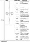
Figure 1.
Schematic of genes involved in ten individuals with 9q22.3 microdeletion and shared phenotypes Adapted from Muller et al [2012]
Cancer and Benign Tumors
Somatic dominant pathogenic loss-of-function variants in PTCH1 have been described in sporadic cancers that are also present in Gorlin syndrome, including medulloblastomas, odontogenic keratocysts, cardiac and ovarian fibromas, and basal cell carcinomas [Kimonis et al 2004].
A somatic nonsense variant in PTCH1 was identified on the non-deleted allele in the Wilms tumor tissue from an individual with germline 9q22.3 microdeletion. The somatic nonsense variant was not present in the normal kidney tissue or blood [Isidor et al 2013].
References
Literature Cited
- Cajaiba MM, Bale AE, Alvarez-Franco M, McNamara J, Reyes-Mugica M. Rhabdomyosarcoma, Wilms tumor, and deletion of the patched gene in Gorlin syndrome. Nat Clin Pract Oncol. 2006;3:575–80. [PubMed: 17019435]
- Chen CP, Lin SP, Wang TH, Chen YJ, Chen M, Wang W. Perinatal findings and molecular cytogenetic analyses of de novo interstitial deletion of 9q (9q22.3-->q31.3) associated with Gorlin syndrome. Prenat Diagn. 2006;26:725–9. [PubMed: 16927391]
- Derwińska K, Smyk M, Cooper ML, Bader P, Cheung SW, Stankiewicz P. PTCH1 duplication in a family with microcephaly and mild developmental delay. Eur J Hum Genet. 2009;17:267–71. [PMC free article: PMC2986050] [PubMed: 18830227]
- Farrell SA, Siegel-Bartelt J, Teshima I. Patients with deletions of 9q22q34 do not define a syndrome: three case reports and a literature review. Clin Genet. 1991;40:207–14. [PubMed: 1773536]
- Fujii K, Ishikawa S, Uchikawa H, Komura D, Shapero MH, Shen F, Hung J, Arai H, Tanaka Y, Sasaki K, Kohno Y, Yamada M, Jones KW, Aburatani H, Miyashita T. High-density oligonucleotide array with sub-kilobase resolution reveals breakpoint information of submicroscopic deletions in nevoid basal cell carcinoma syndrome. Hum Genet. 2007;122:459–66. [PubMed: 17703323]
- Garavelli L, Piemontese MR, Cavazza A, Rosato S, Wischmeijer A, Gelmini C, Albertini E, Albertini G, Forzano F, Franchi F, Carella M, Zelante L, Superti-Furga A. Multiple tumor types including leiomyoma and Wilms tumor in a patient with Gorlin syndrome due to 9q22.3 microdeletion encompassing the PTCH1 and FANC-C loci. Am J Med Genet A. 2013;161A(11):2894–901. [PubMed: 24124115]
- Hahn H, Wicking C, Zaphiropoulous PG, Gailani MR, Shanley S, Chidambaram A, Vorechovsky I, Holmberg E, Unden AB, Gillies S, Negus K, Smyth I, Pressman C, Leffell DJ, Gerrard B, Goldstein AM, Dean M, Toftgard R, Chenevix-Trench G, Wainwright B, Bale AE. Mutations of the human homolog of Drosophila patched in the nevoid basal cell carcinoma syndrome. Cell. 1996;85:841–51. [PubMed: 8681379]
- Isidor B, Bourdeaut F, Lafon D, Plessis G, Lacaze E, Kannengiesser C, Rossignol S, Pichon O, Briand A, Martin-Coignard D, Piccione M, David A, Delattre O, Jeanpierre C, Sévenet N, Le Caignec C. Wilms' tumor in patients with 9q22.3 microdeletion syndrome suggests a role for PTCH1 in nephroblastomas. Eur J Hum Genet. 2013;21:784–7. [PMC free article: PMC3722950] [PubMed: 23169491]
- Kimonis VE, Mehta SG, Digiovanna JJ, Bale SJ, Pastakia B. Radiological features in 82 patients with nevoid basal cell carcinoma (NBCC or Gorlin) syndrome. Genet Med. 2004;6:495–502. [PubMed: 15545745]
- Kroes HY, Tuerlings JH, Hordijk R, Folkers NR, ten Kate LP. Another patient with an interstitial deletion of chromosome 9: case report and a review of six cases with del(9)(q22q32). J Med Genet. 1994;31:156–8. [PMC free article: PMC1049682] [PubMed: 8182726]
- Midro AT, Panasiuk B, Tumer Z, Stankiewicz P, Silahtaroglu A, Lupski JR, Zemanova Z, Stasiewicz-Jarocka B, Hubert E, Tarasow E, Famulski W, Zadrozna-Tolwinska B, Wasilewska E, Kirchhoff M, Kalscheuer V, Michalova K, Tommerup N. Interstitial deletion 9q22.32-q33.2 associated with additional familial translocation t(9;17)(q34.11;p11.2) in a patient with Gorlin-Goltz syndrome and features of Nail-Patella syndrome. Am J Med Genet A. 2004;124A:179–91. [PubMed: 14699618]
- Ming JE, Kaupas ME, Roessler E, Brunner HG, Golabi M, Tekin M, Stratton RF, Sujansky E, Bale SJ, Muenke M. Mutations in PATCHED-1, the receptor for SONIC HEDGEHOG, are associated with holoprosencephaly. Hum Genet. 2002;110:297–301. [PubMed: 11941477]
- Muller EA, Aradhya S, Atkin JF, Carmany EP, Elliott AM, Chudley AE, Clark RD, Everman DB, Garner S, Hall BD, Herman GE, Kivuva E, Ramanathan S, Stevenson DA, Stockton DW, Hudgins L. Microdeletion 9q22.3 syndrome includes metopic craniosynostosis, hydrocephalus, macrosomia and developmental delay. Am J Med Genet A. 2012;158A:391–9. [PubMed: 22190277]
- Nowakowska B, Kutkowska-Kazmierczak A, Stankiewicz P, Bocian E, Obersztyn E, Ou Z, Cheung SW, Cai WW. A girl with deletion 9q22.1-q22.32 including the PTCH and ROR2 genes identified by genome-wide array-CGH. Am J Med Genet A. 2007;143A:1885–9. [PubMed: 17632781]
- Olivieri C, Maraschio P, Caselli D, Martini C, Beluffi G, Maserati E, Danesino C. Interstitial deletion of chromosome 9, int del(9)(9q22.31-q31.2), including the genes causing multiple basal cell nevus syndrome and Robinow/brachydactyly 1 syndrome. Eur J Pediatr. 2003;162:100–3. [PubMed: 12548386]
- Redon R, Baujat G, Sanlaville D, Le Merrer M, Vekemans M, Munnich A, Carter NP, Cormier-Daire V, Colleaux L. Interstitial 9q22.3 microdeletion: clinical and molecular characterisation of a newly recognised overgrowth syndrome. Eur J Hum Genet. 2006;14:759–67. [PubMed: 16570072]
- Ribeiro LA, Murray JC, Richieri-Costa A. PTCH mutations in four Brazilian patients with holoprosencephaly and in one with holoprosencephaly-like features and normal MRI. Am J Med Genet A. 2006;140:2584–6. [PubMed: 17001668]
- Shimkets R, Gailani MR, Siu VM, Yang-Feng T, Pressman CL, Levanat S, Goldstein A, Dean M, Bale AE. Molecular analysis of chromosome 9q deletions in two Gorlin syndrome patients. Am J Hum Genet. 1996;59(2):417–22. [PMC free article: PMC1914731] [PubMed: 8755929]
- Shimojima K, Adachi M, Tanaka M, Tanaka Y, Kurosawa K, Yamamoto T. Clinical features of microdeletion 9q22.3 (pat). Clin Genet. 2009;75:384–93. [PubMed: 19320658]
- Siggberg L, Peippo M, Sipponen M, Miikkulainen T, Shimojima K, Yamamoto T, Ignatius J, Knuutila S. 9q22 deletion - first familial case. Orphanet J Rare Dis. 2011;6:45. [PMC free article: PMC3135502] [PubMed: 21693067]
- Yamamoto K, Yoshihashi H, Furuya N, Adachi M, Ito S, Tanaka Y, Masuno M, Chiyo H, Kurosawa K. Further delineation of 9q22 deletion syndrome associated with basal cell nevus (Gorlin) syndrome: report of two cases and review of the literature. Congenit Anom (Kyoto). 2009;49:8–14. [PubMed: 19243411]
- Ying KL, Curry CJ, Rajani KB, Kassel SH, Sparkes RS. De novo interstitial deletion in the long arm of chromosome 9: a new chromosome syndrome. J Med Genet. 1982;19:68–70. [PMC free article: PMC1048822] [PubMed: 7069749]
Chapter Notes
Revision History
- 2 August 2018 (ma) Chapter retired: non-recurrent deletions or duplications; refers to deletions/duplications of varying size – in contrast to a recurrent deletion/duplication, defined as a deletion/duplication of a specific size (usually mediated by nonallelic homologous recombination) occurring multiple times in the general population
- 20 February 2014 (me) Comprehensive update posted live
- 18 August 2011 (me) Review posted live
- 25 April 2011 (em) Original submission
Publication Details
Author Information and Affiliations
Pediatric Specialties
California Pacific Medical Center
San Francisco, California
Publication History
Initial Posting: August 18, 2011; Last Update: February 20, 2014.
Copyright
GeneReviews® chapters are owned by the University of Washington. Permission is hereby granted to reproduce, distribute, and translate copies of content materials for noncommercial research purposes only, provided that (i) credit for source (http://www.genereviews.org/) and copyright (© 1993-2024 University of Washington) are included with each copy; (ii) a link to the original material is provided whenever the material is published elsewhere on the Web; and (iii) reproducers, distributors, and/or translators comply with the GeneReviews® Copyright Notice and Usage Disclaimer. No further modifications are allowed. For clarity, excerpts of GeneReviews chapters for use in lab reports and clinic notes are a permitted use.
For more information, see the GeneReviews® Copyright Notice and Usage Disclaimer.
For questions regarding permissions or whether a specified use is allowed, contact: ude.wu@tssamda.
Publisher
University of Washington, Seattle, Seattle (WA)
NLM Citation
Muller E II, Hudgins L. 9q22.3 Microdeletion – RETIRED CHAPTER, FOR HISTORICAL REFERENCE ONLY. 2011 Aug 18 [Updated 2014 Feb 20]. In: Adam MP, Feldman J, Mirzaa GM, et al., editors. GeneReviews® [Internet]. Seattle (WA): University of Washington, Seattle; 1993-2024.
