Continuing Education Activity
American physicians Allen and Hines described an abnormal deposition of adipose tissue affecting females in the 1940s in a Mayo Clinic termed lipedema. Lipedema is the disproportionate and symmetrical distribution of adipose tissue involving the lower extremities to a greater degree than the trunk or upper extremities. History and physical examination provide clues regarding the lipedema, which is evaluated more with nuclear medicine lymphangioscintigraphy, ultrasound, or MRI of the legs. To avoid more complications, it should be diagnosed and intervene early. This activity reviews the evaluation and management of lipedema. Treatment starts with conservative management, like diet and exercise, but then surgical options like bariatric surgery and liposuction should be considered.
Objectives:
Identify the etiology, pathophysiology, and epidemiology of lipedema.
Review the evaluation of lipedema.
Summarize the management for lipedema.
Outline some strategies that will improve patient care and outcomes for individuals with lipedema.
Access free multiple choice questions on this topic.
Introduction
Dr. Allen and Dr. Hines pioneered and first described lipedema in the 1940s, a common subcutaneous adipose tissue disorder characterized by enlargement of both lower extremities. Lipedema is not edema; it is a genetically determined disturbance in adipose tissue mass and adipose tissue distribution. In 1951 a second seminal paper provided more description of lipedema. Fat distribution involves the lower extremities, upper arms, hips, buttocks, thighs, sparing trunks, and feet. Lower extremities are characterized by pain, easy bruisability, firm subcutaneous nodules of adipose tissue, and resistance of fat to traditional diet and exercise.[1]
Etiology
Lipedema is considered as genetic inheritance, either X-linked dominant or autosomal dominant. Child et al. studied 330 family members of patients with lipedema and described autosomal inheritance as a major mode of inheritance, with a few instances with X-linked dominant inheritance were found.[2][3] Few cases have been reported with POU1F1/PIT-1 gene mutation in short mothers, but no phenotypic features were noted in the son who carried the gene from short mothers or normal height daughters. Lipedema is also found in cases of Williams syndrome, caused by a microdeletion of around 1.6 million base pairs of chromosomes 7q11.23, which includes ELN (elastin gene).[4]
Epidemiology
It is proposed that the prevalence of lipedema is nearly 1 in 72000 population. Lipedema is often misdiagnosed or underdiagnosed, so these prevalence numbers are likely to be underestimated numbers.[2] Lipedema occurs predominantly in females but has been reported rarely in males. Few reports mentioned the prevalence of lipedema around 11% in adults and around 6.5% in the US, ranging from 15% to 18% in European countries.[5]
Pathophysiology
In 1940, Allen and Hives proposed edema in lipedema probably results from the poor resistance of the fat against the hydrostatic passage of fluid from the capillaries into the interstitium. Presumably, hormonal influence may contribute to its pubertal onset or post-pregnancy lipedema development in females when estrogen levels are high. Lipedema in men is uncommon, but few cases have been reported in liver disease or low testosterone, both of which have high estrogen states. Lipedema is also found in cases with polycystic ovarian disease where testosterone levels are high. The role of estrogen as a causative factor in lipedema is not well established.[6]
Several theories have been postulated to describe lipedema. One theory mentions the loss of elastic tissue and abnormal vasculature. Structures in loose connective tissue include blood vessels, lymph nodes, and connective tissue fascia, containing elastin that helps to hold its shape. Lymphatic vessels do not have any elastic fibers, but they support the closure and opening of lymph vessels in response to the pressure in tissue. Similarly, capillaries do not have any elastic tissue, but loose connective tissue surrounding capillaries contains elastic tissue. Therefore, with the growth of adipose tissue in lipedema, loss of elasticity results in a lack of ability of lymph vessels to open with increased pressure in the extracellular matrix and capillary leakage in the tissue. Subsequently, the development of hypoxia followed by stimulation of vascular endothelial growth factor (VEGF) proliferates stem cells in adipose tissue.[7]
Eventually, lymph vessels lose their function, resulting in microaneurysm formation in the lymph vessels responsible for lymphedema development. Another theory of abnormal lymphatic vasculature depicts a primary defect in lymphatic vessels that results in increased fluid accumulation in an extracellular matrix that enhances permeability in surrounding microvessels. Lipedema is associated with altered left ventricular rotational mechanics and increased aortic stiffness.[8][9]
Histopathology
Diagnosis of lipedema is often difficult to differentiate from obesity. According to the WHO guidelines, people with a BMI of more than 25 kg/m2 are considered overweight, and a BMI of greater than 30 kg/m2 are considered obese. In lipedema patients, BMI is elevated, but differentiating points like diet and exercise favor obesity, whereas disproportionate fat distribution between above and below the trunk, tissue tenderness, and easy bruising favors lipedema. Lipedema can be complicated by obesity or lymphedema later in life.[2] Lymphatic vessels regulate the sodium, chloride, and water in the skin. In the case of lipedema failure of a lymphatic vessel, the function is observed, which is supported by the dilatation of lymphatic vessels in the MRI of the legs. Per Crescenzi et al., increased salt is found in women with lipedema's skin and loose connective tissues.[5] Despite higher BMI, lower prevalence of diabetes, normal blood pressure, and normal lipid levels have been observed in the case of lipedema.
Classification
Lipedema is classified into stages and types. Stage 1 represents the normal skin surface over a thick and nodular hypodermis, but pea-sized fat nodules can be palpated. These nodules are fibrosed and enlarged structures in the extracellular matrix and connective tissue surrounding the fat lobule. In this stage, skin appearance is normal and smooth on palpation, but nodules can be felt and are potentially associated with pain or easy bruising. Stage 2 represents uneven skin surface over hypodermal masses.
Dimpling of the skin, indentations, and mattress-like patterns on the skin is thickening and contraction of connective tissue over increased fat. Stage 3 represents lobular extrusions of skin, fat, and facia tissue. Inflammation and fibrosis of adipofascia result in loss of elasticity; therefore, blood flow and lymph flow out of lobules are reduced. In this stage, the appearance of both lower extremities can be columnar, visible large folds of skin and fat are palpable due to fibrosis in the tissues and loose connection to the underlying hypodermis. This may compromise the integrity of joints, mobility, and balance. Stage 4 is often classified when lipedema is associated with lymphedema, also called lipo-lymphedema.[5][10][11]
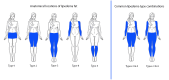
Types of lipedema are classified based on anatomical locations. Type I lipedema fat is distributed from the umbilicus to the hips, including the pelvis and buttocks. Type II includes fat distribution from around the pelvis to the knees. Type III includes from around the pelvis to ankles with a prominent cuff at the ankle sparing dorsum of the feet. Type IV includes from shoulders to wrists. Type V is a rare occurrence that involves fat distribution from or below the knees and extends through the ankles, sparing the dorsum of the feet. Type II & IV or III & IV are common combinations. Involvement of the arm can be varied in fat distribution from elbows to wrists or from shoulders to wrists.[5][12]
History and Physical
Child et al. (2009) mentioned about the age of onset of lipedema during puberty (55%), reduction of fat above the waist only through diet (96%), no effect on lower extremities making it more disproportionate fat distribution, pain upon pressure (71%), easy bruising (82%), varicose veins (39.5%), vein telangiectasia (53%), fat pads (bulging protuberance of fat above or below the knee; pre-tibial fat pads) (58%). Surprisingly, four percent of lipedema patients recorded to have normal BMI (18.5 to 24.99 kg/m2), eleven percent were pre-obese or overweight (BMI 25.0 to 29.99 kg/m2), eight percent obese class I group (BMI 30 to 34.99kg/m2), twenty-seven percent obese class II (BMI 35.0 to 39.99%) and fifty percent obese class III (BMI greater than or equal to 40%).[2]
Adapted from a table proposed by Wold et al. (1951) & Child et al. (2009), Dayan et al. (2017).[2][13][3][14]
Evaluation
Laboratory tests, including liver function, kidney function, thyroid function, lipid profile, and insulin resistance, should be ruled out before proceeding with imaging.[3]
With recent advancements in imaging, nuclear medicine helps differentiate lipedema from lymphedema. In nuclear medicine lymphangioscintigraphy, NMLAS technetium-99m-sulfur colloid is injected dermally. Remarkable asymmetry of the lymphatic system and slower lymphatic flow have been found.[5][15] Bioimpedance spectroscopy reveals increased impedance (extracellular water level) in the lower extremities compared to the upper extremities.[10]
Dual-energy X-ray absorptiometry (DEXA) scan is used to measure bone mineral density; it is very useful to assess fat mass and lean body mass. It differentiates patients without lipedema by a cut-off value of 0.46 for fat mass in the legs (adjusted for BMI).
Ultrasound of lipedema tissue demonstrates thinner skin, increased thickness, and increased hypoechogenicity of subcutaneous fat more prominent towards the medial calf and distal extremities. Based on the thickness of the subcutis, the severity of lipedema has been classified by Marschall and Schwahn-Schreiber: mild: 12 to 15 mm, moderate: 15 to 20 mm, distinct: > 20 mm, and severe: > 30 mm. Ironically, similar values have been identified in the case of obesity as well. Therefore, the proposed value can misdiagnose lipedema in the absence of other symptoms.[5][16]
Magnetic resonance imaging (MRI) of the legs shows a widening of lymphatic vessels up to 2 mm, whereas, in the case of lipedema, the widening can be more than 3 mm.
In transthoracic 2D echocardiography, enlarged left atrial diameter, increased LV end-diastolic diameter and volume, increased LV end-systolic volume, impaired left ventricular apical rotation, left ventricular twist, and aortic stiffness, along with increased ascending aortic systolic and diastolic diameters have been reported. Ejection fraction has been preserved in Lipedema patients. In 3D speckle-tracking echocardiography dilated end-systolic and end-diastolic mitral annulus disjunction, the mitral annulus area has been observed.[8]
Treatment / Management
The diet helps to manage obesity that often accompanies lipedema. Generally, lipedema fat is resistant to weight loss with diet.[5] However, few reports suggest a ketogenic diet helps in lipolysis.[13] Despite exercise body parts, those affected with lipedema continue to grow. Still, graded exercise is recommended for women who have unhealthy lifestyles. A carbohydrate-restricted diet may also be more useful in some cases when lipedema is resistant to the ketogenic diet. Few reports suggested water exercise helps mobilize the fluid and soften the fibrotic tissues, and few suggested that speed walking or bicycle riding can also be helpful. Exercise activates calf and foot muscle to pump that increase the lymphatic drainage and reduces edema formation and risk of additional obesity development.[16]
Compression garments are recommended for cosmetic reasons. Circular knit garments are seamless and better suited for women with less lymphedema, whereas flat knit garments are suited for women with ankle cuffs or unusual shapes.[5] 25% of women with lipedema have venous insufficiency. Conservative treatment should be tried first, as data are lacking to show the correction of venous insufficiency with surgery improves lipedema. Conservative therapy aims to reduce edema, pain, and associated obesity development.[16][5][11] Patient education on understanding the disease is of prime importance to decide the goals of the treatment and expectations of the disease.[3]
Bariatric surgery like gastric bypass or sleeve gastrectomy should be suggested for benefits on overall glucometabolic aspects, especially prediabetes, diabetes, or cardiovascular risk. Women should be counseled before the bariatric surgery that the post-surgery lipedema part would get more accentuated. Compression garments and decongestive therapy should be offered to prevent the development of lymphedema, and a consult with a plastic surgeon should also be recommended for excess skin removal post-bariatric surgery.[17]
Liposuction is a very effective treatment for lipedema when conservative therapies fail. In this procedure, lipedema fat is removed, sparing the lymphatic vessels. Several techniques are used as follows.
Tumescent technique: Klein solution (normal saline or lactated ringer, lidocaine or prilocaine, epinephrine, sodium bicarbonate) is used to saturate the fat. This tumescence provides turgor to tissue that microcannula slide through the fat, and tiny, rapid vibrations break the fat, which is then suctioned out.
Water-jet assisted liposuction (WAL): Klein solution or saline is used as a jet that releases the fat for liposuction. No waiting period is required to tumescent the tissue.
Laser-assisted liposuction: this technique is reserved for areas with fibrosis, like posterior thighs. Liposuction performed without general anesthesia has rapid recovery; also, it is recommended to undergo liposuction in stages rather than removing all fat in one sitting. 3 to 5 sessions of liposuction with a good interval of time in between are suggested.
The quality of life has significantly improved regarding pain, muscle cramps, tightening, itching, edema, bruising, and cosmetic appearance after liposuction. The aim of surgical treatment is mainly to reduce the volume and help in the reduction of mechanical impairment of walking. But liposuction is a very costly therapy; insurance coverage and reimbursement are the main issues.[18][19]
A pneumatic compression device is used for lipedema as well as lymphedema. In treating lymphedema, manual lymphatic drainage like myofascial therapy or deep tissue therapy like Astym therapy, Graston technique, and compression reduction techniques have been shown to reduce fibrosis in lipedema fat and increased blood flow to the tissues.[20][5] In lipedema, poor quality of life, immobility, and stress related to physical appearance can result in depression. Appropriate education about the disease, motivational interviewing, and counseling to focus on healthy eating patterns, activity, and social interaction, including internet groups and national and local groups, significantly impact a positive outcome. Referring to a psychotherapist, physical therapy, and pain management specialist is very useful when needed.[3][21][22]
No specific medication or supplement is used to treat lipedema, but drugs are used as an adjunctive, especially to prevent lymphedema and venous disease; both are complications of lipedema. Sympathomimetics like amphetamine or phentermine binds to adrenergic receptors that induce lipolysis. Adrenergic receptors are located on lymphatic and blood vessels. Metformin and resveratrol are useful to prevent fibrosis and hypoxia-inducible factor (inflammatory factor) development. Diosmin, a bioflavonoid found in citrus fruits, functions as lymphagogue, reduces swelling is helpful in lipedema and associated chronic venous insufficiency. Selenium, a mineral, has improved inflammation and reduced leg volume in lipedema and lymphedema.[5][9][23]
Several drugs that cause fluid retention should be avoided, like calcium channel blockers, thiazolidinediones, gabapentin, oral corticosteroids, and beta-blockers. Furosemide, a diuretic, should also be avoided as it increases the protein concentration in the interstitial organ that halts the fluid.[5][24]
Differential Diagnosis
Lipohypertrophy in obesity, lymphedema, phlebedema, Dercum disease, panniculitis, Cushing syndrome, hypothyroidism, familial multiple lipomatosis, adipose tissue tumors, neurofibromatosis type I, multiple symmetric lipomatosis (Madelung disease, Lanois-Bensaude syndrome), fibromyalgia or chronic polyarthritis, depression with obesity, angiolipomatosis.[25][9]
Prognosis
With the current treatment options available, lipedema is not curable, but the patients can positively impact the course of the disease. Compliance with the treatment, such as regular exercise, compression stocking, and taking part in sports activities, have a better prognosis and milder course than those who do not. The course of the disease is not predetermined as severity is greatly influenced by the associated comorbidities like depression or other psychiatric disorders, obesity, lymphedema, or peripheral artery disease.[16]
Complications
Lipedema can be complicated by lymphedema later in life. Lymphedema can be primary or secondary. Intrinsic abnormality (ie, hypoplasia or dysfunction of lymph vessels) is responsible for primary lymphedema, and extrinsic abnormality (ie, surgical lymphadenectomy) causes secondary lymphedema. It is characterized by bilateral and asymmetric swelling of both lower extremities due to inadequate drainage of fluids, cells, and proteins, resulting in excessive accumulation of interstitial fluid. The inability to pick up the fold of skin (or pinch the skin) at the base of the second toe called the “Stemmer sign” (Described by the scientist Stemmer in 1976) is pathognomonic for lymphedema. Also, pitting is evident in lymphedema. Lipedema is often complicated by secondary lymphedema and is called lipo-lymphedema.[2] Lymphedema increases the risk of cellulitis and wound development.[5]
The association of obesity with lipedema is very common. The important differentiating point between these two conditions is tissue tenderness; easy bruising is not found in obesity. Also, diet and exercise can reduce the fat deposition in obesity. In lipedema, fat distribution is disproportionate between the upper and lower bodies, which is not seen in obesity.[5] Obesity and or polycystic ovarian disease can worsen the lipedema severity. Contributing factors may be adipokines, leptin, or tumor necrosis factor-alpha (TNFα).[5]
Development of depression, appearance-related distress, self-loathing, poor quality of life, and social isolation are highly prevalent in patients with lipedema. Higher rates of suicide in women with lipedema have also been reported. Several other comorbidities and complications are associated with lipedema, like lordosis, genu valgum, arthritis of knees, antalgic gait, ankle pronation, varicose veins, cellulitis, etc.[20][5]
Cardiovascular risk includes increased aortic stiffness index and increased mean systolic and diastolic aortic diameters, while decreased aortic strain and distensibility have been reported. Connective tissue changes in the aorta considered responsible for higher aortic distensibility, aortic strain, and arterial elasticity are possible risk factors for cardiovascular mortality.[20] The use of compression stockings does not improve mitral annulus enlargement and functional impairment.[8][11]
Consultations
Endocrinologists, rheumatologists, orthopedic surgeons, vascular surgeons, physical therapists, and lymphedema specialists.
Deterrence and Patient Education
Lipedema should be recognized prior to weight loss programs so knowledge of the disease and expectations can be discussed. Manual therapies, medicines, supplements, other conservative treatments, and bariatric surgery can be tried; if it fails, liposuction should be considered. A careful diagnosis should be performed to prevent unnecessary frustration and cost of therapy, and only then should therapy goals be formulated.[16]
Associated co-morbidities, genetic and environmental factors should be considered, each patient with lipedema should be considered on a different spectrum, and treatment should be personalized.[5]
Enhancing Healthcare Team Outcomes
Lipedema requires an interprofessional healthcare team that includes clinicians (MDs, DOs, PAs, NPs), specialists, surgeons, nursing staff, and pharmacists. The condition requires a careful diagnosis and strong collaborative effort between a number of medical disciplines. Psychological professionals may also be part of the clinical picture to help the patient with self-esteem issues. Interprofessional cooperation can help drive patient outcomes to a more favorable result. [Level 5]
Lipedema Types Contributed by Vyas et al
References
- 1.
Aksoy H, Karadag AS, Wollina U. Cause and management of lipedema-associated pain.
Dermatol Ther. 2021 Jan;34(1):e14364. [
PubMed: 33001552]
- 2.
Child AH, Gordon KD, Sharpe P, Brice G, Ostergaard P, Jeffery S, Mortimer PS. Lipedema: an inherited condition.
Am J Med Genet A. 2010 Apr;152A(4):970-6. [
PubMed: 20358611]
- 3.
Kruppa P, Georgiou I, Biermann N, Prantl L, Klein-Weigel P, Ghods M. Lipedema-Pathogenesis, Diagnosis, and Treatment Options.
Dtsch Arztebl Int. 2020 Jun 01;117(22-23):396-403. [
PMC free article: PMC7465366] [
PubMed: 32762835]
- 4.
Paolacci S, Precone V, Acquaviva F, Chiurazzi P, Fulcheri E, Pinelli M, Buffelli F, Michelini S, Herbst KL, Unfer V, Bertelli M., GeneOb Project. Genetics of lipedema: new perspectives on genetic research and molecular diagnoses.
Eur Rev Med Pharmacol Sci. 2019 Jul;23(13):5581-5594. [
PubMed: 31298310]
- 5.
Herbst KL. Subcutaneous Adipose Tissue Diseases: Dercum Disease, Lipedema, Familial Multiple Lipomatosis, and Madelung Disease. In: Feingold KR, Anawalt B, Blackman MR, Boyce A, Chrousos G, Corpas E, de Herder WW, Dhatariya K, Dungan K, Hofland J, Kalra S, Kaltsas G, Kapoor N, Koch C, Kopp P, Korbonits M, Kovacs CS, Kuohung W, Laferrère B, Levy M, McGee EA, McLachlan R, New M, Purnell J, Sahay R, Shah AS, Singer F, Sperling MA, Stratakis CA, Trence DL, Wilson DP, editors.
Endotext [Internet]. MDText.com, Inc.; South Dartmouth (MA): Dec 14, 2019. [
PubMed: 31895524]
- 6.
Katzer K, Hill JL, McIver KB, Foster MT. Lipedema and the Potential Role of Estrogen in Excessive Adipose Tissue Accumulation.
Int J Mol Sci. 2021 Oct 29;22(21) [
PMC free article: PMC8583809] [
PubMed: 34769153]
- 7.
Szél E, Kemény L, Groma G, Szolnoky G. Pathophysiological dilemmas of lipedema.
Med Hypotheses. 2014 Nov;83(5):599-606. [
PubMed: 25200646]
- 8.
Nemes A, Kovács Z, Kormányos Á, Domsik P, Kalapos A, Piros GÁ, Kemény L, Forster T, Szolnoky G. The mitral annulus in lipedema: Insights from the three-dimensional speckle-tracking echocardiographic MAGYAR-Path Study.
Echocardiography. 2019 Aug;36(8):1482-1491. [
PubMed: 31318091]
- 9.
Herbst KL. Rare adipose disorders (RADs) masquerading as obesity.
Acta Pharmacol Sin. 2012 Feb;33(2):155-72. [
PMC free article: PMC4010336] [
PubMed: 22301856]
- 10.
Crescenzi R, Donahue PMC, Weakley S, Garza M, Donahue MJ, Herbst KL. Lipedema and Dercum's Disease: A New Application of Bioimpedance.
Lymphat Res Biol. 2019 Dec;17(6):671-679. [
PMC free article: PMC6919257] [
PubMed: 31408408]
- 11.
Buso G, Depairon M, Tomson D, Raffoul W, Vettor R, Mazzolai L. Lipedema: A Call to Action!
Obesity (Silver Spring). 2019 Oct;27(10):1567-1576. [
PMC free article: PMC6790573] [
PubMed: 31544340]
- 12.
Buck DW, Herbst KL. Lipedema: A Relatively Common Disease with Extremely Common Misconceptions.
Plast Reconstr Surg Glob Open. 2016 Sep;4(9):e1043. [
PMC free article: PMC5055019] [
PubMed: 27757353]
- 13.
Keith L, Seo CA, Rowsemitt C, Pfeffer M, Wahi M, Staggs M, Dudek J, Gower B, Carmody M. Ketogenic diet as a potential intervention for lipedema.
Med Hypotheses. 2021 Jan;146:110435. [
PubMed: 33303304]
- 14.
Warren Peled A, Kappos EA. Lipedema: diagnostic and management challenges.
Int J Womens Health. 2016;8:389-95. [
PMC free article: PMC4986968] [
PubMed: 27570465]
- 15.
Shin BW, Sim YJ, Jeong HJ, Kim GC. Lipedema, a rare disease.
Ann Rehabil Med. 2011 Dec;35(6):922-7. [
PMC free article: PMC3309375] [
PubMed: 22506222]
- 16.
Reich-Schupke S, Altmeyer P, Stücker M. Thick legs - not always lipedema.
J Dtsch Dermatol Ges. 2013 Mar;11(3):225-33. [
PubMed: 23231593]
- 17.
Herbst KL, Hansen EA, Cobos Salinas LM, Wright TF, Larson EE, Schwartz JS. Survey Outcomes of Lipedema Reduction Surgery in the United States.
Plast Reconstr Surg Glob Open. 2021 Apr;9(4):e3553. [
PMC free article: PMC8078351] [
PubMed: 33912372]
- 18.
Peprah K, MacDougall D.
Liposuction for the Treatment of Lipedema: A Review of Clinical Effectiveness and Guidelines [Internet]. Canadian Agency for Drugs and Technologies in Health; Ottawa (ON): Jun 07, 2019. [
PubMed: 31479212]
- 19.
Dadras M, Mallinger PJ, Corterier CC, Theodosiadi S, Ghods M. Liposuction in the Treatment of Lipedema: A Longitudinal Study.
Arch Plast Surg. 2017 Jul;44(4):324-331. [
PMC free article: PMC5533060] [
PubMed: 28728329]
- 20.
Torre YS, Wadeea R, Rosas V, Herbst KL. Lipedema: friend and foe.
Horm Mol Biol Clin Investig. 2018 Mar 09;33(1) [
PMC free article: PMC5935449] [
PubMed: 29522416]
- 21.
Halk AB, Damstra RJ. First Dutch guidelines on lipedema using the international classification of functioning, disability and health.
Phlebology. 2017 Apr;32(3):152-159. [
PubMed: 27075680]
- 22.
Schneider R. Low-frequency vibrotherapy considerably improves the effectiveness of manual lymphatic drainage (MLD) in patients with lipedema: A two-armed, randomized, controlled pragmatic trial.
Physiother Theory Pract. 2020 Jan;36(1):63-70. [
PubMed: 29847188]
- 23.
Pfister C, Dawczynski H, Schingale FJ. Selenium Deficiency in Lymphedema and Lipedema-A Retrospective Cross-Sectional Study from a Specialized Clinic.
Nutrients. 2020 Apr 25;12(5) [
PMC free article: PMC7281982] [
PubMed: 32344864]
- 24.
Reich-Schupke S, Schmeller W, Brauer WJ, Cornely ME, Faerber G, Ludwig M, Lulay G, Miller A, Rapprich S, Richter DF, Schacht V, Schrader K, Stücker M, Ure C. S1 guidelines: Lipedema.
J Dtsch Dermatol Ges. 2017 Jul;15(7):758-767. [
PubMed: 28677175]
- 25.
Hansson E, Svensson H, Brorson H. Review of Dercum's disease and proposal of diagnostic criteria, diagnostic methods, classification and management.
Orphanet J Rare Dis. 2012 Apr 30;7:23. [
PMC free article: PMC3444313] [
PubMed: 22546240]
Disclosure: Ankit Vyas declares no relevant financial relationships with ineligible companies.
Disclosure: Ghufran Adnan declares no relevant financial relationships with ineligible companies.