

NCBI Bookshelf. A service of the National Library of Medicine, National Institutes of Health.
Menini A, editor. The Neurobiology of Olfaction. Boca Raton (FL): CRC Press/Taylor & Francis; 2010.

9.1. INTRODUCTION
To detect a myriad of chemical cues signaling potential food, mates, and danger, most species (from worms, insects to mammals) develop sophisticated chemosensory systems. In mammals, the olfactory, gustatory, and trigeminal systems, which are primarily responsible for smell, taste, and somatosensation, respectively, are all involved in chemical senses. The nose, a seemingly unitary organ, consists of multiple olfactory apparatuses, among which the main olfactory epithelium (MOE) and the vomeronasal organ (VNO) have been extensively studied. Both systems comprise several subtypes of sensory cells with specialized morphological, molecular, and/or functional features. The MOE contains ciliated olfactory sensory neurons (OSNs) and microvillar cells. Most ciliated OSNs express G-protein-coupled odorant receptors (GPCRs) (Chapter 7) and employ the canonical cyclic adenosine monophosphate (cAMP) cascade to transform chemical energy into electrical signals (Chapter 8). Some of the ciliated OSNs express distinct chemoreceptors or noncanonical signal transduction machineries and project to specific regions in the olfactory bulb (OB) (see below). Likewise, the VNO contains at least two subsystems (the apical and basal compartments), which express two classes of vomeronasal receptors (V1Rs and V2Rs, respectively) and project to different portions of the accessory olfactory bulb (AOB) (Chapter 6). Additionally, some species (e.g., rodents) develop two spatially segregated clusters of sensory cells in the nasal cavity, forming the septal organ (SO) of Masera and the Grueneberg ganglion (GG) (Figure 9.1A). These chemosensory subsystems detect distinct but overlapping olfactory cues and some neurons may convey other sensory modalities transmitted by mechanical and thermal stimuli. This chapter covers several subsystems within the MOE as well as the SO and the GG. The key features of each subsystem will be discussed, including chemoreceptors, signal transduction cascades, central projections, and functional roles.

FIGURE 9.1
The rodent nose contains multiple chemosensory systems. (A) Schematic illustration of the midsagittal view of the nasal cavity. The olfactory sensory neurons in the main olfactory epithelium (MOE) project to the main olfactory bulb (MOB). The apical and (more...)
9.2. SUBSYSTEMS WITHIN THE MAIN OLFACTORY EPITHELIUM (MOE)
The pseudostratified MOE contains heterogeneous populations of sensory cells, most with cilia but some with microvilla for sensing different signals. The ciliated, bipolar OSNs project their axons to the main olfactory bulb (MOB) and terminate in specific glomeruli based on their odorant receptor (OR) identity. While the ciliated OSNs appear uniform in morphology, they can be divided into subpopulations that express distinct chemoreceptors or signal transduction machineries, including the canonical cAMP cascade, guanylyl cyclase type D (GC-D), trace amine-associated receptors (TAARs), and transient receptor potential channel M5 (TRPM5). The microvillar cells also consist of several subtypes, including the solitary epithelial cells expressing TRPM5 and the bipolar cells expressing another TRP channel, TRPC6. The subsystems utilizing the unconventional receptors or transduction pathways are the focus of this section.
9.2.1. Guanylyl Cyclase Type D Neurons
Guanylyl cyclases (membrane-bound or soluble) are a family of enzymes critically involved in transforming the external stimuli into intracellular signals in many cell types. Membrane-bound guanylyl cyclases are single transmembrane proteins with three functional domains: an extracellular receptor domain, an intracellular regulatory domain, and an intracellular catalytic domain that generates the second messenger, cyclic guanosine monophosphate (cGMP) (Garbers et al. 2006). One of the membrane-bound guanylyl cyclases, GC-D, was originally cloned from the rat olfactory epithelium (Fulle et al. 1995). GC-D-positive neurons are ciliated OSNs and scattered in the MOE (more concentrated in the recesses of the ectoturbinates rather than the endoturbinates) (Fulle et al. 1995; Juilfs et al. 1997; Walz et al. 2007) and the SO (Ma et al. 2003; Walz et al. 2007). These neurons clearly define a unique chemosensory subsystem in the MOE by their distinct signal transduction machineries and central targets in the OB. Unlike the majority of ciliated OSNs, GC-D neurons do not express the key elements in the cAMP-signaling pathway, such as Gaolf, ACIII, CNGA2, PDE1C, and PDE4A (Chapter 8). Instead, they express a cGMP-stimulated phosphodiesterase (PDE2A) and a cGMP-specific CNG channel (CNGA3) in addition to GC-D (Fulle et al. 1995; Juilfs et al. 1997; Meyer et al. 2000). Although GC-D neurons are broadly distributed in the MOE, their axons coalesce onto a small number of “necklace” glomeruli, initially revealed by immunostaining of the axon terminals by a PDE2A antibody (Juilfs et al. 1997; Baker et al. 1999) and then confirmed in genetically labeled GC-D mice (Hu et al. 2007; Leinders-Zufall et al. 2007; Walz et al. 2007). These interconnected glomeruli form a ring that encircles the caudal end of the MOB and the anterior AOB (Figure 9.1B), a region that clearly contains heterogeneous glomeruli (Shinoda et al. 1989, 1993; Ring et al. 1997). Because some GG neurons also express PDE2A, it remains to be determined to what extent the GC-D neuron axons coalesce with the GG axons (Section 9.4).
Two potential functions have been proposed for GC-D neurons based on the identified chemosensory cues. First, GC-D neurons may serve as sensitive C02 sensors. C02 represents an important environmental cue that can be detected by many species, from worms (Hallem and Sternberg 2008), insects (Suh et al. 2004; Jones et al. 2007), to mammals (Youngentob et al. 1991; Hu et al. 2007). GC-D neurons coexpress carbonic anhydrase II (CAII), which catalyzes the rapid conversion of carbon dioxide to bicarbonate and proton. In behavioral tests, CAII null mice showed diminished responses to C02 as compared with their wildtype counterparts. Individual GC-D neurons displayed excitatory responses to C02, which were blocked by carbonic anhydrase or CNG channel blockers. Furthermore, stimulation of the MOE by C02 induced activity in the OB neurons associated with the necklace glomeruli (Hu et al. 2007). Further biochemical assays indicated that bicarbonate directly acts on the intracellular catalytic domain of GC-D to produce cGMP, which opens the CNG channel (Sun et al. 2009). Interestingly, the gene encoding GC-D becomes a pseudogene in many primates including humans (Young et al. 2007) and C02 is odorless to humans.
Second, GC-D neurons may serve as specific detectors for natriuretic peptides and components of urine. Natriuretic peptides, a family of cGMP-regulating agonists, play an essential role in the maintenance of salt and water homeostasis (Forte 2004). GC-D neurons responded to two natriuretic peptides (uroguanylin and guanylin) by displaying an increased firing frequency and an elevated intracellular Ca2+ level, and the responses were eliminated in knockout mice lacking GC-D (Leinders-Zufall et al. 2007). In electro-olfactogram (EOG) recordings, the MOE responded to uroguanylin and guanylin with high sensitivity, and the responses were retained in CNGA2-/- but not in CNGA3-/- mice, supporting the involvement of a cGMP cascade (Leinders-Zufall et al. 2007). The peptide receptors in GC-D neurons are still elusive, but GC-D itself serves as a good candidate because it shares a similar ligand-binding domain as other guanylyl cyclases responding to natriuretic peptides (Forte 2004). Consistent with this notion, uroguanylin (but not guanylin) stimulates rat GC-D in heterologous cells (Duda and Sharma 2008). Since individual GC-D neurons showed different tuning properties, i.e., some responded to both peptides and others responded to only one (Leinders-Zufall et al. 2007), it is possible that some unidentified chemoreceptors are coexpressed in GC-D neurons and are involved in peptide detection. It is possible that detection of C02 and peptide signals by GC-D neurons is integrated at the cellular and behavioral level, but the underlying mechanisms and functional implications remain elusive.
9.2.2. Trace Amine-Associated Receptor (TAAR) Expressing Neurons
The main olfactory system is conventionally thought to detect volatile odors only, but several studies indicate that it also responds to social cues carried via volatile pheromones and small peptides (Lin et al. 2004, 2005; Xu et al. 2005; Spehr et al. 2006; Wang et al. 2006). This raises the possibility that OSNs in the MOE may express chemoreceptors other than GPCRs (Chapter 7). In a large-scale search for additional GPCRs expressed in the olfactory epithelium, Liberles and Buck (2006) discovered a second class of chemosensory receptors, the TAARs. Originally identified in the brain as amine receptors (Borowsky et al. 2001), TAARs share some sequence similarities with receptors of classical biogenic neurotransmitters, such as serotonin and dopamine, but not with ORs. The TAAR family is found in all vertebrates, including fish (>100 members in zebrafish), amphibian (six members in frog), rodent (15 members in mouse), and human (five members) (Gloriam et al. 2005; Liberles and Buck 2006; Hashiguchi and Nishida 2007). The mouse TAARs are divided into nine subtypes (TAAR1 to 9) with each subtype containing one member, except TAAR7 (five highly related members) and TAAR8 (three members). Eight out of the nine subtypes (TAAR2–9) were primarily found in the olfactory epithelium in similar patterns as ORs, i.e., each TAAR member is detected in a small subset of OSNs scattered in discrete zones and is not coexpressed with other TAARs or ORs. Some TAAR members are expressed in the GG neurons (Section 9.4.3). The central targets of the TAAR-expressing OSNs have not been determined. It is not clear whether the OSNs expressing the same TAAR converge their axons onto specific glomeruli. The TAAR-positive OSNs in the MOE coexpress Gaolf, suggesting that these receptors transduce signals via the canonical cAMP pathway. However, this requires verification by direct measurement of the responses from TAAR-expressing OSNs, which has not been achieved. In a heterologous expression system, interestingly but not surprisingly, several TAAR members were found to detect specific amines, including three that are present in the urine. One member (TAAR5) was activated by diluted urine from sexually mature males only, but not from females or prepubescent males (Liberles and Buck 2006). While some TAAR-expressing OSNs may be involved in detecting species-specific social cues, these chemoreceptors are likely to serve more general functions since TAARs are conserved across many species. Further studies on the central targets of these neurons and the behavioral deficits of knocking out these genes will help to unveil the specific function this subsystem serves.
9.2.3. Transient Receptor Potential Channel Expressing Cells
The TRP ion channels are involved in mediating the electrical signals in sensory cells for different modalities, including touch, pain, temperature, sound, pheromones, and taste. The TRP proteins are six-transmembrane (6-TM) cation-permeable channels and opening of these channels leads to depolarization of the cell membrane. On the basis of sequence homology, mammalian TRP proteins can be grouped into six subfamilies: TRPC, TRPV, TRPM, TRPA, TRPP, and TRPML, which are activated by diverse mechanisms (Ramsey et al. 2006; Venkatachalam and Montell 2007). Several TRP channels are present in distinct subpopulations of sensory cells in the nasal cavity.
9.2.3.1. Transient Receptor Potential Channel M5-Positive Cells
TRPM5 was originally identified in taste receptor cells (Perez et al. 2002; Zhang et al. 2003). In a TRPM5-GFP transgenic line (GFP expression is driven by the TRPM5 promoter), surprisingly, GFP cells are found in the nasal epithelium. There are at least two subtypes of TRPM5-expressing cells: a large population of ciliated OSNs and a subset of solitary microvillar cells (Lin et al. 2007, 2008).
TRPM5-positive OSNs are predominantly located in the ventrolateral areas of the olfactory epithelium and project to the ventral regions of the OB. These neurons are positively stained by antibodies against phospholipase C (PLC) (β2 and G protein γl3, two components that are involved in taste transduction in addition to TRPM5 (Huang et al. 1999; Perez et al. 2002; Zhang et al. 2003). Most TRPM5 neurons also express CNGA2, suggesting the coexistence of the cAMP and the phosphoinositide (PI) signaling pathways in individual OSNs (Lin et al. 2007). These neurons are probably involved in the detection of semiochemicals, because the periglomerular cells associated with TRPM5-positive glomeruli are activated by mouse urine and putative pheromones (Lin et al. 2007). With an intact cAMP-signaling cascade, the MOE of TRPM5 null mice did not show reduced odor or pheromone responses in EOG recordings. However, in CNGA2 knockout mice or in the presence of an adenylyl cyclase inhibitor, the PI pathway might mediate the residual odor or pheromone-induced responses (Lin et al. 2004, 2007). TRPM5-expressing neurons represent a subset of OSNs that may detect general odors as well as pheromones, using a dual signal transduction cascade.
TRPM5 is also found in a subset of solitary microvillar cells, which are especially concentrated in the anterior part of the respiratory epithelium in the nasal cavity (Kaske et al. 2007; Lin et al. 2007, 2008). These epithelial cells were originally characterized by the expression of T2R “bitter-taste” receptors, α-gustducin, and PLC(β2 (Finger et al. 2003), and TRPM5 probably serves as a downstream channel in this signal transduction pathway. However, TRPM5-expressing cells do not always express these taste signal transduction machineries, indicating these cells consist of different subpopulations (Lin et al. 2008). These cells form synaptic-like contacts with trigeminal afferent nerve fibers (Finger et al. 2003; Lin et al. 2008), which carry the sensory information into the brain. Irritating odorants at relatively high concentrations induced electrical signals in the anterior respiratory epithelium and caused Ca2+ elevation in dissociated TRPM5-positive cells (Lin et al. 2008). These solitary chemosensory cells are probably involved in sensing harmful or irritant chemicals that trigger protective reflexes (such as sneezing and apnea) mediated by the trigeminal system.
9.2.3.2. Transient Receptor Potential Channel 6-Positive Cells
TRPC6 is expressed in a distinct subtype of microvillar cells in the MOE. Unlike ciliated OSNs, TRPC6+ cells do not express olfactory marker protein (OMP) or the key elements in the canonical cAMP cascade. Instead, they express PLC γ2, TRPC6, and inositol 3,4,5-trisphosphate receptors type III (InsP3R-III) (Elsaesser et al. 2005). These bipolar cells express neuronal marker proteins and possess an axonlike process, which does not extend through the basal lamina. This is consistent with the fact that these cells do not degenerate following bulbectomy, suggesting a local role in chemoreception within the MOE. The chemoreceptors expressed in these cells are undetermined, even though they responded to some odorants by showing an elevated Ca2+ level (Elsaesser et al. 2005). Interestingly, some TRPC6+ cells also express neuropeptide Y, which suggests that they may play a role in the development and/or regeneration of the olfactory epithelium (Montani et al. 2006).
9.2.3.3. Transient Receptor Potential Channel 2-Positive Cells
TRPC2 is a key element of the signal transduction cascade in the VNO neurons. While a TRPC2 antisense probe strongly labels the VNO neurons, it faintly stains a small subset of cells in the adult and embryonic MOE. These TRPC2+ cells are probably immature neurons because of their basal location, and their function is unknown (Liman et al. 1999).
9.3. THE SEPTAL ORGAN (SO)
The SO (also termed Organ of Masera) is a small island of olfactory neuroepithelium lying bilaterally at the ventral base of the nasal septum near the entrance of the nasopharynx (Figure 9.1A and D). It was first observed in newborn mice (Broman 1921) and subsequently described in detail by Rodolfo-Masera (1943). This olfactory apparatus has been observed in many mammals, including rat, mouse, hamster, deer mouse, rabbit, opossum, guinea pig, bandicoot, and koala (Rodolfo-Masera 1943; Adams and McFarland 1971; Bojsen-Moller 1975; Katz and Merzel 1977; Breipohl et al. 1983, 1989; Kratzing 1984a, 1984b; Taniguchi et al. 1993), but not in cat (Breipohl et al. 1983) or ferret (Weiler and Farbman 2003). The SO resembles the MOE in cellular composition and is composed of ciliated OSNs, microvillar cells, supporting cells, and Bowman’s glands (Graziadei 1977; Miragall et al. 1984; Kratzing 1984a, 1984b; Breipohl et al. 1989; Adams 1992; Taniguchi et al. 1993; Giannetti et al. 1995a). However, some morphological differences are observed between these two systems. The SO contains fewer layers of OSNs (two to three layers) as compared to the MOE (six to eight layers in most regions) (Ma et al. 2003; Weiler and Farbman 2003). Furthermore, the SO OSNs have flattened somata, shortened dendrites, and larger dendritic knobs, representing one of the rare differences described among the otherwise uniform morphology of ciliated OSNs in the MOE (Ma et al. 2003).
9.3.1. Odorant Receptors and Signal Transduction
The chemoreceptors expressed in the SO have been described in great detail. Using different degenerate primers in a cDNA cloning approach, two groups have collectively detected more than 120 candidate OR genes in the mouse SO (Kaluza et al. 2004; Tian and Ma 2004). However, the expression levels of individual OR genes vary dramatically, verified by Affymetrix genechips covering all the mouse olfactory receptor genes (a high-density oligonucleotide array suitable for monitoring the expression of a large number of genes simultaneously) (Zhang et al. 2004) and in situ hybridization (a method of detecting transcribed mRNAs of certain genes by specific antisense RNA probes). The SO of young adult mice mainly expresses a few ORs from a repertoire of ~1000, with the most predominant OR (MOR256–3, also known as SR1 or 01M24) in nearly 50% of the cells (Figure 9.2A) and the nine most predominant ORs (MOR256–3, 244–3, 235–1, 0–2, 256–17, 236–1, 160–5, 122–1, and 267–16) together in ~95% of the cells. Notably, the cells expressing different ORs have asynchronous temporal onsets and differential growth rates during pre- and postnatal development (Tian and Ma 2008b). The unusually high density of MOR256–3 cells in the SO raises the question of whether a single cell expresses a single OR type. Experiments with combined OR probes in double in situ hybridization reiterate the one cell-one receptor tenet with few exceptions. Interestingly, the coexpression frequency of MOR256–3 vs a mixture of the remaining eight ORs in single neurons is nearly ten times higher in newborn mice (2.0% at P0 vs 0.2% at postnatal four weeks) or following four-week sensory deprivation. The olfactory epithelium seems to utilize an activity-dependent mechanism to ensure the singular expression pattern in a subset of OSNs (Tian and Ma 2008a). All nine abundant SO ORs are also expressed in the most ventrolateral zone of the MOE, even though the relative abundance does not match that in the SO (Tian and Ma 2004).
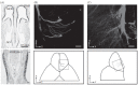
FIGURE 9.2
The Fseptal organ predominantly expresses a few odorant receptors and projects to the main olfactory bulb. (A) Two coronal sections were hybridized with an OMP (upper panel) or an MOR256–3 (lower panel) antisense RNA probe. Approximately 50% of (more...)
Most SO neurons express Gαolf and ACIII, suggesting that signal transduction in these neurons is mediated by the canonical cAMP pathway (Chapter 8), which is further supported by patch-clamp recordings from individual OSNs in this region. An adenylyl cyclase activator and a phosphodiesterase inhibitor mimicked odorant responses, and these responses were blocked by an adenylyl cyclase inhibitor and eliminated in CNGA2 null mice (Ma et al. 2003; Grosmaitre et al. 2007).
9.3.2. Central Projection
SO neurons send off a few axon bundles, which travel across the cribriform plate and terminate in the MOB. In rats, focal injection of horseradish peroxidase (HRP) in the MOB resulted in labeled cells in the SO (Pedersen and Benson 1986), while retrograde injection of HRP in the SO traced the axons to the posterior, ventromedial OB with a few densely labeled and many lightly labeled glomeruli (Astic and Saucier 1988; Giannetti et al. 1992). More recently, neurotracing studies using lipophilic dyes, such as Dil, were carried out in the mouse SO (Levai and Strotmann 2003; Ma et al. 2003). Genetically targeted OMP-GFP mice (the coding region of OMP was replaced by tauGFP; Potter et al. 2001) allow accurate identification and thus precise placement of Dil onto this area (Figure 9.1D) (Levai and Strotmann 2003). The labeled axons projected mainly to the posterior, ventromedial OB and targeted onto a few densely labeled glomeruli, plus many (up to 150) lightly labeled ones (Figure 9.2B and C). The densely labeled glomeruli (so called “septal” glomeruli) receive inputs mainly (if not exclusively) from the SO, while the lightly labeled glomeruli receive mixed inputs from the SO and the MOE. The projection pattern of the SO is consistent with the fact that this region expresses a few ORs at high densities and many other ORs sparsely.
9.3.3. Broad Responsiveness and Mechanosensitivity
Although the SO primarily covers a small fraction of the receptor repertoire, the majority (~70%) of SO neurons were surprisingly responsive to diverse chemicals with different size, shape, and functional groups, as demonstrated by patch-clamp recordings (Figure 9.3A). These neurons were extremely sensitive to some odorants with a nanomolar threshold and a wide dynamic range, which covers 3–4 log units of concentration from threshold to saturation (Figure 9.3B) (Grosmaitre et al. 2007). This is consistent with a previous EOG study in which the SO responded to many chemicals with a lower threshold than the MOE (Marshall and Maruniak 1986).
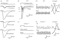
FIGURE 9.3
Most septal organ neurons respond to a wide range of odorants and to mechanical stimulation. (A) A single neuron from a wildtype (WT) animal responded to multiple odorants (except (+) limonene) at 300 μM, recorded with perforated patch-clamp. (more...)
The most dominant receptor, MOR256–3, was then confirmed to confer broad tuning to mouse OSNs using genetically targeted mouse lines. All OSNs expressing this receptor (regardless of their location in the SO or in the MOE) were highly responsive, while genetic deletion of this receptor resulted in more selective OSNs. The broad response spectrum of MOE256–3 was also verified in a heterologous cell line (our unpublished data). The SO, situated in the direct air path, may thus serve as a general odor detector and/or a sensor of the total odor concentration to “alert” the organism, a function originally proposed by Rodolfo-Masera (1943). The potential alerting role was not verified in a behavioral study with surgical removal of the SO (Giannetti et al. 1995b), probably due to the existence of similar OSNs in the MOE. An alternative and complementary hypothesis was suggested: that the SO functions as a “mini-nose” in surveying food odors as well as social cues (Breer et al. 2006).
Another surprising finding that arises from the SO is that many neurons responded not only to odorants, but also to mechanical stimuli delivered by pressure ejections of odorant-free Ringer solution (Figure 9.3A and B). The mechanical responses directly correlated with the pressure intensity and similar mechanosensitivity also existed in ~50% of the neurons in the MOE. The responses occurred with relatively long delays and were completely blocked by an adenylyl cyclase inhibitor, suggesting the involvement of cAMP as a second messenger. Elimination of mechanosensitivity in the OSNs from CNGA2 knockout mice further supports this notion (Figure 9.3C). Thus, the chemical and mechanical responses of the OSNs are mediated by a shared cascade involving cAMP and the CNG channel (Grosmaitre et al. 2007). The mechanosensitivity is probably tied to certain OR types, because all OSNs expressing MOR256–3 showed mechanical responses, while most OSNs with a deleted MOR256–3 gene displayed no mechanosensitivity (Grosmaitre et al. 2009). The SO can thus serve as an airflow sensor in addition to its chemosensory roles.
The mechanosensitivity found in the OSNs is particularly interesting, because these neurons are situated in the nostril and constantly experience episodic pressure changes carried by the airflow. One possible role of the mechanosensitivity is that when the air flows faster in the nose, such as during a powerful sniff, it can enhance the firing probability and frequency of individual OSNs weakly stimulated by odorants. In addition to the critical roles in odor delivery and sampling (Verhagen et al. 2007), sniffing may increase the overall sensitivity of the olfactory system via the mechanosensitivity of the sensory neurons (Grosmaitre et al. 2007). A second role that the mechanosensitivity of the OSNs may play is to synchronize the rhythmic activity (theta-band oscillation) in the OB with the breathing cycles, even in the absence of odorants, priming the system for processing odor information. In CNGA2 null mice, the OSNs failed to exhibit odorant and mechanical responses (Figure 9.3C), and the coupling between the bulb rhythmic activity and respiration was drastically reduced (Figure 9.3D and E). Therefore, the mechanosensitivity of the OSNs, in addition to episodic access to odorants, may cause the respiration-coupled, odorant-induced activity in the olfactory epithelium (Chaput 2000), the OB (Adrian 1951; Ueki and Domino 1961; Macrides and Chorover 1972; Onoda and Mori 1980; Philpot et al. 1997; Luo and Katz 2001; Cang and Isaacson 2003; Spors et al. 2006), and the olfactory cortex (Fontanini et al. 2003; Rennaker et al. 2007). The mechanosensitivity of OSNs provides new insights into the important roles played by sniffing in olfactory perception (Mainland and Sobel 2006). In fact, sniffing alone is sufficient to induce activities in the olfactory cortex in human subjects (Sobel et al. 1998). It remains to be determined how the “odor maps” along the olfactory pathway are modified under different breathing and sniffing patterns.
9.4. THE GRUENEBERG GANGLION (GG)
9.4.1. Central Targets
The GG is located bilaterally to the anterior vestibule of the nasal cavity (Figure 9.1A). Unlike the pseudostratified MOE, VNO, and SO, each GG contains small grape-like clusters of ~500 neurons (Figure 9.1C). This organ was found in all mammals examined, including human, and was originally thought to be nonsensory and part of the terminal nerve system (Grueneberg 1973). However, recent studies rediscovered the GG as a chemosensory organ. Surprisingly, GG neurons express OMP (a specific marker for mature chemosensory neurons) and, consequently, they are readily visible in genetically targeted OMP-GFP mice (Figure 9.1C). The GG starts to form around embryonic day 16 and becomes fully developed at birth. The axons of GG neurons fasciculate into a single or a few nerve bundles and project to the caudal MOB (Fuss et al. 2005; Koos and Fraser 2005; Fleischer et al. 2006a; Roppolo et al. 2006; Storan and Key 2006). Neurotracing by Dil reveals that GG axons innervate ~10 glomeruli (including one or two large ones) in the same region as the necklace glomeruli (the central targets of GC-D neurons) (Figure 9.1B). Because a subset of GG neurons (V2r83-positive, see below) express PDE2A, but not GC-D or CAII (two specific proteins in GC-D neurons) (Fuss et al. 2005; Fleischer et al. 2008), the PDE2A-positive glomeruli apparently contain heterogeneous populations. It is possible that individual PDE2A glomeruli converge inputs from GC-D and GG neurons, since all PDE2A glomeruli are innervated by GC-D neurons (Leinders-Zufall et al. 2007). However, the central targets of these two systems should be segregated at least to some extent, because some necklace glomeruli are homogenously innervated by GC-D fibers (Walz et al. 2007). Similar to the sensory neurons in other nasal organs, GG neurons undergo degeneration after axotomy, suggesting that the survival of these neurons depends on their central connections (Roppolo et al. 2006; Brechbuhl et al. 2008).
9.4.2. Morphology of Grueneberg Ganglion (GG) Neurons
GG neurons appear to lack the typical chemoreceptive structures, such as cilia or microvilli, under light or confocal microscopy (Fuss et al. 2005; Fleischer et al. 2006a; Roppolo et al. 2006; Storan and Key 2006). Using scanning electron microscopy, a recent study demonstrates that clusters of GG cells are located in a fibroblast meshwork between the nasal septum and a keratinized epithelium (KE) (Figure 9.4). Further examination by transmission electron microscopy reveals that each GG cell possesses 30–40 nonmotile primary cilia (15 μm long and 0.2 μm thick). These cilia are profoundly invaginated into the cytoplasm and are found in the extracellular matrix. Moreover, the GG also contains glial cells (immunostained by glial markers), which wrap around the OMP-positive neurons and trap most cilia within the ganglion. Since the cilia of GG neurons do not cross the KE layer to reach the nasal cavity, it raises the question of whether chemical compounds can reach these neurons. In standard skin permeability assays, the KE appears to be leaky, suggesting that water-soluble chemicals can have access to GG neurons (Brechbuhl et al. 2008).
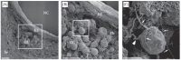
FIGURE 9.4
GG neurons contain ciliary processes. (A) A scanning EM micrograph shows a GG coronal section. Clusters of GG cells (GC) are located along the nasal septum (Se) underneath the keratinized epithelium (KE). NC, nasal cavity. Square detailed in (B). (B) (more...)
9.4.3. Chemoreceptors and Signal Transduction
Identification of chemoreceptors in GG neurons further supports their role in chemoreception. By combining RT-PCR and in situ hybridization, a substantial portion of OMP-positive GG neurons is found to express V2r83 (or V2R2), a V2R subtype expressed in all basal VNO neurons (Fleischer et al. 2006b). The remaining OMP-positive, V2r83-negative cells express several subtypes of TAARs (Fleischer et al. 2007). Certain ORs are transiently expressed in the GG. For instance, MOR256–17 is expressed in very few cells at the embryonic stages, but its expression disappears at postnatal stages (Fleischer et al. 2006b). Some signal transduction elements are identified in GG neurons, including Gα0 and Gαi2, which are probably coexpressed in single neurons (Fleischer et al. 2006b). Interestingly, the V2r83-positive neurons coexpress a transmembrane guanylyl cyclase subtype (GC-G) and PDE2A (Fleischer et al. 2008), indicating the existence of a cGMP-mediated signal transduction pathway.
9.4.4. Functions
9.4.4.1. Sensing Coolness
The number of GG neurons is particularly high in perinatal stages, followed by a decline in postnatal development, suggesting a more important function of this organ in newborns (Fuss et al. 2005; Fleischer et al. 2007). A subset of the necklace glomeruli (MGC: modified glomerular complex, Figure 9.1B) in rodent pups is activated during suckling behavior (Teicher et al. 1980; Greer et al. 1982), which leads to the hypothesis that the GG may play a role in mother/child interaction. To test this possibility, Mamasuew et al. (2008) examined c-Fos expression in the GG neurons of neonatal mouse pups in the presence and absence of the dam. Surprisingly, these neurons were only activated in the absence of the mother, and the activation was independent on olfactory cues revealed by naris closure. Cool ambient temperatures (but not warmer temperatures) were then confirmed to induce strong activity in V2r83-positive GG neurons (Figure 9.5A and B). The responses were significantly reduced in older stages, suggesting that these GG neurons serve as a thermosensor in newborns.
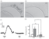
FIGURE 9.5
The Grueneberg ganglion fulfils multiple functions. (A, B) Placing isolated pups at 22°C (A) but not at 30°C (B) for 3 h induced c-Fos expression in the GG neurons. (C) Alarm pheromones (AP), but not general pheromones (GP), induced a (more...)
V2r83-positive GG neurons do not express TRPM8 (Fleischer et al. 2009), an ion channel essential for cold thermosensation (Bautista et al. 2007). These neurons instead coexpress several key components of the cGMP cascade (such as GC-G and PDE2A) (Fleischer et al. 2009), similar to the chemo- and thermosensitive AWC neurons in Caenorhabditis elegans (Kuhara et al. 2008). The molecular mechanisms underlying thermosensing in GG neurons remain to be determined.
9.4.4.2. Sensing Alarm Signals
Another group extensively researched the chemostimulus for GG neurons using calcium imaging on coronal tissue slices (Brechbuhl et al. 2008). A variety of stimuli, including mouse milk, mammary secretions from lactating female mice, a mix of odorants, some known pheromones, mouse urine, and C02, did not enhance the fluorescence of the GG neurons. Strikingly, alarm pheromones collected during the killing of mice with C02 induced transient calcium signals in almost all GG neurons from newborn and adult mice (Figure 9.5C). The calcium ions were preferentially released from the internal stores, because the signals were present in divalent-free solutions, but abolished by depletion of the internal calcium stores. The identity of the effective compounds in the alarm pheromones is still elusive.
The same group also examined the behavioral relevance of alarm pheromone sensing via GG cells by sectioning the GG axon bundles. Alarm pheromones elicited a stereotyped freezing reaction in rodents (Kikusui et al. 2001). Control mice displayed similar freezing behavior after exposure to alarm pheromones, which induced calcium signals in GG neurons. However, after the GG axotomy, the freezing behavior was replaced by exploring activity (Figure 9.5D). The behaviors of these mice were no longer affected by the same alarm pheromones. These results strongly support a role for GG neurons in alarm sensing. The GG apparently serves multiple functions and it is not clear if coolness and alarm signals are detected by the same set of GG neurons. Humans also possess this organ (Grueneberg 1973), but whether it has the same function(s) is not clear.
9.5. CONCLUDING REMARKS
Organization of the mammalian nose is more complicated than previously appreciated. Each of the four physically segregated apparatuses (MOE, VNO, SO, and GG) contains heterogeneous cell types with distinct chemoreceptors, transduction machineries, and/or central targets. Each organ can convey sensory information of multiple modalities and serve multiple functions. More subsystems may still emerge with new molecular markers and more detailed anatomical/functional analysis. Cutting-edge technology applied in modern neuroscience from various disciplines holds great hopes for revealing the specific roles played by each subsystem.
The advantage of having multiple olfactory subsystems is manifold. The different chemoreceptors expressed in these subsystems can expand the overall detection capacity of the olfactory system for chemicals and other stimulations. In addition, critical information can be processed in parallel by multiple subsystems, which send signals to different brain regions for further integration and execution. The diversity and complexity of the chemosensory systems allow the organisms to accurately perceive their chemical surroundings and respond appropriately by adjusting their behaviors, emotions, and hormones.
REFERENCES
- Adams D.R. Fine structure of the vomeronasal and septal olfactory epithelia and of glandular structures. Microsc Res Tech. 1992;23:86–97. [PubMed: 1392074]
- Adams D.R., McFarland L.Z. Septal olfactory organ in Peromyscus. Comp Biochem Physiol A. 1971;40:971–74. [PubMed: 4400102]
- Adrian E.D. The role of air movement in olfactory stimulation. J Physiol. 1951;114:4–5. [PubMed: 14861801]
- Astic L., Saucier D. Topographical projection of the septal organ to the main olfactory bulb in rats: Ontogenetic study. Brain Res. 1988;470:297–303. [PubMed: 2464410]
- Baker H., Cummings D.M., Munger S.D., Margolis J.W., Franzen L., Reed R.R., Margolis F.L. Targeted deletion of a cyclic nucleotide-gated channel subunit (OCNC1): Biochemical and morphological consequences in adult mice. J Neurosci. 1999;19:9313–21. [PMC free article: PMC6782908] [PubMed: 10531436]
- Bautista D.M., Siemens J., Glazer J.M., Tsuruda P.R., Basbaum A.I., Stucky C.L., Jordt S.E., Julius D. The menthol receptor TRPM8 is the principal detector of environmental cold. Nature. 2007;448:204–8. [PubMed: 17538622]
- Bojsen-Moller F. Demonstration of terminalis, olfactory, trigeminal and perivascular nerves in the rat nasal septum. J Comp Neurol. 1975;159:245–56. [PubMed: 1112913]
- Borowsky B., Adham N., Jones K.A., Raddatz R., Artymyshyn R., Ogozalek K.L., Durkin M.M., Lakhlani P.P., Bonini J.A., Pathirana S., Boyle N., Pu X., Kouranova E., Lichtblau H., Ochoa F.Y., Branchek T.A., Gerald C. Trace amines: Identification of a family of mammalian G protein-coupled receptors. Proc Natl Acad Sci USA. 2001;98:8966–71. [PMC free article: PMC55357] [PubMed: 11459929]
- Brechbuhl J., Klaey M., Broillet M.C. Grueneberg ganglion cells mediate alarm pheromone detection in mice. Science. 2008;321:1092–95. [PubMed: 18719286]
- Breer H., Fleischer J., Strotmann J. The sense of smell: Multiple olfactory subsystems. Cell Mol Life Sci. 2006;63:1465–75. [PubMed: 16732429]
- Breipohl W., Naguro T., Miragall F. Morphology of the Masera organ in NMRI mice (combined morphometric, freeze-fracture, light- and scanning electron microscopic investigations. Verh Anat Ges. 1983;77:741–43.
- Breipohl W., Naguro T., Walker D.G. The postnatal development of Maresa’s organ in the rat. Chem Senses. 1989;14:649–62.
- Broman I. Über die Entwickelung der konstanten grösseren NasenhöhlendrÜsen der Nagetiere. Anat Entw Gesch. 1921;60:439–586.
- Cang J., Isaacson J.S. In vivo whole-cell recording of odor-evoked synaptic transmission in the rat olfactory bulb. J Neurosci. 2003;23:4108–16. [PMC free article: PMC6741073] [PubMed: 12764098]
- Chaput M.A. EOG responses in anesthetized freely breathing rats. Chem Senses. 2000;25:695–701. [PubMed: 11114147]
- Duda T., Sharma R.K. ONE-GC membrane guanylate cyclase, a trimodal odorant signal transducer. Biochem Biophys Res Commun. 2008;367:440–45. [PubMed: 18178149]
- Elsaesser R., Montani G., Tirindelli R., Paysan J. Phosphatidyl-inositide signalling proteins in a novel class of sensory cells in the mammalian olfactory epithelium. Eur J Neurosci. 2005;21:2692–700. [PubMed: 15926917]
- Finger T.E., Bottger B., Hansen A., Anderson K.T., Alimohammadi H., Silver W.L. Solitary chemoreceptor cells in the nasal cavity serve as sentinels of respiration. Proc Natl Acad Sci USA. 2003;100:8981–86. [PMC free article: PMC166424] [PubMed: 12857948]
- Fleischer J., Hass N., Schwarzenbacher K., Besser S., Breer H. A novel population of neuronal cells expressing the olfactory marker protein (OMP) in the anterior/dorsal region of the nasal cavity. Histochem Cell Biol. 2006a;125:337–49. [PubMed: 16273384]
- Fleischer J., Mamasuew K., Breer H. Expression of cGMP signaling elements in the Grueneberg ganglion. Histochem Cell Biol. 2009;131:75–88. [PubMed: 18830617]
- Fleischer J., Schwarzenbacher K., Besser S., Hass N., Breer H. Olfactory receptors and signalling elements in the Grueneberg ganglion. J Neurochem. 2006b;98:543–54. [PubMed: 16805845]
- Fleischer J., Schwarzenbacher K., Breer H. Expression of trace amine-associated receptors in the Grueneberg ganglion. Chem Senses. 2007;32:623–31. [PubMed: 17556730]
- Fontanini A., Spano P., Bower J.M. Ketamine-xylazine-induced slow (<1.5 Hz) oscillations in the rat piriform (olfactory) cortex are functionally correlated with respiration. J Neurosci. 2003;23:7993–8001. [PMC free article: PMC6740491] [PubMed: 12954860]
- Forte L.R. Jr. Uroguanylin and guanylin peptides: Pharmacology and experimental therapeutics. Pharmacol Ther. 2004;104:137–62. [PubMed: 15518884]
- Fulle H.J., Vassar R., Foster D.C., Yang R.B., Axel R., Garbers D.L. A receptor guanylyl cyclase expressed specifically in olfactory sensory neurons. Proc Natl Acad Sci USA. 1995;92:3571–75. [PMC free article: PMC42209] [PubMed: 7724600]
- Fuss S.H., Omura M., Mombaerts P. The Grueneberg ganglion of the mouse projects axons to glomeruli in the olfactory bulb. Eur J Neurosci. 2005;22:2649–54. [PubMed: 16307607]
- Garbers D.L., Chrisman T.D., Wiegn P., Katafuchi T., Albanesi J.P., Bielinski V., Barylko B., Redfield M.M., Burnett J.C. Jr. Membrane guanylyl cyclase receptors: An update. Trends Endocrinol Metab. 2006;17:251–58. [PMC free article: PMC2647281] [PubMed: 16815030]
- Giannetti N., Pellier V., Oestreicher A.B., Astic L. Immunocytochemical study of the differentiation process of the septal organ of Masera in developing rats. Brain Res Dev Brain Res. 1995a;84:287–93. [PubMed: 7743649]
- Giannetti N., Saucier D., Astic L. Organization of the septal organ projection to the main olfactory bulb in adult and newborn rats. J Comp Neurol. 1992;323:288–98. [PubMed: 1383286]
- Giannetti N., Saucier D., Astic L. Analysis of the possible altering function of the septal organ in rats: A lesional and behavioral study. Physiol Behav. 1995;58:837–45. [PubMed: 8577878]
- Gloriam D.E., Bjarnadottir T.K., Yan Y.L., Postlethwait J.H., Schioth H.B., Fredriksson R. The repertoire of trace amine G-protein-coupled receptors: Large expansion in zebrafish. Mol Phylogenet Evol. 2005;35:470–82. [PubMed: 15804416]
- Graziadei P.P.C. Functional anatomy of the mammalian chemoreceptor system. In: Muller-Schwarze D., Mozell M.M., editors. Chemical Signals in Vertebrates. Plenum, New York: 1977. pp. 435–54.
- Greer C.A., Stewart W.B., Teicher M.H., Shepherd G.M. Functional development of the olfactory bulb and a unique glomerular complex in the neonatal rat. J Neurosci. 1982;2:1744–59. [PMC free article: PMC6564375] [PubMed: 7143049]
- Grosmaitre X., Santarelli L.C., Tan J., Luo M., Ma M. Dual functions of mammalian olfactory sensory neurons as odor detectors and mechanical sensors. Nat Neurosci. 2007;10:348–54. [PMC free article: PMC2227320] [PubMed: 17310245]
- Gruneberg H. A ganglion probably belonging to the N. terminalis system in the nasal mucosa of the mouse. Z Anat Entwicklungsgesch. 1973;140:39–52. [PubMed: 4749131]
- Hallem E.A., Sternberg P.W. Acute carbon dioxide avoidance in Caenorhabditis elegans. Proc Natl Acad Sci USA. 2008;105:8038–43. [PMC free article: PMC2430355] [PubMed: 18524955]
- Hashiguchi Y., Nishida M. Evolution of trace amine-associated receptor (TAAR) gene family in vertebrates: Lineage-specific expansions and degradations of a second class of vertebrate chemosensory receptors expressed in the olfactory epithelium. Mol Biol Evol. 2007;24:2099–107. [PubMed: 17634392]
- Hu J., Zhong C., Ding C., Chi Q., Walz A., Mombaerts P., Matsunami H., Luo M. Detection of near-atmospheric concentrations of CO2 by an olfactory subsystem in the mouse. Science. 2007;317:953–57. [PubMed: 17702944]
- Huang L., Shanker Y.G., Dubauskaite J., Zheng J.Z., Yan W., Rosenzweig S., Spielman A.I., Max M., Margolskee R.F. Ggamma13 colocalizes with gustducin in taste receptor cells and mediates IP3 responses to bitter denatonium. Nat Neurosci. 1999;2:1055–62. [PubMed: 10570481]
- Jones W.D., Cayirlioglu P., Kadow I.G., Vosshall L.B. Two chemosensory receptors together mediate carbon dioxide detection in Drosophila. Nature. 2007;445:86–90. [PubMed: 17167414]
- Juilfs D.M., Fulle H.J., Zhao A.Z., Houslay M.D., Garbers D.L., Beavo J.A. A subset of olfactory neurons that selectively express cGMP-stimulated phosphodiesterase (PDE2) and guanylyl cyclase-D define a unique olfactory signal transduction pathway. Proc Natl Acad Sci USA. 1997;94:3388–95. [PMC free article: PMC20380] [PubMed: 9096404]
- Kaluza J.F., Gussing F., Bohm S., Breer H., Strotmann J. Olfactory receptors in the mouse septal organ. J Neurosci Res. 2004;76:442–52. [PubMed: 15114616]
- Kaske S., Krasteva G., Konig P., Kummer W., Hofmann T., Gudermann T., Chubanov V. TRPM5, a taste-signaling transient receptor potential ion-channel, is a ubiquitous signaling component in chemosensory cells. BMC Neurosci. 2007;8 [PMC free article: PMC1931605] [PubMed: 17610722]
- Katz S., Merzel J. Distribution of epithelia and glands of the nasal septum mucosa in the rat. Acta Anat (Basel). 1977;99:58–66. [PubMed: 70949]
- Kikusui T., Takigami S., Takeuchi Y., Mori Y. Alarm pheromone enhances stress-induced hyperthermia in rats. Physiol Behav. 2001;72:45–50. [PubMed: 11239980]
- Koos D.S., Fraser S.E. The Grueneberg ganglion projects to the olfactory bulb. Neuroreport. 2005;16:1929–32. [PubMed: 16272881]
- Kratzing J.E. The structure and distribution of nasal glands in four marsupial species. J Anat. 1984a;139:553–64. [PMC free article: PMC1165068] [PubMed: 6490535]
- Kratzing J.E. The anatomy and histology of the nasal cavity of the koala (Phascolarctos cinereus). J Anat. 1984;138:55–65. [PMC free article: PMC1164310] [PubMed: 6706839]
- Kuhara A., Okumura M., Kimata T., Tanizawa Y., Takano R., Kimura K.D., Inada H., Matsumoto K., Mori I. Temperature sensing by an olfactory neuron in a circuit controlling behavior of C. elegans. Science. 2008;320:803–7. [PubMed: 18403676]
- Leinders-Zufall T., Cockerham R.E., Michalakis S., Biel M., Garbers D.L., Reed R.R., Zufall F., Munger S.D. Contribution of the receptor guanylyl cyclase GC-D to chemosensory function in the olfactory epithelium. Proc Natl Acad Sci USA. 2007;104:14507–12. [PMC free article: PMC1964822] [PubMed: 17724338]
- Levai O., Strotmann J. Projection pattern of nerve fibers from the septal organ: DiI-tracing studies with transgenic OMP mice. Histochem Cell Biol. 2003;120:483–92. [PubMed: 14628145]
- Liberles S.D., Buck L.B. A second class of chemosensory receptors in the olfactory epithelium. Nature. 2006;442:645–50. [PubMed: 16878137]
- Liman E.R., Corey D.P., Dulac C. TRP2: A candidate transduction channel for mammalian pheromone sensory signaling. Proc Natl Acad Sci USA. 1999;96:5791–96. [PMC free article: PMC21939] [PubMed: 10318963]
- Lin D.Y., Zhang S.Z., Block E., Katz L.C. Encoding social signals in the mouse main olfactory bulb. Nature. 2005;434:470–77. [PubMed: 15724148]
- Lin W., Arellano J., Slotnick B., Restrepo D. Odors detected by mice deficient in cyclic nucleotide-gated channel subunit A2 stimulate the main olfactory system. J Neurosci. 2004;24:3703–10. [PMC free article: PMC6729751] [PubMed: 15071119]
- Lin W., Margolskee R., Donnert G., Hell S.W., Restrepo D. Olfactory neurons expressing transient receptor potential channel M5 (TRPM5) are involved in sensing semiochemicals. Proc Natl Acad Sci USA. 2007;104:2471–76. [PMC free article: PMC1892929] [PubMed: 17267604]
- Lin W., Ogura T., Margolskee R.F., Finger T.E., Restrepo D. TRPM5-expressing solitary chemosensory cells respond to odorous irritants. J Neurophysiol. 2008;99:1451–60. [PubMed: 18160424]
- Luo M., Katz L.C. Response correlation maps of neurons in the mammalian olfactory bulb. Neuron. 2001;32:1165–79. [PubMed: 11754845]
- Ma M. Encoding olfactory signals via multiple chemosensory systems. Crit Rev Biochem Mol Biol. 2007;42:463–80. [PubMed: 18066954]
- Ma M., Grosmaitre X., Iwema C.L., Baker H., Greer C.A., Shepherd G.M. Olfactory signal transduction in the mouse septal organ. J Neurosci. 2003;23:317–24. [PMC free article: PMC2227318] [PubMed: 12514230]
- Macrides F., Chorover S.L. Olfactory bulb units: Activity correlated with inhalation cycles and odor quality. Science. 1972;175:84–87. [PubMed: 5008584]
- Mainland J., Sobel N. The sniff is part of the olfactory percept. Chem Senses. 2006;31:181–96. [PubMed: 16339268]
- Mamasuew K., Breer H., Fleischer J. Grueneberg ganglion neurons respond to cool ambient temperatures. Eur J Neurosci. 2008;28:1775–85. [PubMed: 18973593]
- Marshall D.A., Maruniak J.A. Masera’s organ responds to odorants. Brain Res. 1986;366:329–32. [PubMed: 3697687]
- Meyer M.R., Angele A., Kremmer E., Kaupp U.B., Muller F. A cGMP-signaling pathway in a subset of olfactory sensory neurons. Proc Natl Acad Sci USA. 2000;97:10595–600. [PMC free article: PMC27070] [PubMed: 10984544]
- Miragall F., Breipohl W., Naguro T., Voss-Wermbter G. Freeze-fracture study of the plasma membranes of the septal olfactory organ of Masera. J Neurocytol. 1984;13:111–25. [PubMed: 6707707]
- Montani G., Tonelli S., Elsaesser R., Paysan J., Tirindelli R. Neuropeptide Y in the olfactory microvillar cells. Eur J Neurosci. 2006;24:20–24. [PubMed: 16800866]
- Onoda N., Mori K. Depth distribution of temporal firing patterns in olfactory bulb related to air-intake cycles. J Neurophysiol. 1980;44:29–39. [PubMed: 7420137]
- Pedersen P.E., Benson T.E. Projection of septal organ receptor neurons to the main olfactory bulb in rats. J Comp Neurol. 1986;252:555–62. [PubMed: 3782515]
- Perez C.A., Huang L., Rong M., Kozak J.A., Preuss A.K., Zhang H., Max M., Margolskee R.F. A transient receptor potential channel expressed in taste receptor cells. Nat Neurosci. 2002;5:1169–76. [PubMed: 12368808]
- Philpot B.D., Foster T.C., Brunjes P.C. Mitral/tufted cell activity is attenuated and becomes uncoupled from respiration following naris closure. J Neurobiol. 1997;33:374–86. [PubMed: 9322155]
- Potter S.M., Zheng C., Koos D.S., Feinstein P., Fraser S.E., Mombaerts P. Structure and emergence of specific olfactory glomeruli in the mouse. J Neurosci. 2001;21:9713–23. [PMC free article: PMC2570017] [PubMed: 11739580]
- Ramsey I.S., Delling M., Clapham D.E. An introduction to TRP channels. Annu Rev Physiol. 2006;68:619–47. [PubMed: 16460286]
- Rennaker R.L., Chen C.F., Ruyle A.M., Sloan A.M., Wilson D.A. Spatial and temporal distribution of odorant-evoked activity in the piriform cortex. J Neurosci. 2007;27:1534–42. [PMC free article: PMC2291208] [PubMed: 17301162]
- Ring G., Mezza R.C., Schwob J.E. Immunohistochemical identification of discrete subsets of rat olfactory neurons and the glomeruli that they innervate. J Comp Neurol. 1997;388:415–34. [PubMed: 9368850]
- Rodolfo-Masera T. Su 1’esistenza di un particolare organo olfattivo nel setto nasale della cavia e di altri roditori. Arch Ital Anat Embryol. 1943;48:157–212.
- Roppolo D., Ribaud V., Jungo V.P., Luscher C., Rodriguez I. Projection of the Gruneberg ganglion to the mouse olfactory bulb. Eur J Neurosci. 2006;23:2887–94. [PubMed: 16819977]
- Shinoda K., Ohtsuki T., Nagano M., Okumura T. A possible functional necklace formed by placental antigen X-P2-immunoreactive and intensely acetylcholinesterase-reactive (PAX/IAE) glomerular complexes in the rat olfactory bulb. Brain Res. 1993;618:160–66. [PubMed: 8402170]
- Shinoda K., Shiotani Y., Osawa Y. ‘Necklace olfactory glomeruli’ form unique components of the rat primary olfactory system. J Comp Neurol. 1989;284:362–73. [PubMed: 2754040]
- Sobel N., Prabhakaran V., Desmond J.E., Glover G.H., Goode R.L., Sullivan E.V., Gabrieli J.D. Sniffing and smelling: Separate subsystems in the human olfactory cortex. Nature. 1998;392:282–86. [PubMed: 9521322]
- Spehr M., Kelliher K.R., Li X.H., Boehm T., Leinders-Zufall T., Zufall F. Essential role of the main olfactory system in social recognition of major histocompatibility complex peptide ligands. J Neurosci. 2006;26:1961–70. [PMC free article: PMC6674934] [PubMed: 16481428]
- Spors H., Wachowiak M., Cohen L.B., Friedrich R.W. Temporal dynamics and latency patterns of receptor neuron input to the olfactory bulb. J Neurosci. 2006;26:1247–59. [PMC free article: PMC6674558] [PubMed: 16436612]
- Storan M.J., Key B. Septal organ of Gruneberg is part of the olfactory system. J Comp Neurol. 2006;494:834–44. [PubMed: 16374816]
- Suh G.S., Wong A.M., Hergarden A.C., Wang J.W., Simon A.F., Benzer S., Axel R., Anderson D.J. A single population of olfactory sensory neurons mediates an innate avoidance behaviour in Drosophila. Nature. 2004;431:854–59. [PubMed: 15372051]
- Sun L., Wang H., Hu J., Han J., Luo M. Guanylyl cyclase-D in the olfactory CO2 neurons is activated by bicarbonate. Proc Natl Acad Sci USA. 2009;106:2041–46. [PMC free article: PMC2644160] [PubMed: 19181845]
- Taniguchi K., Arai T., Ogawa K. Fine structure of the septal olfactory organ of Masera and its associated gland in the golden hamster. J Vet Med Sci. 1993;55:107–16. [PubMed: 8461403]
- Teicher M.H., Stewart W.B., Kauer J.S., Shepherd G.M. Suckling pheromone stimulation of a modified glomerular region in the developing rat olfactory bulb revealed by the 2-deoxyglucose method. Brain Res. 1980;194:530–35. [PubMed: 7388629]
- Tian H., Ma M. Molecular organization of the olfactory septal organ. J Neurosci. 2004;24:8383–90. [PMC free article: PMC2227317] [PubMed: 15385621]
- Tian H., Ma M. Activity plays a role in eliminating olfactory sensory neurons expressing multiple odorant receptors in the mouse septal organ. Mol Cell Neurosci. 2008a;38:484–88. [PMC free article: PMC2574655] [PubMed: 18538580]
- Tian H., Ma M. Differential development of odorant receptor expression patterns in the olfactory epithelium: A quantitative analysis in the mouse septal organ. Dev Neurobiol. 2008b;68:476–86. [PMC free article: PMC2266684] [PubMed: 18214836]
- Ueki S., Domino E.F. Some evidence for a mechanical receptor in olfactory function. J Neurophysiol. 1961;24:12–25. [PubMed: 13778965]
- Venkatachalam K., Montell C. TRP channels. Annu Rev Biochem. 2007;76:387–417. [PMC free article: PMC4196875] [PubMed: 17579562]
- Verhagen J.V., Wesson D.W., Netoff T.I., White J.A., Wachowiak M. Sniffing controls an adaptive filter of sensory input to the olfactory bulb. Nat Neurosci. 2007;10:631–39. [PubMed: 17450136]
- Walz A., Feinstein P., Khan M., Mombaerts P. Axonal wiring of guanylate cyclase-D-expressing olfactory neurons is dependent on neuropilin 2 and semaphorin 3F. Development. 2007;134:4063–72. [PubMed: 17942483]
- Wang Z., Balet Sindreu, C., Li V., Nudelman A., Chan G.C., Storm D.R. Pheromone detection in male mice depends on signaling through the type 3 adenylyl cyclase in the main olfactory epithelium. J Neurosci. 2006;26:7375–79. [PMC free article: PMC6674185] [PubMed: 16837584]
- Weiler E., Farbman A.I. The septal organ of the rat during postnatal development. Chem Senses. 2003;28:581–93. [PubMed: 14578120]
- Xu F., Schaefer M., Kida I., Schafer J., Liu N., Rothman D.L., Hyder F., Restrepo D., Shepherd G.M. Simultaneous activation of mouse main and accessory olfactory bulbs by odors or pheromones. J Comp Neurol. 2005;489:491–500. [PubMed: 16025460]
- Young J.M., Waters H., Dong C., Fulle H.J., Liman E.R. Degeneration of the olfactory guanylyl cyclase D gene during primate evolution. PLoS ONE. 2007;2 [PMC free article: PMC1964805] [PubMed: 17849013]
- Youngentob S.L., Hornung D.E., Mozell M.M. Determination of carbon dioxide detection thresholds in trained rats. Physiol Behav. 1991;49:21–26. [PubMed: 1901996]
- Zhang X., Rogers M., Tian H., Zou D.J., Liu J., Ma M., Shepherd G.M., Firestein S.J. High-throughput microarray detection of olfactory receptor gene expression in the mouse. Proc Natl Acad Sci USA. 2004;101:14168–73. [PMC free article: PMC521132] [PubMed: 15377787]
- Zhang Y., Hoon M.A., Chandrashekar J., Mueller K.L., Cook B., Wu D., Zuker C.S., Ryba N.J. Coding of sweet, bitter, and umami tastes: Different receptor cells sharing similar signaling pathways. Cell. 2003;112:293–301. [PubMed: 12581520]
- Review Vomeronasal Receptors and Signal Transduction in the Vomeronasal Organ of Mammals.[Neurobiology of Chemical Commu...]Review Vomeronasal Receptors and Signal Transduction in the Vomeronasal Organ of Mammals.Francia S, Pifferi S, Menini A, Tirindelli R. Neurobiology of Chemical Communication. 2014
- Review Encoding olfactory signals via multiple chemosensory systems.[Crit Rev Biochem Mol Biol. 2007]Review Encoding olfactory signals via multiple chemosensory systems.Ma M. Crit Rev Biochem Mol Biol. 2007 Nov-Dec; 42(6):463-80.
- Review Odor and pheromone sensing via chemoreceptors.[Adv Exp Med Biol. 2012]Review Odor and pheromone sensing via chemoreceptors.Ma M. Adv Exp Med Biol. 2012; 739:93-106.
- Review Engineering Aspects of Olfaction.[Neuromorphic Olfaction. 2013]Review Engineering Aspects of Olfaction.Persaud KC. Neuromorphic Olfaction. 2013
- Review Signaling mechanisms and behavioral function of the mouse basal vomeronasal neuroepithelium.[Front Neuroanat. 2014]Review Signaling mechanisms and behavioral function of the mouse basal vomeronasal neuroepithelium.Pérez-Gómez A, Stein B, Leinders-Zufall T, Chamero P. Front Neuroanat. 2014; 8:135. Epub 2014 Nov 26.
- Multiple Olfactory Subsystems Convey Various Sensory Signals - The Neurobiology ...Multiple Olfactory Subsystems Convey Various Sensory Signals - The Neurobiology of Olfaction
Your browsing activity is empty.
Activity recording is turned off.
See more...