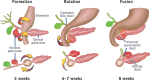
The pancreas first appears at approximately 5 weeks of gestation as two outpouchings of the endodermal lining of the duodenum just distal to the forming stomach (Figure 5). The outpouchings are the ventral and dorsal pancreas. The dorsal pancreas grows more rapidly than the ventral pancreas. In addition, the ventral pancreas rotates toward the dorsal pancreas as it is “carried” by the common bile duct. Finally, the ventral and dorsal pancreas join and the ductal systems fuse so that secretions from the ventral pancreas enter the shared ductal system of the ventral pancreas and common bile duct. In the final anatomic arrangement, the head of the pancreas originates from both the dorsal pancreas and the ventral pancreas. The ventral pancreas portion is called the uncinate process. The body and tail of the pancreas originate from the dorsal pancreas.
The signaling pathways underlying the development process include the Hedgehog system, the homeobox gene Pdx1 and Notch signaling [22, 23]. Inhibition of Hedgehog signaling leads to ectopic budding of pancreatic structures in the stomach and the duodenum [24]. Pdx1 expression in the duodenum during development marks the location of pancreatic bud development [23]. Notch signaling inhibits endocrine cell differentiation and promotes exocrine cell differentiation [25]. Inhibition of Notch signaling results in marked endocrine cell expansion with blockade of exocrine cell development.
Publication Details
Copyright
Publisher
Morgan & Claypool Life Sciences, San Rafael (CA)
NLM Citation
Pandol SJ. The Exocrine Pancreas. San Rafael (CA): Morgan & Claypool Life Sciences; 2010. Pancreatic Embryology and Development.
