Introduction
The appendicular skeleton is one of two major bone groups in the body, the other being the axial skeleton. The appendicular skeleton is comprised of the upper and lower extremities, which include the shoulder girdle and pelvis. The shoulder girdle and pelvis provide connection points between the appendicular skeleton and the axial skeleton to where mechanical loads transfer. Of the 206 bones in the adult human body, a total of 126 bones form the appendicular skeleton. The bones that contribute to the appendicular skeleton include the bones of the hands, feet, upper extremity, lower extremity, shoulder girdle, and pelvic bones.[1]
A single upper extremity includes 14 phalanges (proximal, intermediate, and distal), five metacarpals, eight carpal bones, two forearm bones (radius and ulna), the humerus, and the shoulder girdle (scapula and clavicle).[2] A single lower extremity contains 14 phalanges (proximal, intermediate, and distal), five metatarsals, seven tarsal bones, two leg bones (fibula, tibia), the femur, and the hip bone or coxal bone (ilium, ischium, and pubis).[3][4]
These bones articulate with each other and are joined by a multitude of ligaments, cartilage, and tendons to form the appendicular skeleton. There are also bony prominences and protuberances that serve as muscle attachment sites on the surfaces of these bones. The appendicular skeleton is structured for a greater range of motion and locomotion generation when compared to the axial skeleton.[5]
Structure and Function
There are 126 named bones of the appendicular skeleton (all bones exist in pairs) [1]:
Upper Limb
Shoulder Girdle:
Arm
Metacarpals x5
Phalanx x14
Scaphoid
Lunate
Triquetrum
Pisiform
Trapezium
Trapezoid
Capitate
Hamate
Radius
Ulna
Humerus
Forearm
Wrist or Carpal Bones
Hand
Lower Limb
Ilium
Ischium
Pubis
Femur
Tibia
Fibula
Talas
Calcaneus
Cuboid
Medial, intermediate, and lateral cuneiform
Navicular
Metatarsals x5
Phalanx x14
There are also various sesamoid bones not included in the list, such as the largest of the sesamoid bones, the patella, which protects the knee joint, and important attachment points for the ligaments that allow knee extension.[6][7]
There are two bilateral joints where the appendicular skeleton directly articulates with the axial skeleton. The first of these articulations is the sternoclavicular joint, where the sternum of the axial skeleton articulates with the clavicle of the appendicular skeleton. The sternoclavicular joint is a synovial joint.[8]
The second point where the appendicular skeleton directly articulates with the axial skeleton is the sacroiliac joint, where the sacrum articulates with the ilium. The sacroiliac joint is both a synovial joint and a syndesmosis. The connection between the sacrum and the ilium is important to transfer the load of the axial skeleton to the lower limb of the appendicular skeleton.[9]
The thoracoscapular articulation is a second articulation between the upper limb of the appendicular skeleton and the axial skeleton. This articulation is not an actual joint and does not have a synovial membrane. The thoracoscapular articulation forms between the anterior surface of the scapula and the posterior ribs 2 through 7.[10]
The bones of the foot function to form a base where the skeleton contacts the ground while standing. During the gait cycle, the articulations between the bones of the foot combined with the fascia and ligaments allow for deformation of the arches, which create spring-like properties in the foot utilized during walking and running.[11]
Embryology
The appendicular skeleton first appears as limb buds near the end of the first month of embryogenesis. There are two upper limb buds and two lower limb buds. These form when the lateral plate mesoderm grows outwards. As these limb buds grow outwards, chondrification forms hyaline cartilage around the sixth week and continues cartilage growth in the limb buds. This chondrification continues rapidly in a proximal to distal manner.[12] Around the tenth week, the ossification of the cartilage begins.[13] Ossification continues following birth with secondary and ultimately complete ossification that is ongoing until around 20 years of age.[14]
Blood Supply and Lymphatics
The blood supply to the lower extremity of the appendicular skeleton originates from the common iliac arteries, which are the terminal branches of the descending aorta. The common iliac artery branches into the internal and external iliac arteries, supplying all the structures of the pelvis and the lower extremities.[15] The external iliac artery continues into the lower extremity to become the femoral artery as it passes under the inguinal ligament.[16]
A major branch of the femoral artery is the deep femoral artery. The deep femoral artery supplies blood to the femur. The medial femoral circumflex artery and lateral femoral circumflex artery are early branches of the deep femoral artery that vascularize the hip joint.[16] The femoral artery continues posteriorly to the knee as the popliteal artery, then continues into the lower leg, dividing into the anterior and posterior tibial arteries. The posterior tibial artery then bifurcates into the posterior tibial and fibular arteries, which distally contribute to the vasculature of the foot.[17][18][19]
The blood supply to the upper extremity of the appendicular skeleton comes from the subclavian artery. The subclavian artery is a branch of the brachiocephalic trunk on the right or a branch directly off the aortic arch on the left. The clavicle receives vascular supply from the suprascapular artery, thoracoacromial artery, and the internal thoracic artery.[20]
The subclavian artery becomes the axillary artery after the lateral edge of the first rib. It then becomes the brachial artery after passing the inferior border of the teres minor muscle. The brachial artery bifurcates near the elbow into the radial and ulnar arteries, which distally contribute to the vasculature of the hands.[21][22]
The upper and lower limb lymphatics primarily follow the major blood vessels.[23]
Nerves
The upper extremity nerves originate from the brachial plexus. The brachial plexus is composed of roots, trunks, divisions, cords, and, ultimately, the five named branches. Spinal nerve roots C5 to T1 contribute to the brachial plexus. The terminal branches of the brachial plexus include the musculocutaneous, axillary, median, radial, and ulnar nerves. These named branches provide innervation to the upper limb.[24]
The lower extremity innervation originates from the lumbar plexus and the sacral plexus, which is formed by spinal nerve roots T12 to S3.[25] Part of the lumbosacral plexus forms the sciatic nerve, which contributes most of the innervation to the lower limb. The sciatic nerve divides into the tibial and fibular nerves, which continue distally to innervate the lower limb.[26]
Physiologic Variants
There are several physiologic variants of the appendicular skeleton. The text below briefly describes several of the anomalies.
Polymelia is a congenital duplication of a limb or appendage. This anomaly is rare in humans but often appears in animals; it seems to occur due to the incomplete separation of monozygotic twins. The gene responsible for polymelia may be related to the disorganization Ds gene seen in mice.[27]
Syndactyly, also known as webbed digits, is the partial or total conjoining of digits in the upper or lower limb; this is one of the most common limb malformations with a prevalence of 3 to 10 per 10,000 births. Syndactyly can present unilateral or bilateral. These are categorized as partial or complete based on the extent of webbing or fusion. Syndactyly can then further sub-classify as simple or complex. Simple syndactyly only involves the fusion of soft tissue, compared to complex, which involves bone fusion. Correcting syndactyl requires surgical intervention.[28][29]
Polydactyly is where a hand or foot has an extra digit. The condition can manifest as anything from a small raised lump or partially formed digit to a fully formed and functioning extra digit. There are three classifications of polydactyly for the hand. The most common, postaxial polydactyly, is where the extra digit arises on the ulnar side of the hand. Preaxial polydactyly is where the extra digit arises on the radial side of the hand. The final classification, central polydactyly, is where the digit arises somewhere in between.[30]
Polydactyly of the foot utilizes a similar classification system, with preaxial on the medial side of the foot, postaxial on the lateral side of the foot, and central somewhere between the previous two.[31] These malformations are often surgically removed at a young age.[32]
Another digit malformation is triphalangeal thumbs, which is a rare congenital anomaly. In this condition, the thumb has three phalanges (proximal, intermediate, and distal) instead of the usual two phalanges (proximal and distal). This anomaly often makes the thumb appear “finger-like” due to its increased length. Like the other digit malformations, triphalangeal thumbs are repairable surgically.[33][34]
A supracondylar process is a bony projection on the anterior surface of the humerus. This process points downward towards the medial epicondyle. Struthers ligament is a ligament that can accompany this bony malformation. The ligament attaches from the supracondylar process to the medial epicondyle. These variants are typically asymptomatic, although there are reports of cases where Struthers ligament entraps structures such as the median nerve.[35][36][37]
Clinical Significance
The appendicular skeleton is clinically relevant in many areas of medicine. External forces applied to the appendicular skeleton from traumas can lead to fractured bones. In the upper limb, the most common fracture is a distal radius and ulna fracture. The second most common fracture site is the phalanges and metacarpals of the hand.[38]
Repetitive smaller forces acting on the appendicular skeleton can also lead to stress fractures. A study of lower extremity stress fractures in the United States military found that the tibia and fibula were the most common location of stress fractures.[39]
Bones of the appendicular skeleton can also be a primary site for malignancy, such as multiple myeloma or osteosarcoma.[40][41] The joints of the appendicular skeleton are also susceptible to a wide variety of pathologies, including osteoarthritis, rheumatoid arthritis, and gout, to name a few. The bones of the appendicular skeleton often undergo imaging through various modalities, including X-ray, computed tomography scan, and magnetic resonance imaging. The imaging technique selected is dependent on the pathology that is being imaged.[42]
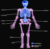
Appendicular skeleton Image courtesy S Bhimji MD
References
- 1.
Docherty B. Skeletal system: part four--the appendicular skeleton.
Nurs Times. 2007 Feb 20-26;103(8):26-7. [
PubMed: 17333873]
- 2.
Panchal-Kildare S, Malone K. Skeletal anatomy of the hand.
Hand Clin. 2013 Nov;29(4):459-71. [
PubMed: 24209945]
- 3.
Wobser AM, Adkins Z, Wobser RW.
StatPearls [Internet]. StatPearls Publishing; Treasure Island (FL): Jul 24, 2023. Anatomy, Abdomen and Pelvis: Bones (Ilium, Ischium, and Pubis) [
PubMed: 30137809]
- 4.
Ficke J, Byerly DW.
StatPearls [Internet]. StatPearls Publishing; Treasure Island (FL): Aug 7, 2023. Anatomy, Bony Pelvis and Lower Limb: Foot. [
PubMed: 31536304]
- 5.
Brockett CL, Chapman GJ. Biomechanics of the ankle.
Orthop Trauma. 2016 Jun;30(3):232-238. [
PMC free article: PMC4994968] [
PubMed: 27594929]
- 6.
Fox AJ, Wanivenhaus F, Rodeo SA. The basic science of the patella: structure, composition, and function.
J Knee Surg. 2012 May;25(2):127-41. [
PubMed: 22928430]
- 7.
Luo TD, Marino DV, Pilson H.
StatPearls [Internet]. StatPearls Publishing; Treasure Island (FL): Jul 31, 2023. Patella Fractures. [
PubMed: 30020702]
- 8.
Dhawan R, Singh RA, Tins B, Hay SM. Sternoclavicular joint.
Shoulder Elbow. 2018 Oct;10(4):296-305. [
PMC free article: PMC6134528] [
PubMed: 30214497]
- 9.
Vleeming A, Schuenke MD, Masi AT, Carreiro JE, Danneels L, Willard FH. The sacroiliac joint: an overview of its anatomy, function and potential clinical implications.
J Anat. 2012 Dec;221(6):537-67. [
PMC free article: PMC3512279] [
PubMed: 22994881]
- 10.
Frank RM, Ramirez J, Chalmers PN, McCormick FM, Romeo AA. Scapulothoracic anatomy and snapping scapula syndrome.
Anat Res Int. 2013;2013:635628. [
PMC free article: PMC3863500] [
PubMed: 24369502]
- 11.
McKeon PO, Hertel J, Bramble D, Davis I. The foot core system: a new paradigm for understanding intrinsic foot muscle function.
Br J Sports Med. 2015 Mar;49(5):290. [
PubMed: 24659509]
- 12.
Al-Qattan MM, Kozin SH. Update on embryology of the upper limb.
J Hand Surg Am. 2013 Sep;38(9):1835-44. [
PubMed: 23684522]
- 13.
Ortega N, Behonick DJ, Werb Z. Matrix remodeling during endochondral ossification.
Trends Cell Biol. 2004 Feb;14(2):86-93. [
PMC free article: PMC2779708] [
PubMed: 15102440]
- 14.
Verbruggen SW, Nowlan NC. Ontogeny of the Human Pelvis.
Anat Rec (Hoboken). 2017 Apr;300(4):643-652. [
PubMed: 28297183]
- 15.
Yiming A, Baqué P, Rahili A, Mayer J, Braccini AL, Fontaine A, Leplatois A, Clavé A, Bourgeon A, de Peretti F. Anatomical study of the blood supply of the coxal bone: radiological and clinical application.
Surg Radiol Anat. 2002 May;24(2):81-6. [
PubMed: 12197024]
- 16.
Zlotorowicz M, Czubak-Wrzosek M, Wrzosek P, Czubak J. The origin of the medial femoral circumflex artery, lateral femoral circumflex artery and obturator artery.
Surg Radiol Anat. 2018 May;40(5):515-520. [
PMC free article: PMC5937904] [
PubMed: 29651567]
- 17.
Hirtler L, Lübbers A, Rath C. Vascular coverage of the anterior knee region - an anatomical study.
J Anat. 2019 Aug;235(2):289-298. [
PMC free article: PMC6637446] [
PubMed: 31070789]
- 18.
Hyland S, Sinkler MA, Varacallo M.
StatPearls [Internet]. StatPearls Publishing; Treasure Island (FL): Jul 25, 2023. Anatomy, Bony Pelvis and Lower Limb: Popliteal Region. [
PubMed: 30422486]
- 19.
Olewnik Ł, Łabętowicz P, Podgórski M, Polguj M, Ruzik K, Topol M. Variations in terminal branches of the popliteal artery: cadaveric study.
Surg Radiol Anat. 2019 Dec;41(12):1473-1482. [
PMC free article: PMC6853856] [
PubMed: 31134299]
- 20.
Knudsen FW, Andersen M, Krag C. The arterial supply of the clavicle.
Surg Radiol Anat. 1989;11(3):211-4. [
PubMed: 2588097]
- 21.
McCausland C, Sawyer E, Eovaldi BJ, Varacallo M.
StatPearls [Internet]. StatPearls Publishing; Treasure Island (FL): Aug 8, 2023. Anatomy, Shoulder and Upper Limb, Shoulder Muscles. [
PubMed: 30521257]
- 22.
Haładaj R, Wysiadecki G, Dudkiewicz Z, Polguj M, Topol M. The High Origin of the Radial Artery (Brachioradial Artery): Its Anatomical Variations, Clinical Significance, and Contribution to the Blood Supply of the Hand.
Biomed Res Int. 2018;2018:1520929. [
PMC free article: PMC6016218] [
PubMed: 29992133]
- 23.
Ma CX, Pan WR, Liu ZA, Zeng FQ, Qiu ZQ, Liu MY. Deep lymphatic anatomy of the upper limb: An anatomical study and clinical implications.
Ann Anat. 2019 May;223:32-42. [
PubMed: 30716466]
- 24.
Orebaugh SL, Williams BA. Brachial plexus anatomy: normal and variant.
ScientificWorldJournal. 2009 Apr 28;9:300-12. [
PMC free article: PMC5823154] [
PubMed: 19412559]
- 25.
Di Benedetto P, Pinto G, Arcioni R, De Blasi RA, Sorrentino L, Rossifragola I, Baciarello M, Capotondi C. Anatomy and imaging of lumbar plexus.
Minerva Anestesiol. 2005 Sep;71(9):549-54. [
PubMed: 16166916]
- 26.
Dupont G, Unno F, Iwanaga J, Oskouian RJ, Tubbs RS. A Variant of the Sciatic Nerve and its Clinical Implications.
Cureus. 2018 Jun 25;10(6):e2874. [
PMC free article: PMC6110408] [
PubMed: 30155377]
- 27.
Montalvo N, Redrobán L, Espín VH. Incomplete duplication of a lower extremity (polymelia): a case report.
J Med Case Rep. 2014 Jun 12;8:184. [
PMC free article: PMC4077643] [
PubMed: 24920152]
- 28.
Malik S. Syndactyly: phenotypes, genetics and current classification.
Eur J Hum Genet. 2012 Aug;20(8):817-24. [
PMC free article: PMC3400728] [
PubMed: 22333904]
- 29.
Kvernmo HD, Haugstvedt JR. Treatment of congenital syndactyly of the fingers.
Tidsskr Nor Laegeforen. 2013 Aug 20;133(15):1591-5. [
PubMed: 23970273]
- 30.
Comer GC, Potter M, Ladd AL. Polydactyly of the Hand.
J Am Acad Orthop Surg. 2018 Feb 01;26(3):75-82. [
PubMed: 29309292]
- 31.
Belthur MV, Linton JL, Barnes DA. The spectrum of preaxial polydactyly of the foot.
J Pediatr Orthop. 2011 Jun;31(4):435-47. [
PubMed: 21572282]
- 32.
Kyriazis Z, Kollia P, Grivea I, Varitimidis SE, Constantoulakis P, Dailiana ZH. Thumb duplication: molecular analysis of different clinical types.
Eur J Orthop Surg Traumatol. 2019 Feb;29(2):421-426. [
PubMed: 30498907]
- 33.
Hovius SER, Potuijt JWP, van Nieuwenhoven CA. Triphalangeal thumb: clinical features and treatment.
J Hand Surg Eur Vol. 2019 Jan;44(1):69-79. [
PMC free article: PMC6297898] [
PubMed: 30223699]
- 34.
Potuijt JWP, Galjaard RH, van der Spek PJ, van Nieuwenhoven CA, Ahituv N, Oberg KC, Hovius SER. A multidisciplinary review of triphalangeal thumb.
J Hand Surg Eur Vol. 2019 Jan;44(1):59-68. [
PMC free article: PMC6297887] [
PubMed: 30318985]
- 35.
Shon HC, Park JK, Kim DS, Kang SW, Kim KJ, Hong SH. Supracondylar process syndrome: two cases of median nerve neuropathy due to compression by the ligament of Struthers.
J Pain Res. 2018;11:803-807. [
PMC free article: PMC5907893] [
PubMed: 29713193]
- 36.
Opanova MI, Atkinson RE. Supracondylar process syndrome: case report and literature review.
J Hand Surg Am. 2014 Jun;39(6):1130-5. [
PubMed: 24862112]
- 37.
Gamble JG, Krygier JE. Fracture of the Supracondylar Process in a Child: A Case Report and Review of the Literature.
JBJS Case Connect. 2019 Dec;9(4):e0396. [
PubMed: 31633496]
- 38.
Karl JW, Olson PR, Rosenwasser MP. The Epidemiology of Upper Extremity Fractures in the United States, 2009.
J Orthop Trauma. 2015 Aug;29(8):e242-4. [
PubMed: 25714441]
- 39.
Waterman BR, Gun B, Bader JO, Orr JD, Belmont PJ. Epidemiology of Lower Extremity Stress Fractures in the United States Military.
Mil Med. 2016 Oct;181(10):1308-1313. [
PubMed: 27753569]
- 40.
Eslick R, Talaulikar D. Multiple myeloma: from diagnosis to treatment.
Aust Fam Physician. 2013 Oct;42(10):684-8. [
PubMed: 24130968]
- 41.
Rogozhin DV, Bulycheva IV, Konovalov DM, Talalaev AG, Roshchin VY, Ektova AP, Bogoroditsky YS, Strykov VA, Kazakova AN, Olshanskaya YV, Kachanov DY, Tereshchenko GV. [Classical osteosarcoma in children and adolescent].
Arkh Patol. 2015 Sep-Oct;77(5):68-74. [
PubMed: 27077157]
- 42.
Wong AK. A comparison of peripheral imaging technologies for bone and muscle quantification: a technical review of image acquisition.
J Musculoskelet Neuronal Interact. 2016 Dec 14;16(4):265-282. [
PMC free article: PMC5259568] [
PubMed: 27973379]
Disclosure: Bradley Anderson declares no relevant financial relationships with ineligible companies.
Disclosure: John Ekblad declares no relevant financial relationships with ineligible companies.
Disclosure: Asa Black declares no relevant financial relationships with ineligible companies.
Disclosure: Bruno Bordoni declares no relevant financial relationships with ineligible companies.