Continuing Education Activity
Hypothyroidism results from low levels of thyroid hormone with varied etiology and manifestations. The drug of choice for the treatment of hypothyroidism is thyroid hormone replacement. Untreated hypothyroidism increases morbidity and mortality. This activity reviews etiology, clinical presentation, diagnosis, and management of hypothyroidism.
Objectives:
Review the causes of hypothyroidism.
Describe the laboratory parameters of hypothyroidism.
Summarize the treatment for hypothyroidism.
Explain strategies to optimize care coordination among interprofessional team members to improve outcomes for patients affected by hypothyroidism.
Access free multiple choice questions on this topic.
Introduction
Hypothyroidism results from low levels of thyroid hormone with varied etiology and manifestations. Untreated hypothyroidism increases morbidity and mortality. In the United States, autoimmune thyroid disease (Hashimoto thyroiditis) is the most common cause of hypothyroidism, but globally lack of iodine in the diet is the most common cause. The patient presentation can vary from asymptomatic disease to myxedema coma. Today, the diagnosis of hypothyroidism is easily made with simple blood tests and can be treated with exogenous thyroid hormone.
Etiology
Hypothyroidism is majorly divided into two categories, primary and secondary (central) hypothyroidism. Hypothyroidism is termed primary when the thyroid gland itself is not able to produce adequate amounts of thyroid hormone. The less common, secondary, or central hypothyroidism is labeled when the thyroid gland itself is normal, and the pathology is related to the pituitary gland or hypothalamus.
The most prevalent etiology of primary hypothyroidism is an iodine deficiency in iodine-deficient geographic areas worldwide. Autoimmune thyroid diseases are the leading causes of hypothyroidism in the United States and the iodine-sufficient regions. Hashimoto thyroiditis is the most common etiology in the United States, and it has a strong association with lymphoma. Etiology can be influenced locally by iodine fortification and the emergence of new iodine-deficient areas.[1]
Other common causes of hypothyroidism include:
Drugs such as amiodarone, thalidomide, oral tyrosine kinase inhibitors (sunitinib, imatinib) stavudine, interferon, bexarotene, perchlorate, rifampin, ethionamide, phenobarbital, phenytoin, carbamazepine, interleukin-2, and lithium,
[1] Thyroid radioactive iodine therapy
Thyroid surgery
Radiotherapy to head or neck area
Central hypothyroidism from neoplastic, infiltrative, inflammatory, genetic, or iatrogenic disorders of the pituitary or hypothalamus.
[2] A new class of cancer medications such as anti-CTLA-4 and anti-PD-L1/PD-1 therapy has been associated with both primary and/or secondary hypothyroidism.
Postpartum thyroiditis affects nearly 10% of women and often presents 8-20 weeks after the delivery of the infant. Only a few women require treatment with thyroid hormone. However, some women are at high risk for permanent hypothyroidism or recurrent postpartum thyroiditis in future pregnancies.
The use of radioactive iodine to manage Grave disease usually results in permanent hypothyroidism in about 80-90% of the patients within 8-20 weeks after treatment. Radiation treatment to the head and neck area can also result in hypothyroidism.
A relatively uncommon cause of primary hypothyroidism is subacute granulomatous thyroiditis, also known as de Quervain disease. It commonly occurs in middle-aged women and is usually a self-limiting disease.
Autoimmune polyendocrinopathy type-1 results from a mutation in the AIRE gene and is a constellation of Addison disease, hypoparathyroidism, and mucocutaneous candidiasis.
Secondary and tertiary hypothyroidism, also known as central hypothyroidism is caused by a defect in the hypothalamic-pituitary axis. Its causes include the following:
Pituitary tumors
Tumors compressing hypothalamus
Sheehan syndrome
Thyroid releasing hormone (TRH) resistance
TRH deficiency
Lymphocytic hypophysitis
Radiation therapy to the brain
Drugs such as dopamine, prednisone, or opioids
[3]
Epidemiology
The NHANESIII (National Health and Nutrition Examination Survey) study found the prevalence of overt hypothyroidism among adults in the United States (12 years of age and older) to be 0.3% and subclinical hypothyroidism 4.3%. Female gender and increasing age were associated with higher thyroid-stimulating hormone (TSH) and the prevalence of antithyroid antibodies.[4]
Hypothyroidism is more prevalent in women with small stature at birth and low body mass index in childhood.
Pathophysiology
The most common cause of hypothyroidism is the inability of the thyroid gland to produce a sufficient amount of thyroid hormone; however, less commonly pituitary and hypothalamus may also result in thyroid dysfunction. The hypothalamus secretes thyrotropin-releasing hormone (TRH) that stimulates the pituitary gland to produce thyroid-stimulating hormone (TSH). Thyroid-stimulating hormone stimulates the thyroid gland to produce and secrete mainly T4 (approximately 100-125 nmol daily) and smaller quantities of T3. The half-life of T4 is 7-10 days, and eventually, T4 is converted to T3 peripherally by 5'-deiodination. Levels of T3 majorly and T4, to some extent, in turn, exert negative feedback on the production of TRH and TSH. Alteration in the structure and function of any of these organs or pathways can result in hypothyroidism.
The decline in the production of T4 results in an increase in the secretion of TSH by the pituitary gland, causing hypertrophy and hyperplasia of the thyroid parenchyma, thereby leading to increased T3 production.
Histopathology
Autoimmune thyroiditis causes an increase in the turnover of iodine and impaired organification. Chronic inflammation of the parenchyma leads to predominant T-cell lymphocytic infiltration.[2] If this persists, the initial lymphocytic hyperplasia and vacuoles are replaced by dense fibrosis and atrophic thyroid follicles. Co-existing or associated malignancy, such as papillary thyroid cancer, can also be seen.[5]
History and Physical
It is important to maintain a high index of suspicion for hypothyroidism since the signs and symptoms can be mild and nonspecific and different symptoms may be present in different patients. Typical features such as cold intolerance, puffiness, decreased sweating and skin changes may not be present always.
Inquire about dry skin, voice changes, hair loss, constipation, fatigue, muscle cramps, cold intolerance, sleep disturbances, menstrual cycle abnormalities, weight gain, and galactorrhea.[2] Also obtain a complete medical, surgical, medication, and family history.
History of adverse pregnancy and neonatal outcomes should also be sought.[6]
Symptoms of depression, anxiety, psychosis, cognitive impairments such as memory loss can be present.[7] Rarely patients can present with ascites,[8] rhabdomyolysis, and pericardial effusion.[9]
Patients can also present with carpal tunnel syndrome, sleep apnea, hyponatremia, hypercholesterolemia, congestive heart failure, and prolonged QT interval.[2]
Hashimoto disease is difficult to differentiate clinically; however, some features are specific for this condition such as:
A careful physical examination may reveal some clues since the signs of hypothyroidism are very subtle. Physical examination may be significant for the following:
However, most of the patients have normal thyroid examinations.
Evaluation
Serum TSH level is used to screen for primary hypothyroidism in most patients. In overt hypothyroidism, TSH levels are elevated, and free T4 levels are low. In subclinical hypothyroidism, TSH levels are elevated, and free T4 levels are normal.[2]
Central hypothyroidism is of pituitary or hypothalamic origin. TSH produced can be biologically inactive and can affect the levels of bioactive TSH, hence the diagnosis of central hypothyroidism should be based on free T4 rather than TSH.[2]
Labs should include evaluation for autoimmune thyroid diseases with levels of anti-thyroid antibodies such as the thyroid peroxidase antibodies. [2]
Patients with subclinical hypothyroidism and thyroid peroxidase antibody positivity have a greater risk of developing overt hypothyroidism[2]. The studies have shown that 50% of the patients will develop primary hypothyroidism in the course of 20 years. The decision to follow up periodically with clinical evaluation as well as lab tests is based on clinical judgment as there are no clear-cut guidelines in this regard.
Hospitalized patients should undergo TSH testing only when thyroid dysfunction is suspected.[2] Slight abnormalities of TSH in sick patients during their hospital stay should hint towards euthyroid sickness. However, if the values of TSH are very high, it does suggest hypothyroidism. "Reverse T3" will be elevated when the patient has euthyroid sickness; however, it is not routinely checked in clinical practice.
Laboratory workup may reveal hyperlipidemia, elevated serum CK, elevated hepatic enzymes, and anemia.[2] BUN, creatinine, and uric acid levels can also be elevated.[10]
Imaging studies (ultrasound) of the neck are not routinely recommended for hypothyroidism.
Screening for Hypothyroidism
While there are no universal guidelines on screening the public for thyroid disease, the American Thyroid Association recommends that screening should commence at the age of 35 and should continue every five years. Individuals at high risk for hypothyroidism include the following:
Women over the age of 60
Pregnancy
Patients with a prior history of head and neck irradiation
Patients with autoimmune disorders and/or type 1 diabetes
Positive thyroid peroxidase antibodies
Family history
Treatment / Management
Hypothyroidism is mainly treated with levothyroxine monotherapy.[11]
Thyroid replacement treatment can exacerbate co-existing adrenal insufficiency. Patients with known or suspected adrenal insufficiency should be tested and treated for adrenal insufficiency while awaiting results.[2] Adrenal insufficiency can also be associated with subclinical hypothyroidism that is reversible with the treatment of adrenal insufficiency.[12] In patients who have confirmed adrenal insufficiency consider a reassessment of thyroid tests following an adequate treatment of adrenal insufficiency. It is important to rule out or treat adrenal insufficiency when a patient has severe hypothyroidism as in myxedema coma.
Replacement levothyroxine dose is 1.6 mcg/kg per day; however, in elderly and atrial fibrillation patients, it is important to reduce the dose[11]. In order to help the absorption, levothyroxine should be taken 30-45 minutes before breakfast and at least 3 hours post-meal at bedtime which are the convenient times for most patients. Moreover, elemental supplements such as calcium, magnesium, to name a few, do affect the absorption of levothyroxine. Commonly used medications such as proton pump inhibitors also have a negative impact on levothyroxine absorption. Maintaining a consistent formulation or brand of levothyroxine is essential.[11] There can be slight variations in the dose of the generic formulations, which can have a clinical impact in a small sub-set of very sensitive hypothyroid patients.
Switching to the intravenous (IV) form in the hospitals is indicated when a patient is unable to take thyroid replacement orally, or there is suspected myxedema coma. The dose of levothyroxine is reduced to generally 50% of the oral dose. The conversion is somewhat controversial in terms of the exact dose as different experts use different conversion percentages.
Gel formulations of thyroid hormone replacements, such as Triocent, are being used in malabsorption syndromes. Medications such as sucralfate, calcium preparations, and bile acid sequestrants can interfere with the absorption of levothyroxine.[2] However, the levothyroxine absorption test is done to prove that a patient can not absorb levothyroxine.
Based on the 2012 Clinical Practice Guidelines for Hypothyroidism in Adults by the American Association of Clinical Endocrinologists and the American Thyroid Association, therapy should be monitored and titrated based on TSH measurements. Serum-free T4 can also be used. Labs should be drawn every 4 to 8 weeks until target levels are achieved after starting the treatment, after any dose changes, changes in formulation or brand of levothyroxine,[2] after starting or stopping of any medications that may affect levels. If stable, then the monitoring interval can be extended to 6 months, and afterward if still, it is stable then, further monitoring can be extended to 12 months or can be done at shorter intervals on a case-to-case basis along with clinical evaluation.[2] Central hypothyroidism should be monitored based on free T4 rather than TSH.[2]
Patients with cardiac disease should be monitored for the development of any symptoms of angina and atrial fibrillation.[2] If a patient is overly treated with thyroid replacement for an extended period of time, screening for osteoporosis is warranted.[11]
Effective treatment helps to achieve a clinical improvement of signs and symptoms, along with an improved sense of patient well-being and normal TSH (or free T4 levels as applicable).[13] However, since the symptoms of hypothyroidism are non-specific, if a patient's labs are normalized while on thyroid replacement treatment, it signifies that symptoms are not from hypothyroidism. This is a difficult situation that providers need to deal with and strong counseling skills are of great help.
A comprehensive workup for other differentials is recommended for unresolved symptoms in the presence of biochemical euthyroidism. There is a lack of strong evidence supporting the routine inclusion of triiodothyronine (T3) preparations with levothyroxine in the treatment of hypothyroidism.[14] FDA has approved treatment options such as armor or nature thyroid; however, it is important to understand that these formulations increase risks of cardiac arrhythmias. Moreover, these formulations are not approved for pregnant patients due to the T3 component as well as in thyroid cancer patients, where strict TSH goals are required.
If symptoms persist despite normalization of TSH/free T4 levels, then non-endocrine etiologies should be considered.
Differential Diagnosis
Owing to the subtle signs and symptoms of hypothyroidism, the list of differential diagnoses is extensive. Differential diagnosis is based on signs and symptoms; for instance, fatigue can point to iron deficiency anemia, sleep apnea, depression, and rheumatological diseases.[14] The following disorders may have to be considered in the differentials:
Prognosis
Without treatment, hypothyroidism may have a risk of high morbidity and mortality. It can eventually lead to coma or even death. In children, failure to treat hypothyroidism can result in severe mental retardation. A leading cause of death in adults is heart failure. With treatment, most patients have a good prognosis, and the symptoms usually reverse in a few weeks or months.
Complications
Severe hypothyroidism may present as myxedema coma and is an endocrine emergency. Prompt recognition and early treatment in the intensive care unit (ICU) are essential, and even then, mortality reaches 25% to 60%.[15]
Myxedema crisis should be suspected in cases where there is encephalopathy, hypothermia, seizures, hyponatremia, hypoglycemia, arrhythmias, cardiogenic shock, respiratory failure, and fluid retention.[15]
Factors leading to an increased risk of myxedema crisis include inadequate doses of thyroid hormone, interruption in treatment, undiagnosed hypothyroidism, or presence of acute illness such as sepsis [15], perhaps due to increased metabolic demands.
Supportive treatment should be provided in the intensive care unit with fluid and electrolyte management, ventilator support, vasopressors, treatment of coexisting acute illness, and hypothermia.[15]
Thyroid replacement treatment is with intravenous hydrocortisone at stress doses followed by intravenous levothyroxine then switched to oral levothyroxine after clinical improvement. The reason to give steroids is that these patients may have adrenal insufficiency, which can lead to an Addisonian crisis if the thyroid deficiency is replaced without addressing adrenal insufficiency. It is recommended to check for Adrenal insufficiency but wait for the results and start treatment with steroids.
If the treatment is effective, this should result in cardiopulmonary and cognitive improvement.[11] There should also be an associated improvement in laboratory derangements, including an up-trending of free T4, which should be measured every 1 to 2 days during the initial treatment period. Low-dose intravenous liothyronine (T3) can be considered until initial improvement.[11] TSH may not reflect changes in such cases as it can take up to 4 weeks to normalize, hence it may not be helpful.
Endocrinology consultation should be considered.
Enhancing Healthcare Team Outcomes
Hypothyroidism affects multiple organ systems across all age groups and affects patient well-being and ability to function on a daily basis. This disorder is best managed by the primary care physician or endocrinologist. Treatment is with levothyroxine monotherapy (Grade A, Best Evidence Level 1).[2]
Effective treatment calls for a team-based and patient-centered approach. When patient symptoms are not adequately controlled despite normalization of thyroid labs, it is important to rule out nonendocrine pathologies for the nonspecific symptoms.
Endocrinology consultation is also recommended in complex scenarios such as preconception, pregnancy, congenital and pediatric hypothyroidism, failure of treatment, thyroid replacement absorption issues, co-existing cardiac or other endocrine disorders, difficulty in interpretation of thyroid test results, drug-induced hypothyroidism.[2] Other specialists that may be needed are psychiatrists, obstetrician-gynecologist, pediatricians, cardiologists, and intensivists.
It is helpful to work closely with a pharmacist to determine medication and food interactions, the effect of changes in levothyroxine formulations, and to investigate the causes for the requirement of unusually high doses of levothyroxine or fluctuating TSH levels. Prompt notification of unusually high levels of TSH by laboratory personnel, and close monitoring of vital signs, and mental status by nurses can facilitate early treatment and better outcomes, especially in the inpatient setting such as in myxedema coma. Rapid response teams can be effectively utilized when severe long-term hypothyroidism causes hemodynamic instability from myxedema coma. Close interprofessional communication with all the involved teams is essential to improve patient outcomes. [Level 5]
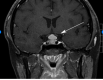
Pituitary Hyperplasia. The hyperplasia is due to primary hypothyroidism. Siddiqi AI, Grieve J, Baldeweg SE, Miszkiel K. Tablets or scalpel: pituitary hyperplasia due to primary hypothyroidism. Radiol Case Rep. 2016;10(2):1099. doi: 10.2484/rcr.v10i2.1099. (more...)
References
- 1.
Taylor PN, Albrecht D, Scholz A, Gutierrez-Buey G, Lazarus JH, Dayan CM, Okosieme OE. Global epidemiology of hyperthyroidism and hypothyroidism.
Nat Rev Endocrinol. 2018 May;14(5):301-316. [
PubMed: 29569622]
- 2.
Garber JR, Cobin RH, Gharib H, Hennessey JV, Klein I, Mechanick JI, Pessah-Pollack R, Singer PA, Woeber KA., American Association Of Clinical Endocrinologists And American Thyroid Association Taskforce On Hypothyroidism In Adults. Clinical practice guidelines for hypothyroidism in adults: cosponsored by the American Association of Clinical Endocrinologists and the American Thyroid Association.
Thyroid. 2012 Dec;22(12):1200-35. [
PubMed: 22954017]
- 3.
Khoury T, Kadah A, Mari A, Sbeit W, Drori A, Mahamid M. Thyroid Dysfunction is Prevalent in Autoimmune Hepatitis: A Case Control Study.
Isr Med Assoc J. 2020 Feb;22(2):100-103. [
PubMed: 32043327]
- 4.
Hollowell JG, Staehling NW, Flanders WD, Hannon WH, Gunter EW, Spencer CA, Braverman LE. Serum TSH, T(4), and thyroid antibodies in the United States population (1988 to 1994): National Health and Nutrition Examination Survey (NHANES III).
J Clin Endocrinol Metab. 2002 Feb;87(2):489-99. [
PubMed: 11836274]
- 5.
Anand A, Singh KR, Kushwaha JK, Hussain N, Sonkar AA. Papillary Thyroid Cancer and Hashimoto's Thyroiditis: An Association Less Understood.
Indian J Surg Oncol. 2014 Sep;5(3):199-204. [
PMC free article: PMC4235865] [
PubMed: 25419066]
- 6.
Hou J, Yu P, Zhu H, Pan H, Li N, Yang H, Jiang Y, Wang L, Wang B, Wang Y, You L, Chen S. The impact of maternal hypothyroidism during pregnancy on neonatal outcomes: a systematic review and meta-analysis.
Gynecol Endocrinol. 2016;32(1):9-13. [
PubMed: 26527131]
- 7.
Samuels MH. Psychiatric and cognitive manifestations of hypothyroidism.
Curr Opin Endocrinol Diabetes Obes. 2014 Oct;21(5):377-83. [
PMC free article: PMC4264616] [
PubMed: 25122491]
- 8.
Khalid S, Asad-Ur-Rahman F, Abbass A, Gordon D, Abusaada K. Myxedema Ascites: A Rare Presentation of Uncontrolled Hypothyroidism.
Cureus. 2016 Dec 05;8(12):e912. [
PMC free article: PMC5215814] [
PubMed: 28083456]
- 9.
Zare-Khormizi MR, Rahmanian M, Pourrajab F, Akbarnia S. Massive pericardial effusion and rhabdomyolysis secondary to untreated severe hypothyroidism: the first report.
Acta Clin Belg. 2014 Oct;69(5):375-8. [
PubMed: 25056490]
- 10.
Saini V, Yadav A, Arora MK, Arora S, Singh R, Bhattacharjee J. Correlation of creatinine with TSH levels in overt hypothyroidism - a requirement for monitoring of renal function in hypothyroid patients?
Clin Biochem. 2012 Feb;45(3):212-4. [
PubMed: 22061337]
- 11.
Jonklaas J, Bianco AC, Bauer AJ, Burman KD, Cappola AR, Celi FS, Cooper DS, Kim BW, Peeters RP, Rosenthal MS, Sawka AM., American Thyroid Association Task Force on Thyroid Hormone Replacement. Guidelines for the treatment of hypothyroidism: prepared by the american thyroid association task force on thyroid hormone replacement.
Thyroid. 2014 Dec;24(12):1670-751. [
PMC free article: PMC4267409] [
PubMed: 25266247]
- 12.
Abdullatif HD, Ashraf AP. Reversible subclinical hypothyroidism in the presence of adrenal insufficiency.
Endocr Pract. 2006 Sep-Oct;12(5):572. [
PubMed: 17002934]
- 13.
Guglielmi R, Frasoldati A, Zini M, Grimaldi F, Gharib H, Garber JR, Papini E. ITALIAN ASSOCIATION OF CLINICAL ENDOCRINOLOGISTS STATEMENT-REPLACEMENT THERAPY FOR PRIMARY HYPOTHYROIDISM: A BRIEF GUIDE FOR CLINICAL PRACTICE.
Endocr Pract. 2016 Nov;22(11):1319-1326. [
PubMed: 27482609]
- 14.
Hennessey JV, Espaillat R. Current evidence for the treatment of hypothyroidism with levothyroxine/levotriiodothyronine combination therapy versus levothyroxine monotherapy.
Int J Clin Pract. 2018 Feb;72(2) [
PMC free article: PMC5873391] [
PubMed: 29381251]
- 15.
Mathew V, Misgar RA, Ghosh S, Mukhopadhyay P, Roychowdhury P, Pandit K, Mukhopadhyay S, Chowdhury S. Myxedema coma: a new look into an old crisis.
J Thyroid Res. 2011;2011:493462. [
PMC free article: PMC3175396] [
PubMed: 21941682]
Disclosure: Nikita Patil declares no relevant financial relationships with ineligible companies.
Disclosure: Anis Rehman declares no relevant financial relationships with ineligible companies.
Disclosure: Ishwarlal Jialal declares no relevant financial relationships with ineligible companies.