U.S. flag

An official website of the United States government

NCBI Bookshelf. A service of the National Library of Medicine, National Institutes of Health.

National Collaborating Centre for Women's and Children's Health (UK). Antenatal Care: Routine Care for the Healthy Pregnant Woman. London: RCOG Press; 2008 Mar. (NICE Clinical Guidelines, No. 62.)

  • January 2019: Recommendation 1.8.8.1 on screening for German measles (rubella) has been withdrawn by NICE as this is no longer offered by the NHS. December 2018: Recommendations 1.3.9.1 -1.3.9.3 have been stood down and replaced with a link to up-to-date advice from the UK Chief Medical Officer. January 2017: A footnote was added to recommendation 1.6.2.2 linking to the NICE diagnostics guidance on high-throughput non-invasive prenatal testing for fetal RHD genotype (DG25). March 2016: Recommendations 1.9.1.1 -1.9.1.3 have been deleted as the guideline they were taken from has since been updated. For guidance on assessing risk of gestational diabetes, see the section on risk assessment in the NICE guideline on diabetes in pregnancy. December 2014: Recommendations 1.5.6.1 to 1.5.6.4 were replaced by recommendations in the NICE guideline on antenatal and postnatal mental health. November 2014: Recommendation 1.3.2.4 was updated to take into account NICE's guideline on vitamin D: increasing supplement use among at-risk groups. June 2010: The recommendations about smoking in pregnancy in section 1.3.10 of this guideline have been further developed in how to stop smoking in pregnancy and following childbirth NICE guideline PH26. We have removed the following recommendation from the antenatal care guideline, as well as the quick reference guide and information for the public: 1.3.10.7 Women who are unable to quit smoking during pregnancy should be encouraged to reduce smoking.

January 2019: Recommendation 1.8.8.1 on screening for German measles (rubella) has been withdrawn by NICE as this is no longer offered by the NHS. December 2018: Recommendations 1.3.9.1 -1.3.9.3 have been stood down and replaced with a link to up-to-date advice from the UK Chief Medical Officer. January 2017: A footnote was added to recommendation 1.6.2.2 linking to the NICE diagnostics guidance on high-throughput non-invasive prenatal testing for fetal RHD genotype (DG25). March 2016: Recommendations 1.9.1.1 -1.9.1.3 have been deleted as the guideline they were taken from has since been updated. For guidance on assessing risk of gestational diabetes, see the section on risk assessment in the NICE guideline on diabetes in pregnancy. December 2014: Recommendations 1.5.6.1 to 1.5.6.4 were replaced by recommendations in the NICE guideline on antenatal and postnatal mental health. November 2014: Recommendation 1.3.2.4 was updated to take into account NICE's guideline on vitamin D: increasing supplement use among at-risk groups. June 2010: The recommendations about smoking in pregnancy in section 1.3.10 of this guideline have been further developed in how to stop smoking in pregnancy and following childbirth NICE guideline PH26. We have removed the following recommendation from the antenatal care guideline, as well as the quick reference guide and information for the public: 1.3.10.7 Women who are unable to quit smoking during pregnancy should be encouraged to reduce smoking.

Cover of Antenatal Care

Antenatal Care: Routine Care for the Healthy Pregnant Woman.

Show details

9Screening for fetal anomalies

9.1. Screening for structural anomalies

Clinical question

What is the diagnostic value and effectiveness of the following screening methods in identifying serious structural abnormalities?

  • ultrasound undertaken in first and second trimesters
  • nuchal translucency measurement
  • serum screening – alpha-fetoprotein (AFP).

Previous NICE guidance (for the updated recommendations see below)

Pregnant women should be offered an ultrasound scan to screen for structural anomalies, ideally between 18 to 20 weeks of gestation, by an appropriately trained sonographer and with equipment of an appropriate standard as outlined by the National Screening Committee. [A]

9.1.1. Introduction and background

Since routine ultrasonography has been introduced into antenatal care women have had the opportunity to visualise the fetus at an early stage of pregnancy. The ultrasound scan has been used by health professionals to assess gestational age more accurately, to diagnose multiple births and to detect fetal anomalies. Improvements in technology have enabled health professionals to identify fetal structures, both normal and abnormal, and also to identify minor anomalies of uncertain significance, known as ‘soft markers’.

Detection of fetal anomalies on antenatal ultrasound offers women and their partners information that may help them better prepare for the birth of their child, the option of delivery in a setting that will permit rapid access to specialist surgical or medical care, and the possibility of considering pregnancy termination or palliative care in the newborn period. Routine antenatal ultrasound has therefore presented women and their partners with difficult decisions and an abnormal result on ultrasound imaging has the potential to cause great anxiety throughout the remaining weeks of pregnancy. These are important considerations with regard to the timing of routine ultrasound screening and the potential for false positive results or detection of ‘soft markers’.

Since the introduction of ultrasound in the 1970s, ultrasound technology has greatly improved. Modern equipment is now far superior and obstetric ultrasound is firmly established in routine practice, allowing identification of fetal anomalies and fetal growth problems. With this technology it is essential that healthcare professionals and clinicians who perform the scans are trained correctly to perform the examination and also understand and interpret the findings of the ultrasound scan correctly.302

This section of the guideline highlights the areas in which ultrasound screening is thought to have a role in the prenatal diagnosis of fetal anomalies.

Aim of screening for fetal structural anomalies

The overall aim of fetal anomaly screening is to identify potential problems so that parents can make an informed choice and to improve the safety of birth.

Specifically, antenatal screening to identify fetal anomalies should allow women and their partners:

  • reproductive choice (a choice about continuing with the pregnancy or choosing termination of pregnancy)
  • time to prepare (for termination of pregnancy/postnatal treatment or palliative care/infant disability)
  • managed delivery in specialist centre
  • intrauterine therapy.

The criteria laid out by Wilson and Jungner (1968)1020 to appraise the validity of any screening programme are that:

  • disorders to be screened for should be clinically well defined
  • the incidence of the conditions (individual malformations) should be known
  • disorders to be screened for should be associated with significant morbidity or mortality
  • effective treatment should be available, e.g. intrauterine treatment, birth managed in a specialist centre, or termination of pregnancy
  • there should be a period before onset of the disorder (the antenatal period) during which intervention is possible to improve outcome or allow informed choice
  • there should be an ethical, safe, simple and robust screening test, e.g. ultrasound appears safe, ethical and acceptable
  • screening should be cost-effective.

However, it is important to note that many of the studies of antenatal screening for fetal anomalies evaluate ultrasound as a suitable test rather than examine the benefits for women and babies of screening for a range of fetal anomalies during pregnancy.

9.1.2. Diagnostic value of routine ultrasound in the second trimester

The diagnostic value of routine ultrasound in the second trimester, including both multi-stage and single-stage ultrasound screening, was reviewed in this section.

Description of included studies

One systematic review297 including 11 studies, and an additional 12 studies727–741 were identified from the search. The 12 studies were critically appraised against the same criteria applied to the systematic review. Six studies were excluded either because of incomplete data or irrelevant study populations (e.g. high-risk populations). Details of the inclusion/exclusion process are provided on the accompanying CD-ROM. A new systematic review of all identified primary 17 studies, 11 studies in the systematic review and six newly identified studies, was conducted by the NCC-WCH. [EL = II]

Data from one RCT, nine prospective cohort studies and seven retrospective cohort studies were extracted. Four studies were conducted in the UK, while four were in the USA, four in Scandinavia, two in Belgium, two in Greece and one in South Korea. Details of the included studies are shown in Table 9.1. Meta-analyses of 11 studies on positive and negative likelihood ratios are presented in Figures 9.1 to 9.4.

Table 9.1. Description of included studies and detection rates of structural anomalies by antenatal ultrasound (first and second trimester).

Table 9.1

Description of included studies and detection rates of structural anomalies by antenatal ultrasound (first and second trimester).

Figure 9.1. Meta-analysis of positive likelihood ratios by routine ultrasound to detect fetal anomalies before 24 weeks.

Figure 9.1

Meta-analysis of positive likelihood ratios by routine ultrasound to detect fetal anomalies before 24 weeks.

Figure 9.2. Meta-analysis of negative likelihood ratios by routine ultrasound to detect fetal anomalies before 24 weeks.

Figure 9.2

Meta-analysis of negative likelihood ratios by routine ultrasound to detect fetal anomalies before 24 weeks.

Figure 9.3. Meta-analysis of overall positive likelihood ratios by routine ultrasound to detect fetal anomalies.

Figure 9.3

Meta-analysis of overall positive likelihood ratios by routine ultrasound to detect fetal anomalies.

Figure 9.4. Meta-analysis of overall negative likelihood ratios by routine ultrasound to detect fetal anomalies.

Figure 9.4

Meta-analysis of overall negative likelihood ratios by routine ultrasound to detect fetal anomalies.

Findings

Overall sensitivity (detection rate), specificity and likelihood ratios

The results of each study are presented in Table 9.1 and Figures 9.1 to 9.4. The sensitivity and specificity of detecting fetal structural anomalies before 24 weeks of gestation reported from the included studies were 24.1% (range 13.5% to 85.7%) and 99.92% (range 99.40% to 100.00%), respectively, while overall sensitivity and specificity were 35.4% (range 15.0% to 92.9%) and 99.86% (range 99.40% to 100.00%), respectively. Meta-analysis of likelihood ratios showed positive and negative likelihood ratios before 24 weeks of 541.54 (95% CI 430.80 to 680.76) and 0.56 (95% CI 0.54 to 0.58), respectively. Meta-analysis of likelihood ratios showed overall positive and negative likelihood ratios were 242.89 (95% CI 218.35 to 270.18) and 0.65 (95% CI 0.63 to 0.66), respectively.

Detection by RCOG category

Sensitivity (detection rate) for each condition according to the RCOG category742 was also sought, and is presented in Table 9.2. Overall sensitivity for lethal anomalies was 83.6%, that for possible survival and long-term morbidity was 50.6%, that for anomalies amenable to intrauterine therapy was 100.0%, and that for anomalies associated with possible short-term/immediate morbidity was 16.1%. The sensitivity varies depending upon each condition.

Table 9.2. Prevalence and detection of congenital anomalies at second-trimester antenatal ultrasound according to RCOG subgroup.

Table 9.2

Prevalence and detection of congenital anomalies at second-trimester antenatal ultrasound according to RCOG subgroup.

Evidence summary

Second-trimester ultrasound seems to show high specificity but poor sensitivity for identifying fetal structural anomalies. Similarly, this test showed good summary value for positive likelihood ratio but poor negative likelihood ratio. However, these values ranged widely by centre and condition. The 100% detection rate for conditions amenable to intrauterine treatment is anomalous and arises from the fact that these conditions had to be identified before treatment could be considered.

9.1.3. Diagnostic value of routine ultrasound in the first trimester

The diagnostic value of routine ultrasound in the first trimester to detect fetal structural anomalies was reviewed in this section.

Description of included studies

One review of literature included in an HTA297 and additional four studies300,743–746 were identified. However, only one300,743 from the additional studies was included in this review owing to methodological weakness and incomplete data. [EL = III]

Findings

The review showed that there were relatively few data on screening an unselected or low-risk population, as most studies report results of screening in high-risk populations.297 Results on nuchal translucency measurement are presented later in the soft markers section. The review included five studies of first-trimester anomaly screening but could not draw any conclusion because of the methodological weakness of these studies.

The additional study was published in 1999, although the study did not specify the time when it was conducted.300,743 Details of the study are presented in Table 9.1. This was a prospective cross-sectional study at a university hospital in the UK, and included 6634 unselected women carrying 6443 fetuses. All women underwent either transabdominal or transvaginal sonography at 11–14 weeks. Nuchal translucency and an anatomical survey were performed. There were six clinicians undertaking these examinations. The incidence of anomalous fetuses was 1.4%, and sensitivity (detection rate) was 59.0% (37/63 (95% CI 46.5% to 72.4%)). The specificity was 99.9%. Positive and negative likelihood ratios were 624.5 and 0.41. When first- and second-trimester scans were combined, the sensitivity was 81.0% (51/63 (95% CI 67.7% to 89.2%).

Evidence summary

There were only a few good-quality studies conducted which examine the diagnostic value of routine ultrasound in the first trimester. Although high specificity and positive likelihood ratio were reported, the sensitivity and negative likelihood ratio reported from a single centre in the UK were at a moderate level.

9.1.4. Effectiveness of routine ultrasound in pregnancy

The clinical effectiveness of routine use of ultrasound compared with no routine use was reviewed in this section.

Routine versus selective ultrasound before 24 weeks

Description of included studies

One systematic review that examined the effectiveness of routine ultrasound in early pregnancy (before 24 weeks), compared with selective ultrasound, was identified and included.57 [EL = 1+] The systematic review included eight RCTs and one quasi-randomised controlled trial, involving 34 251 women. The quality of these trials was generally good.

Findings

Routine ultrasound screening for fetal anomalies showed an increase in termination of pregnancy for fetal abnormality (four trials, OR 3.19, 95% CI 1.54 to 6.60), and a reduction in the number of undiagnosed twins (at 20 weeks, one trial, OR 0.12, 95% CI 0.03 to 0.56; at 26 weeks, six trials, OR 0.08 95% CI 0.04 to 0.16) and number of inductions for ‘post-term’ pregnancy (six trials, OR 0.61, 95% CI 0.52 to 0.72) compared with selective ultrasound. There is borderline evidence of the effect of routine ultrasound in reducing the number of children admitted to special care (five trials, OR 0.86, 95% CI 0.74 to 1.00) and with poor spelling at school (one trial, OR 0.73, 95% CI 0.53 to 1.00), compared with selective ultrasound. There was no evidence of difference in other outcomes.

Evidence summary

There is high-level evidence that routine, rather than selective, ultrasound in early pregnancy before 24 weeks enables better gestational age assessment, earlier detection of multiple pregnancies and improved detection of fetal anomalies with resulting higher rate of termination of affected pregnancies. There is no good-quality evidence on long-term outcomes for women and their children.

Routine versus no/concealed/selective ultrasound after 24 weeks

Description of included studies

One systematic review that examined effectiveness of routine ultrasound in late pregnancy (after 24 weeks), compared with no/concealed/selective ultrasound, was identified and included.574 [EL = 1+] The systematic review included five RCTs and one quasi-randomised controlled trial, involving 22 202 women. Among them, three trials offered routine ultrasound in the second and third trimester versus selective ultrasound. In one New Zealand trial, all women had a second-trimester scan and only the study group had a further third-trimester scan. In one UK trial, all women were offered second- and third-trimester scans but the results of the third-trimester scan were revealed only for those in the study group. In another UK trial, all women had routine second- and third-trimester scans, although placental grading at the third-trimester scans was revealed only for those in the study group. The quality of these trials was generally good.

Findings

Routine ultrasound screening for fetal anomalies after 24 weeks of gestation showed a reduction in post-term birth after 42 weeks (two trials, OR 0.69, 95% CI 0.58 to 0.81) but the timing and manner of gestational age assessment differed between the two trials. There was no difference in the overall perinatal mortality (six trials, OR 1.03, 95% CI 0.75 to 1.42), stillbirths (four trials, OR 1.15, 95% CI 0.74 to 1.79) or neonatal mortality (four trials, OR 1.04 (95% CI 0.58 to 1.86) between the two groups. After exclusion of babies with congenital anomalies, a statistically significant reduction was observed only for stillbirths (two trials, OR 0.13, (95% CI 0.04 to 0.50)), but one of the trials had incorporated placental grading into the routine third-trimester scan. There was no evidence of difference in other clinically important outcomes including obstetric and neonatal interventions.

Evidence summary

Results show a reduction in the number of post-term births and stillbirths (for normal babies) with routine third-trimester ultrasound, but the evidence is not of high quality. There is no evidence of difference for other clinically important outcomes, including obstetric and neonatal interventions and neonatal outcomes, between routine and no routine ultrasound after 24 weeks.

Routine versus no/concealed/selective Doppler ultrasound in pregnancy

Description of included studies

One systematic review that examined the effectiveness of routine Doppler ultrasound in pregnancy, compared with no/concealed/selective use of Doppler ultrasound, was identified and included.575 [EL = 1+] The systematic review included four RCTs involving 11 504 women. In one included UK trial, two different protocols were used for high- and low-risk populations, with the high-risk group having serial Doppler examinations and the low-risk group having Doppler examination on two occasions (19–22 weeks and 32 weeks). The data for each population were not reported separately and it was not possible to analyse separately. Three included trials only studied umbilical artery Doppler and reported different parameters.

Findings

Meta-analysis of the four trials showed no evidence of difference in antenatal admissions, obstetric interventions or neonatal interventions between routine and no routine use of Doppler ultrasound during pregnancy. Although one UK trial reported significantly increased perinatal mortality in the routine Doppler group compared with the no Doppler routine group, there was no evidence of difference in overall perinatal mortality.

Evidence summary

There was no evidence of difference in antenatal admissions, obstetric interventions, neonatal interventions or overall perinatal mortality between routine and no routine use of Doppler ultrasound during pregnancy.

Serial ultrasound plus Doppler versus selective ultrasound in pregnancy

Description of included studies

Two systematic reviews297,574 compared serial ultrasound plus Doppler with selective ultrasound. Both reviews included the same trial that compared effectiveness between serial ultrasound plus Doppler and selective ultrasound in pregnancy. [EL = 1+] This trial compared combined intensive repeated ultrasound assessment of the fetus plus Doppler study of the umbilical and uterine arteries versus selective ultrasound. The trial included 2834 women.

Findings

The included trial reported significantly more infants with intrauterine growth restriction in the routine serial and Doppler ultrasound than in the selective ultrasound group (birthweight < 10th centile, OR 1.41, 95% CI 1.11 to 1.78; birthweight < 3rd centile, OR 1.67, 95% CI 1.11 to 2.53), but otherwise no evidence of difference in antenatal and obstetric interventions, neonatal interventions or neonatal mortality/morbidity.

Evidence summary

There is little evidence on the effectiveness of routine use of combined serial and Doppler ultrasound compared with selective ultrasound and there is no evidence of difference in antenatal and obstetric interventions, neonatal interventions or neonatal mortality/morbidity.

First- versus second-trimester routine ultrasound in pregnancy

Description of included studies

One RCT was identified.747,748 [EL = 1+] The trial compared the antenatal detection rate of malformations in chromosomally normal fetuses between the policy of offering one routine ultrasound examination at 12 weeks, including nuchal translucency measurement, and one routine ultrasound examination at 18 weeks. The trial was conducted in eight hospitals in Sweden, involving 39 572 unselected women. A repeat scan was offered in the 12 week scan group if the fetal anatomy could not be adequately seen at 12–14 weeks or if nuchal translucency thickness was 3.5 mm or greater in a fetus with normal or unknown chromosome status.

Findings

The sensitivity of detecting fetuses with a major malformation was 38% (66/176) in the 12 week scan group, while that in the 18 week scan group was 47% (72/152) (P = 0.06). In the 12 week scan group, 69% of fetuses with a lethal anomaly were detected at a scan at 12–14 weeks.

The sensitivity of detecting fetuses with a major heart malformation was 11% (7/61) in the 12 week scan group, while that in the 18 week scan group was 15% (9/60) (P = 0.60). The proportion of women whose routine ultrasound was the starting point for further investigation resulting in a prenatal diagnosis was 6.6% in the 12 week group (4/61) and 15% in the 18 week group (9/60) (P = 0.15).

Evidence summary

There is little evidence of the effectiveness of a routine first-trimester scan for detecting major fetal malformation compared with a routine second-trimester scan. The available evidence showed no evidence of difference in any clinical outcomes.

9.1.5. Fetal echocardiography

The diagnostic value and clinical effectiveness of fetal echocardiography to detect fetal cardiac anomalies was reviewed in this section.

Diagnostic value of fetal echocardiography

Description of included studies

Studies examining the diagnostic value of fetal echocardiography on low-risk or unselected populations were reviewed. One systematic review including five studies was identified plus two additional studies.749–751 A description of these studies is presented in Table 9.3.

Table 9.3. Diagnostic value of fetal echocardiography: description of included studies and reported sensitivity and specificity.

Table 9.3

Diagnostic value of fetal echocardiography: description of included studies and reported sensitivity and specificity.

Findings

The sensitivity of detecting major cardiac anomalies from included studies ranged from 16.7% to 94.0%, and that for minor cardiac anomalies ranged from 3.6% to 82.1%. The overall sensitivity of detecting cardiac anomalies ranged from 4.5% to 86.1% and the specificity was reported as 99.9% throughout.

Evidence summary

The reported sensitivity of fetal echocardiography is widely ranged by centre and condition, although reported specificity was generally high.

Effectiveness of routine use of fetal echocardiography

Description of included studies

Neither RCTs nor quasi-randomised trials were identified to address this question. Two observational studies were identified,752,753 neither of which controlled for the background severity of conditions.

Findings

One cohort study in France752 compared outcome of babies between antenatally and postnatally diagnosed transposition of the great arteries (TGA). The study reported significantly lower preoperative mortality (postnatal diagnosis: 15/250 (6.0%) versus antenatal diagnosis 0/68 (0.0%); P < 0.05) and postoperative mortality (postnatal diagnosis: 20/235 (8.5%) versus 0/68 (0.0%); P < 0.01) for antenatally diagnosed TGA, although there was no evidence of difference in postoperative morbidity (postnatal diagnosis 25/235 (10.6%); antenatal diagnosis 6/68 (8.8%); P > 0.05). [EL = 2+]

Another population-based study in France753 compared detection rates of TGA and mortality for babies with TGA between three study periods. Between 1983 and 1988, antenatally diagnosed TGA was 12.5% and mortality for babies with TGA was 23.5%, between 1989 and 1994 the detection rate was 48.1% and mortality 12.0%, and between 1995 and 2000 the detection rate was 72.5% and mortality 5%.

A similar trend was reported in babies with hypoplastic left heart syndrome. [EL = 3]

Evidence summary

There was low-level evidence that showed babies with antenatally diagnosed TGA had reduced mortality compared with those diagnosed after birth.

9.1.6. Soft markers

The diagnostic value and clinical effectiveness of ultrasound soft markers including nuchal translucency measurement to detect fetal cardiac anomalies was reviewed in this section. Nuchal translucency measurement to detect Down’s syndrome was reviewed in Section 9.2.

Nuchal translucency measurement

Description of included studies

Studies examining the diagnostic value of nuchal translucency measurement of low-risk or unselected populations on detecting cardiac anomalies were reviewed. One systematic review including eight studies and four additional studies was identified.754–758 Since studies used different cut-off points, meta-analysis of these twelve studies to obtain summary likelihood ratios was conducted (Table 9.4 and Figures 9.5 and 9.6) Neither RCTs nor quasi-randomised controlled trials were identified to address the effectiveness of routine use of this measurement on clinical outcomes of women and their babies.

Table 9.4. Diagnostic value of nuchal translucency measurement on fetal cardiac anomaly.

Table 9.4

Diagnostic value of nuchal translucency measurement on fetal cardiac anomaly.

Figure 9.5. Meta-analysis of positive likelihood ratios by nuchal translucency measurement to detect fetal cardiac anomalies.

Figure 9.5

Meta-analysis of positive likelihood ratios by nuchal translucency measurement to detect fetal cardiac anomalies.

Figure 9.6. Meta-analysis of negative likelihood ratios by nuchal translucency measurement to detect fetal cardiac anomalies.

Figure 9.6

Meta-analysis of negative likelihood ratios by nuchal translucency measurement to detect fetal cardiac anomalies.

Findings

Meta-analysis of the included 11 studies showed a positive likelihood ratio of 5.01 (95% CI 4.42 to 5.68) and a negative likelihood ratio of 0.70 (95% CI 0.65 to 0.75).

Evidence summary

The reported sensitivity and likelihood ratios of nuchal translucency measurement to detect cardiac anomalies ranged widely by centre and condition, and generally the technique seems to have poor diagnostic value.

9.1.7. Use of maternal serum alpha-fetoprotein to detect structural anomalies

The diagnostic value and clinical effectiveness of biochemical markers including maternal serum alpha-fetoprotein to detect neural tube defects was reviewed in this section.

Alpha-fetoprotein to detect neural tube defects

Description of included studies

Two studies were identified.759,760 One study in the USA investigated the value of alpha-fetoprotein in screening for neural tube defects. The other was a case–control study in the USA comparing the ability of routine ultrasound and maternal serum alpha-fetoprotein levels to detect neural tube defects.

Findings

The first study,759 which investigated maternal serum alpha-fetoprotein as a screening test, was conducted between 1991 and 1994 in the USA and involved 27 140 women. The prevalence of neural tube defects was reported as 1.03 per 1000. Sensitivity, specificity and positive and negative likelihood ratios were reported as 85.7%, 97.6%, 35.16 and 0.15, respectively.

In the case–control study,760 an integrated database of 219 000 consecutive pregnancies between 1995 and 2002 was used. Among 189 identified fetuses with neural tube defects, 102 had received maternal serum alpha-fetoprotein screening, and 25% of 102 cases were test negative. Of the 186 neural tube defects identified prenatally, 62% were initially detected by routine second-trimester ultrasound, 37% were detected by targeted ultrasound prompted by high maternal serum alpha-fetoprotein level, and the remaining 1% were diagnosed by pathology examination after miscarriage.

Evidence summary

There were only two studies dealing with the diagnostic value and effectiveness of maternal serum alpha-fetoprotein level as a screening test. Results from a single study indicate maternal serum alpha-fetoprotein level to have good diagnostic value in predicting and ruling out structural anomalies, but evidence from another study shows it to have less value as a screening test than routine ultrasound. There is no evidence assessing the diagnostic value and effectiveness of combining maternal serum alpha-fetoprotein and routine ultrasound.

9.1.8. Women’s views on screening for structural anomalies

Three studies on women’ views regarding ultrasound screening during pregnancy, their responses to detection of soft markers, and antenatal counselling by specialist staff have been included under this section.

Description of included studies

The first study was a review297 [EL = 2++] which focused on women’s views and experiences of antenatal ultrasound. As the topic was very wide, it was decided to limit the review to studies where antenatal ultrasound was used for any purpose and direct data were obtained from pregnant women. Studies and reviews about prenatal screening and diagnosis were excluded. After a broad initial search to identify material related to women’s views in all screening and diagnostic tests, studies related to antenatal ultrasound use were selected after going through their abstracts. A series of six questions was prepared, targetting: (i) women’s knowledge about ultrasound and what a scan can do; (ii) women’s value about scans; (iii) her views about how ultrasound is conducted; (iv) impact of the result; (v) psychological impact of ultrasound; and (vi) wider impact of ultrasound on society. Studies were tabulated according to the question asked and data entered accordingly.

In the second study761 qualitative interviews were conducted to determine women’s experiences and responses to detection of a minor structural variant, the choroid plexus cyst (CPC), in their fetuses on prenatal ultrasound. Thirty-four pregnant women with isolated CPC detected during a mid-trimester scan who had already been counselled by their physicians regarding the findings at a university-based hospital in the USA were enrolled for the study. Interviews lasting approximately 15 minutes were conducted by a trained research assistant or nurse clinician at 24 weeks of pregnancy, and no information was given about CPCs by the research team. The interview included both open-ended and more specific questions, and all were audiotaped and transcribed verbatim. Common themes were identified, and several categories of responses identified for each theme. Initial validation was undertaken by an independent qualitative study consultant not involved in the research. The t-test was used for comparing means and χ2 for categorical variables. The results are reported as mean ± standard deviation. [EL = 3]

The aim of the third study762 was to evaluate parental anxiety after diagnosis of a congenital malformation and to assess whether counselling by a consultant paediatric surgeon and a neonatal nurse practitioner could decrease parents’ psychological distress. Participants were all parents attending a fetal medicine unit in the UK with an antenatal diagnosis of surgical anomaly (principally abdominal wall defects and gastrointestinal and thoracic anomalies). Women unable to read English and those booked to give birth somewhere else were excluded. Anonymous questionnaires were used to gain information as well as the Spielberger State-Trait Anxiety Inventory (STAI) for measuring anxiety levels. The STAI consists of two parts – the STAI-S score measuring anxiety at the time of completing the inventory, and the STAI-T score measuring the inherent trait anxiety levels. Participants were asked to complete STAI after ultrasound at the fetal centre. Then each couple had a detailed consultation with the paediatric consultant and the clinical nurse specialist. Before leaving, the subjects were given a second STAI and asked to complete and return within 1 week. A control group comprising pregnant women with a normal ultrasound scan and uncomplicated pregnancy was recruited and asked to complete STAI as the other group. Non-parametric tests were used for comparison, and data are quoted as medians and interquartile ranges (IQRs). [EL = 3]

Findings

In the first study,297 a total of 82 reports representing 64 studies were selected (including five studies which were added later). There was wide variation among the selected studies in terms of questions addressed, methods used, and when and where they were conducted. The studies were not graded in terms of research quality or removed because of poor quality, although many had problems of design and reporting. This was done because, in spite of poor quality, these studies gave useful information. The main findings of the review are discussed below.

Antenatal ultrasound is very attractive to pregnant women and their partners as it provides early visual confirmation of pregnancy, direct contact with their baby and reassurance about fetal wellbeing. At the same time, these features may augment the potential for feelings of anxiety, shock and disappointment when the scan shows a problem.

Recent trends in the use of ultrasound have led to more findings of uncertain clinical importance, and this is likely to have important psychological and social consequences for women.

Although it was reported in earlier studies that some women feared that ultrasound might harm their babies, there is a paucity of evidence about it from the later studies.

Reports of a reduction in anxiety after ultrasound examination are likely to reflect increased anxiety before the scan rather than a real benefit.

No reliable evidence is available for any positive health behaviour (e.g. reduced smoking) as a consequence of antenatal ultrasound.

None of the trials comparing ultrasound use with no ultrasound use has looked at its social and psychological impact on parents and babies.

In general, participants in the second study761 were college educated (mean years of education 16.6 ± 2.5), married (85.7%), employed (100%) and had private insurance (97%). The mean maternal age was 32.2 ± 5.2 years. About 60% were primiparous and 80% had a planned pregnancy. Women’s responses have been organised into categories as listed below.

  • Diagnostic situation. Mean gestational age at CPC detection was 18.86 ± 1.29 weeks. The majority of the participants (71%) were informed about CPC by an attending or local obstetrician at the conclusion of the ultrasound examination, and 35% of women were shown the CPC on ultrasound.
  • Accuracy of knowledge. Most of the women (79%) had never heard of CPC before the diagnosis. When asked about the significance of the CPC, 82% felt that it was probably benign, 71% expressed it is a marker for trisomy, and 53% mentioned that it could be both. Among those who expressed it as a marker for trisomy, 79% understood that other factors (maternal age, serum markers) also influenced the probability of trisomy. Women with positive serum screening results were less likely to describe CPC as benign compared with women with a normal serum screen (OR 0.04, 95% CI 0.004 to 0.36; P < 0.001). No statistically significant difference was observed between the older women (> 34 years) and younger ones.
  • Information seeking. Seventy-seven percent of women reported seeking additional information about CPCs beyond that given by their provider at the original scan, with the most common source being the internet. When asked about the usefulness of this additional information, 62% found it more useful than the primary information given at the time of ultrasound screening.
  • Subsequent testing. The majority of women (65%) already had a serum screening test before detection of CPCs. After detection of an isolated CPC and in spite of accurate counselling about low risk, three women (9%) sought diagnostic tests purely for reassurance.
  • Affective responses. When asked in an open-ended way to describe their emotions, 88% of women described an intensely negative immediate reaction, with most (68%) reporting their initial reaction as temporary. But only half of the women with a reassuring serum screen and none with an abnormal serum screen described their reaction as temporary. Sixty-eight percent of women revealed that they continued experiencing negative emotions even after receiving the diagnostic tests results, but neither increased maternal age nor visualisation of CPC on ultrasound were associated with persistence of the initial negative response. The later emotional responses included anxiety (23.5%), shock/grief (26.5%), decreased attachment (14.7%), decreased pleasure in pregnancy (14.7%), and thoughts of abortion/miscarriage (11.8%), confusion (8.8%), guilt (2.9%) and fear (5.9%).

Fifty-six pregnant women (subjects 26, control 30) completed the questionnaire in the third study.762 The most common congenital malformation present was gastroschisis followed by diaphragmatic hernia and cystic adenomatoid malformation. Maternal age was significantly lower in subjects (median 26.5 years) than in the control group (median 32 years) (P = 0.006).

No significant difference was found between STAI-T scores of subjects and controls. No correlation was found between the score and maternal age or social class, or between maternal and paternal scores.

STAI-S scores of subjects were significantly higher than those of controls before paediatric consultation (P = 0.0004), but not after (P = 0.31). There was a significant reduction in the anxiety levels of both subjects (mothers and fathers) after consultation (on comparing their scores before and after paediatric consultation) (P = 0.01 for mothers, P = 0.006 for fathers). After grouping the subjects into fetal diagnostic groups, a significant decrease in anxiety levels was found for those with anterior abdominal defects but not with cystic adenomatoid malformation. No correlation was found between the scores and maternal age.

The study showed that there was a high anxiety state in both prospective mothers and fathers of fetuses diagnosed with congenital malformations on ultrasound which is over and above that associated with pregnancy. Counselling by specialist staff reduced levels of parental anxiety significantly.

Evidence summary

Results from a well-conducted structured review show that visual confirmation of fetal wellbeing is the primary reason why women seek ultrasound during pregnancy. There is a lack of evidence regarding its other benefits and harms.

Evidence from a qualitative study indicates that detection of an isolated choroid plexus cyst on antenatal ultrasound leads to negative emotions and anxiety in the majority of women, who then seek additional information from other sources. In spite of reassurance in the form of a negative serum screening test for Down’s syndrome, a few women also opt for an invasive test for confirmation.

Detection of surgically treatable congenital anomalies on antenatal ultrasound led to increased anxiety levels in the parents but counselling by specialist staff helped to alleviate it significantly.

Health economics evidence

In the NICE clinical guideline on diabetes in pregnancy636 an economic model was developed to compare the cost-effectiveness of screening for congenital cardiac malformations using a four chamber ultrasound scan versus the four chamber plus outflow tracts view. This was considered to be important because women with diabetes are at increased risk of having a baby with a cardiac malformation. It was felt that this model was also relevant for the antenatal care guideline and therefore it was adapted for the antenatal care population. The results are summarised here; futher details are provided in Appendix E.

The baseline analysis suggested that the four chamber plus outflow tracts view has an ICER of £24,000 relative to the four chamber view alone. This falls within the borderline cost-effectiveness range of £20,000 to £30,000 per QALY used by NICE.

For the health economics evidence for the combined Down’s syndrome and structural anomalies screening, please see Section 9.2 (Screening for Down’s syndrome)

GDG interpretation of evidence (screening for structural anomalies)

Routine ultrasound screening

Ultrasound appears to be acceptable to women. Prenatal ultrasound scanning for fetal anomalies is now undertaken at around 20 weeks (rather than 18 weeks). However, the screening window should be between 18 weeks 0 days and 20 weeks 6 days. Screening later than 20 weeks 6 days may delay the diagnosis of an abnormality to a point where termination of an affected pregnancy becomes problematic and may involve additional procedures such as feticide. However, it should be remembered that where women are very overweight, performing the ultrasound scan can be very difficult and time-consuming. There is also a potential for an increase in repetitive strain injury (RSI)-related problems if sonographers are expected to complete all anomaly scans by 20 weeks. For this reason, the recommendation uses the word ‘normally’ in recognition of these potential difficulties.

Screening for congenital cardiac anomalies using the four chamber plus outflow tracts view has been shown to have an ICER of £24,000 relative to the four chamber view alone. There are likely to be further benefits of this method for detecting congenital cardiac malformations over and above that of TGA detection (the main focus of the model).

It is noted that some of the reviewed literature is from the 1980s and 1990s when scanning equipment was less well developed. The literature on scanning for fetal heart anomalies is more recent, however. It is also important to note that detection rates very much depend on the expertise of the person scanning as well as gestation and standard of equipment. Detection rates have improved in certain areas but this is due to further training as well as to advances in technology.

The prevalence of fetal anomalies and their detection rates can be evaluated either individually or after categorising them into four groups based on the RCOG criteria – lethal anomalies, anomalies with possible survival and long-term morbidity, anomalies amenable to intrauterine therapy, and anomalies with possible short-term or immediate morbidity (Table 9.2). Ultrasound cannot reassure women that their baby is normal, as many anomalies are missed. Ultrasound may not offer improved outcomes despite antenatal diagnosis, but may offer reproductive choices and the opportunity to plan intrauterine therapy or managed delivery.

Evidence from a single study shows that a first-trimester scan with nuchal translucency measurement is equally effective as the second-trimester scan in detecting fetal malformation overall. However, this may not be true for individual conditions, for example spina bifida is more likely to be detected by the second-trimester scan, while anencephaly and anterior abdominal wall defects may be detected in the earlier scans.

There is insufficient evidence that routine ultrasound between 10 and 24 weeks improves long-term outcomes after birth.

There is no evidence to support the use of selective rather than routine ultrasound scanning for fetal anomalies, gestational age determination and the diagnosis of multiple pregnancies.

Findings from an HTA review suggest a second-trimester scan is the most cost-effective strategy for screening for fetal anomalies. However, there is also evidence that each different method of screening has its advantages and disadvantages, and these often seem to balance out. No one screening method stands out as being much more cost-effective than any other.

Diagnostic accuracy of fetal echocardiography

The sensitivity of fetal echocardiography for detecting major malformations varies widely (from 17% to 94%) depending on gestation, skill of the operator and the equipment. However, there is some evidence that better training leads to improved performance of fetal cardiac screening and some limited evidence that antenatal diagnosis of TGA leads to better outcome for the babies.

Diagnostic accuracy of the nuchal test: soft markers

Studies evaluating nuchal translucency as a marker of cardiac anomaly found it to have poor sensitivity. Different cut-off points across centres and for different cardiac defects affected sensitivity and false positive rates, which are important considerations for women undergoing this test.

Diagnostic accuracy of AFP

AFP has lower diagnostic value than routine ultrasound in screening for neural tube defects. There is no evidence for effect on outcomes. However, the introduction of screening using AFP has led to a reduction in the number of affected babies born at term with neural tube defects.

Women’s views on screening for structural anomalies

Ultrasound screening provides reassurance if no anomaly is detected but heightens anxiety if a possible problem is identified

Recommendations on screening for fetal anomalies

Ultrasound screening for fetal anomalies should be routinely offered, normally between 18 weeks 0 days and 20 weeks 6 days.

At the first contact with a healthcare professional, women should be given information about the purpose and implications of the anomaly scan to enable them to make an informed choice as to whether or not to have the scan. The purpose of the scan is to identify fetal anomalies and allow:

  • reproductive choice (termination of pregnancy)
  • parents to prepare (for any treatment/disability/palliative care/termination of pregnancy)
  • managed birth in a specialist centre
  • intrauterine therapy.

Women should be informed of the limitations of routine ultrasound screening and that detection rates vary by the type of fetal anomaly, the woman’s body mass index and the position of the unborn baby at the time of the scan.

If an anomaly is detected during the anomaly scan pregnant women should be informed of the findings to enable them to make an informed choice as to whether they wish to continue with the pregnancy or have a termination of pregnancy.

Fetal echocardiography involving the four chamber view of the fetal heart and outflow tracts is recommended as part of the routine anomaly scan.

Routine screening for cardiac anomalies using nuchal translucency is not recommended.

When routine ultrasound screening is performed to detect neural tube defects, alpha-fetoprotein testing is not required.

Participation in regional congenital anomaly registers and/or UK National Screening Committee-approved audit systems is strongly recommended to facilitate the audit of detection rates.

Research recommendation on screening for fetal anomalies

Research should be undertaken to elucidate the relationship between increased nuchal translucency and cardiac defects.

9.2. Screening for Down’s syndrome

Clinical question

What is the diagnostic value and effectiveness of the following screening methods in identifying babies with Down’s syndrome?

  • blood tests
  • nuchal translucency
  • maternal age
  • ultrasound – soft markers (choroid plexus cyst, thickened nuchal fold, echogenic intracardiac focus, echogenic bowel, renal pyelectasis, humeral and femoral shortening)
  • ultrasound – nasal bone
  • different timings include:

    first trimester

    second trimester

    integrated

Previous NICE guidance (for the updated recommendations see below)

Pregnant women should be offered screening for Down’s syndrome with a test that provides the current standard of a detection rate above 60% and false positive rate of less than 5%.

By April 2007, pregnant women should be offered screening for Down’s syndrome with a test which provides a detection rate above 75% and false positive rate of less than 3%. These performance measures should be age standardised and based on a cut-off of 1/250 at term.

Pregnant women should be given information about the detection rates and false positive rates of any Down’s syndrome screening test being offered and about further diagnostic tests that may be offered. The woman’s right to accept or decline the test should be made clear.

9.2.1. Introduction and background

Down’s syndrome, also termed trisomy 21, is a congenital syndrome that arises when the affected baby has an extra copy of chromosome 21. In the absence of antenatal screening, about 1 in 700 babies born would be affected. The birth incidence of Down’s syndrome in England and Wales was 1.1 per 1000 live births in 2005 (represents 753 live births) (National Down’s syndrome register). Down’s syndrome causes learning disability, often profound, but the majority of children with the condition learn to walk, talk, read and write, although will meet these developmental milestones later than other children. It is also associated with increased incidence of congenital malformations (particularly cardiac and gastrointestinal anomalies) as well as an increased incidence of thyroid disorders, childhood leukaemias, and hearing, ophthalmic and respiratory problems. About half of children with Down’s syndrome are born with cardiac defects that require surgery, but survival rates are high. Average life expectancy for someone with the condition is 50–60 years.

Screening for Down’s syndrome should start with the provision of unbiased, evidence-based information about the condition, enabling women to make autonomous, informed decisions. Ideally, this information should be made available early in the pregnancy so that women have enough time to carefully consider the options and seek further information if needed. Screening for Down’s syndrome is part of an integrated screening programme and all staff involved should be familiar with the care pathways and their role within them.

Screening for Down’s syndrome takes place during either the first or second trimester by either ultrasound or maternal serum biochemistry, or a combination of both. Screening tests include the following:

Once a screening test has been performed, the chance of the fetus having Down’s syndrome is calculated taking into account maternal age and gestation. Results are classified as either ‘screen positive’ if the chance is equal to or greater than a nationally agreed cut-off level. This is often expressed numerically to indicate the likelihood that a woman has a baby with Down’s syndrome when a positive screening result is returned, for example a 1/250 chance that a pregnant woman is carrying an affected baby. When a screen-positive result is returned, the woman will usually be offered a diagnostic test, either chorionic villus sampling (following a first-trimester screening test) or amniocentesis (following a second-trimester screening test). Invasive diagnostic testing and karyotyping by either chorionic villus sampling or amniocentesis is the gold standard test for confirming the diagnosis but is associated with an excess risk of fetal loss of approximately 1% compared with women with no invasive testing. When a woman is offered a diagnostic test after a positive screening result, she should be informed of the risks associated with the invasive testing and that other chromosomal abnormalities, not just Down’s syndrome, may be identified and that in some cases the prognosis for the fetus may not be clear.

9.2.2. Diagnostic accuracy

Some studies have presented data on the screening performance as observed directly, while others have estimated diagnostic accuracy based on the study results. Where possible, results have been presented using a fixed false positive rate (FPR) of 5% (wherever calculated) in order to allow comparison between the findings, but the unadjusted results are also given.

The included studies have been stratified according to:

  1. the timing of the screening test, that is, conducted in the first trimester only, in the second trimester only, or both
  2. the type of abnormality detected – babies with Down’s syndrome only or both Down’s syndrome and other chromosomal anomalies.

First-trimester studies

Description of included studies

A total of 15 studies have been included under first-trimester screening. Initially, nine studies were identified for inclusion – all prospective cohort studies, including six multicentre ones. The objectives in all studies were clearly defined. Three studies comprised an unselected population, one study included both selected and unselected, and five selected population only. Except for a single study,767 the screening test and the quality measures used to monitor the study were adequately explained. All the studies used a validated reference test (karyotyping or postnatal assessment of babies or pregnancy records). The screening tests were performed before the reference tests in most studies, but it is difficult to ascertain blinding of the reference test operator. As the three studies on nasal bone gave conflicting results, six more studies were reviewed. All these studies were prospective cohorts but the quality of the studies was not good (all are EL III studies either owing to selected population, incomplete follow-up or inadequate quality control).

Findings

The first-trimester studies have been divided into the anomalies they looked at.

Down’s syndrome and other chromosomal anomalies

Three studies evaluated the serum combined test768–770 and three fetal nasal bone on ultrasound.771–773 These studies have been tabulated in Tables 9.5 and 9.6, respectively. The additional six studies on evaluation of fetal nasal bone771,773–777 are given in Table 9.7.

Table 9.5. First-trimester screening for Down’s syndrome and other chromosomal anomalies using the serum combined test.

Table 9.5

First-trimester screening for Down’s syndrome and other chromosomal anomalies using the serum combined test.

Table 9.6. First-trimester screening for Down’s syndrome and other chromosomal anomalies using nasal bone evaluation.

Table 9.6

First-trimester screening for Down’s syndrome and other chromosomal anomalies using nasal bone evaluation.

Table 9.7. First-trimester screening for Down’s syndrome using nasal bone evaluation – additional studies.

Table 9.7

First-trimester screening for Down’s syndrome using nasal bone evaluation – additional studies.

Results from a good-quality cohort with large sample size768 showed the serum combined test to have a detection rate of 92.6% at a false positive rate of 5.2% for the detection of Down’s syndrome, and a slightly lower detection rate for trisomy 18 or 13 and other chromosomal anomalies. Similar results were observed in another study,770 while the third study769 showed a lower detection rate but higher FPR for the combined test.

Conflicting results were seen for the diagnostic accuracy of fetal nasal bone evaluation (Table 9.6). While one study772 showed fetal nasal bone to increase the detection rate of Down’s syndrome from 90% to 93% (fixed FPR 5%) compared with using the combined test only, the other study771 showed it to have very poor diagnostic value. The third study773 had variable diagnostic accuracy results for the selected and unselected population.

Results from the additional six studies evaluated for fetal nasal bone were also inconclusive and wide variation was observed in them (Table 9.7). In two studies779,780 it improved the detection rate compared with using the serum combined test alone, but in one study775 there was a reduction in the detection rate. The sensitivity and detection rate of fetal nasal bone alone in the rest of the studies varied from 32% to 70%.

From these nine included studies on nasal bone characteristics, various factors have been identified which seem to influence the finding of absent nasal bone on first-trimester ultrasound. These factors are experience and training of the ultrasound operator, gestational age at which ultrasound is conducted (ideally CRL to be more than 45 mm as ossification of nasal bone starts after this), type of population screened (low-risk or high-risk), and marker used for diagnosis (complete absence or hypoplasia of the nasal bone).

Down’s syndrome only

The diagnostic accuracy results of the three included studies for the serum combined test were similar (Table 9.8). While one multicentre study778 found a detection rate of 79.6% at an FPR of 2.9%, the other two showed detection rates of 90.3% and 82% at a fixed FPR of 5%.

Table 9.8. First-trimester screening for Down’s syndrome only.

Table 9.8

First-trimester screening for Down’s syndrome only.

Second-trimester screening

Compared with the first trimester only and first and second trimester together, few studies were found relating to serum screening tests done exclusively in the second trimester. Good-quality serum marker studies comparing both the first- and second-trimester tests have been grouped under the next section on combined first- and second-trimester screening. A number of studies were identified which evaluated the use of ultrasound for identifying ‘soft markers’ – nuchal fold thickening, choroid plexus cyst, echogenic intracardiac foci, renal pyelectasis and shortening of femur, but the general quality was low (EL = III).

Five studies were selected for inclusion in this section – three meta-analyses, one prospective cohort study and one retrospective cohort study. As these studies were quite different from each other, their data could not be tabulated and they have been described in a narrative manner.

The second-trimester studies have been further divided into the anomalies they looked at:

a. Down’s syndrome and other chromosomal anomalies
Description of included studies

A single retrospective cohort782 study with evaluation of maternal serum screening (MSS) using quadruple test for Down’s syndrome, trisomy 18, and neural tube defects (NTD) was carried out in an Australian state using record linkage and manual follow-up. As initially the quadruple test used free alpha-hCG instead of inhibin A, data from that period were not used for analysis. The period covered was 1998 to 2000. Increased risk result was defined as > 1 : 250 for Down’s syndrome, and > 1 : 200 for trisomy 18. Levels of AFP > 2.5 MoM were considered as high risk for NTD. Three databases were used for record linkage – the state’s MSS database, register of births held at the Perinatal Data Collection Unit, and the Birth Defects Register. No mention was made about monitoring of test quality. An automated probabilistic record linkage technique was used to link these databases. The DR, FPR and PPV were calculated for each condition [EL = II]

Findings

In this retrospective cohort study, pregnancy outcome information was ascertained for 99.2% of all pregnancies screened during the period. The study population was 19 143 and 154 pregnancies were lost to follow-up. Mean maternal age was 30.3 years (range 14–51 years) and 20.1% were above 35 years. The sample size for analysis was 16 607 (86.7%) for Down’s syndrome and trisomy 18, and 17 288 (90.3%) for NTD. The sample size for Down’s syndrome and trisomy 18 was smaller owing to exclusion of pregnancies where alpha-hCG was used before inhibin A was introduced. The prevalence of Down’s syndrome, trisomy 18 and NTD was 0.16%, 0.05% and 0.08%, respectively.

The observed performance of the quadruple testing was as follows:

DRFPRPPV
For Down’s syndrome
Quadruple test (risk ≥ 1 : 250)85% (95% CI 72 to 99)6.8%2%
Quadruple test (FPR fixed at 5%)78%5.0%2.5%
For trisomy 18
Quadruple test (risk ≥ 1 : 200)44% (95% CI 12 to 77)0.5%4.7%
For NTD (AFP2.5 MoM)
All NTD73%1.1%5.6%
Spina bifida50%1.1%2.1%
Anencephaly100%1.1%3.1%
b. Down’s syndrome only

Four studies (three meta-analyses and one prospective cohort study) were identified. Meta-analysis studies were related to use of ultrasonographic soft markers, effectiveness of triple marker, and evaluation of intracardiac echogenic foci. The fourth study is a good-quality prospective study evaluating the screening performance of fetal pyelectasis detected on ultrasound.

Description of included studies

A meta-analysis315 was conducted to evaluate accuracy of second-trimester ultrasound in detecting Down’s syndrome. It included all the studies of ‘soft markers’ – choroid plexus cyst, thickened nuchal fold, echogenic intracardiac focus, echogenic bowel, renal pyelectasis, and humeral and femoral shortening. Exclusion criteria were well defined but quality assessment of studies was not specified. Studies were independently reviewed, selected and abstracted by two reviewers. Retrospective studies were included provided that the original ultrasound interpretation was used. Sensitivity, specificity and 95% CI was calculated for each ultrasound finding individually. A summary measure (sensitivity, specificity, LR+, LR−, PPV) with 95% CI and fetal loss per case diagnosed was calculated for each marker when identified as an isolated abnormality. [EL = II]

Another meta-analysis320 evaluated effectiveness of triple marker screen for Down’s syndrome. Only cohort studies were considered. Inclusion and exclusion criteria were well defined. Quality assessment criteria included selection of study subjects, description of methods, estimates of sensitivity, screen-positive rate and FPR, cut-offs used, blinding of outcome assessors, follow-up, and accuracy estimated independently of test threshold. Studies were independently reviewed, selected and abstracted by two reviewers. Results of sensitivity and FPR from different subgroups of study sample were compared by using summary ROC analysis. [EL = III]

A third meta-analysis783 was conducted to evaluate the diagnostic performance of intracardiac echogenic foci. Both prospective and retrospective studies (including case–control) were considered. Eligibility criteria for studies were availability of adequate information about both chromosomally normal and abnormal fetuses (so that a 2 × 2 table could be made), fetal karyotype unknown at the time of ultrasound, and chromosomal status of fetuses confirmed by either karyotyping or postnatal clinical examination. Studies were independently reviewed, selected and abstracted by two reviewers. Diagnostic performance was assessed in two different settings – ‘combined’ which included women regardless of whether they had other ultrasound findings, and ‘isolated’ where women did not have any other ultrasound finding. Weighted sensitivity and specificity values were calculated and summary ROC analysis performed using both the fixed and random effects model separately for both the settings. [EL = II]

A prospective cohort study784 was carried out (1998–2002) in a single medical centre in Italy with the aim of determining whether isolated pyelectasis is a risk factor for Down’s syndrome. The study population was low risk and the centre served the needs of a group of 30 obstetricians. Inclusion criteria were well defined and a thorough ultrasound examination was carried out for all the soft markers between 16 and 23 weeks of gestation. Monitoring of the quality of ultrasound was not specified. Complete follow-up was obtained of the study population by karyotyping, postnatal records or information from mother. The sensitivity, specificity, PPV, NPV, LR+ and LR− (with 95% CI) were calculated separately for an ‘isolated’ finding, and in association with other anomalies. The sample size was 12 672 (77.8%) after excluding high-risk and referred women. None of the women had a first-trimester aneuploidy screen. [EL = II]

Findings

The first meta-analysis315 included 56 studies involving 1930 babies with Down’s syndrome and 130 365 unaffected fetuses. Forty-nine studies were carried out in high-risk women. Overall prevalence of Down’s syndrome was 1.5%, and outcome was assessed by karyotyping in 53 studies. There was marked heterogeneity in the results for all ultrasound findings. Two factors were found to be responsible for heterogeneity: (i) study design (retrospective or prospective); and (ii) whether the marker was seen in isolation or together with other fetal structural anomalies. The sensitivity for Down’s syndrome detection with an isolated ultrasound finding was low (1% for choroid plexus cyst to a maximum of 16% for shortened femur). The specificity for each marker when seen individually was greater than 95%. Except for nuchal fold thickness (LR+ of 17), the LR+ for others was lower.

The summary measures (with 95% CI) for ultrasound markers when seen individually are given below:

MarkerSensitivitySpecificityLR+LR−Fetal loss per case
Thickened nuchal fold0.04 (0.02–0.10)0.99 (0.99–0.99)17 (8–38)0.97 (0.94–1.00)0.6
Choroid plexus cyst0.01 (0–0.03)0.99 (0.97–1.00)1.00 (0.12–9.4)1.00 (0.97–1.00)4.3
Femur length0.16 (0.05–0.40)0.96 (0.94–0.98)2.7 (1.2–6.0)0.87 (0.75–1.00)1.2
Humerus length0.09 (0–0.60)0.97 (0.91–0.99)7.5 (4.7–12)0.87 (0.67–1.1)1.9
Echogenic bowel0.04 (0.01–0.24)0.99 (0.97–1.00)6.1 (3.0–12.6)1.00 (0.98–1.00)1.0
Echogenic intracardiac focus0.11 (0.06–0.18)0.96 (0.94–0.97)2.8 (1.5–5.5)0.95 (0.89–1.00)2.0
Renal pyelectasis0.02 (0.01–0.06)0.99 (0.98–1.00)1.9 (0.7–5.1)1.00 (1.00–1.00)2.6

The second meta-analysis involving the triple marker320 included 20 cohort studies involving a total of 194 326 pregnant women. There was strong evidence of study-to-study variation, implying heterogeneity (P < 0.001). The cut-offs used in these studies ranged from 1 : 190 to 1 : 380. No study reported on the independence of assessment. Only four studies obtained fetal karyotypes (validated reference test) for all the women studied. In other studies, chorionic villus sampling or amniocentesis was offered to screen-positive women and the proportion of women accepting prenatal diagnostic testing ranged from 67% to 92%. Follow-up information on pregnancy outcome was incomplete in eight studies. The mean maternal age varied between 24.5 and 33.5 years. The triple marker had a high sensitivity for women older than 35 years, but did not perform well in the younger age group.

The summary sensitivity and FPR (with ranges) based on various cut-offs and maternal ages are given below:

SensitivityFPR
Cut-off 1 : 190–200
Maternal age ≥ 35 years0.89 (0.78–1.00)0.25 (0.20–0.29)
All ages0.67 (0.48–0.91)0.04 (0.03–0.07)
Cut-off 1 : 250–295
Maternal age ≥ 35 years0.80 (0.75–1.00)0.21 (0.20–0.21)
Maternal age < 35 years0.57 (0.53–0.58)0.04 (0.03–0.06)
All ages0.71 (0.48–0.80)0.06 (0.04–0.07)
Cut-off 1 : 350–380
All ages0.73 (0.70–0.80)0.08 (0.07–0.13)

The third meta-analysis concerning an echogenic focus in the heart783 included 11 studies (five retrospective including two case–controls). Eight studies gave data on combined setting, while seven gave data on isolated setting independently. The data included 51 831 fetuses with 333 Down’s syndrome cases (‘combined’: 27 360 with 321 Down’s syndrome cases; ‘isolated’: 39 360 with 130 Down’s syndrome cases). The mean age of mothers ranged between 29 and 35 years, and seven studies had high-risk women as their study population. Regarding sensitivity, there was no statistically significant heterogeneity as the confidence intervals were widely overlapping. For specificity, there was significant between-study heterogeneity (P < 0.001).

The weighted sensitivity and specificity estimates (with 95% CI) using the two models, random effects model (REM) and fixed effects model (FEM), are given below:

Random effects modelFixed effects model
SensitivitySpecificitySensitivitySpecificity
‘Combined’ setting0.26 (0.19–0.35)0.963 (0.937–0.979)0.30 (0.25–0.36)0.927 (0.924–0.931)
‘Isolated’ setting0.22 (0.14–0.33)0.959 (0.910–0.982)0.22 (0.15–0.30)0.964 (0.961–0.966)
All0.26 (0.19–0.34)0.958 (0.922–0.978)0.30 (0.25–0.36)0.940 (0.937–0.942)

It was further estimated that the probability of Down’s syndrome (assuming LR+ of 6.2) after an intracardiac echogenic foci has been detected would be 0.44% in a population with prevalence of 1 : 1400, 0.62% with prevalence of 1 : 1000, and 1.03% with prevalence of 1 : 600. The probability of a case of Down’s syndrome being detected was equal to the probability of an unnecessary miscarriage caused by amniocentesis when the background prevalence of Down’s syndrome was 1 : 770.

In the prospective cohort study on pyelectasis784 the mean maternal age was 27.2 ± 5.5 years and the prevalence of Down’s syndrome was 0.09% (11 cases). In the study population, the prevalence of pyelectasis was 2.9%, with 83.3% of these as an isolated finding. Only one case of Down’s syndrome was identified with pyelectasis. The presence of isolated pyelectasis had sensitivity 9.1% (95% CI 1.62 to 37.4%), specificity 97.6% (95% CI 97.32 to 97.85%), PPV 0.33%, NPV 99.9%, LR+ 3.8 (95% CI 0.58 to 24.61) and LR− 0.9 (95% CI 0.77 to 112).

Among fetuses with pyelectasis and other associated markers, the sensitivity, specificity, PPV, NPV and LR+ were 9.1%, 99.5%, 1.6%, 99.9% and 19.2 (95% CI 2.91 to 126.44), respectively.

Combined first- and second-trimester studies

Description of included studies

Four good-quality studies were included: three prospective cohort studies785–787 and one nested case–control study.316 All the studies were multicentred with clearly defined objectives. One of the two studies with a selected population had first-trimester screen-positive and screen-negative women together in its sample population.787 In all studies the screening test and monitoring of quality measures were adequately explained. The reference test in all was a validated one (karyotyping/postnatal assessment/pregnancy records) (Table 9.9).

Table 9.9. First- and second-trimester screening for Down’s syndrome only.

Table 9.9

First- and second-trimester screening for Down’s syndrome only.

Findings

All the selected studies looked at Down’s syndrome only. The best-quality study785 showed the integrated test to have the best DR of 96% at a fixed FPR of 5%, followed by the serum integrated test (DR 88%), combined test (DR 87%) and the quadruple test (DR 81%). Similar results were observed in the nested case–control study.316 Another study786 found the serum integrated test to have better diagnostic accuracy compared with the second-trimester serum triple and quadruple tests. In the last study,787 sequential screening using the triple test after a first-trimester combined test had a DR of 85.7% at FPR of 8.9%.

9.2.3. Implementation of the integrated test

One study1021 was identified which evaluated the implementation of the integrated test as a new method of screening for Down’s syndrome. The integrated test was conducted in a tertiary referral hospital in the UK. Prior to the introduction of the integrated test, local GPs and midwives were given information about this two-stage screening test. All women with singleton pregnancies booked before 14 weeks were offered the test, and the results of the first-trimester screening were not disclosed; however, women were offered further screening (combined test or integrated test) or invasive prenatal testing if NT was ≥ 3.5 mm. Women with NT < 3.5 mm were asked to return at 15 weeks for the second-trimester component of the integrated test, and a reminder letter was sent at 17 weeks to all those who failed to attend the second blood analysis. Women who did not have an NT measurement underwent testing by the serum integrated test, while the combined test and the quadruple test were also used depending on individual preference/timing of booking. All the data were entered in the computerised database. The cut-off values used for computing risk of Down’s syndrome were ≥ 1 in 150 (at term) for the integrated test, serum integrated test and NT + quadruple test, and ≥ 1 in 250 (at term) for the combined test and the quadruple test. [EL = 3]

During the 18 month study period, the overall uptake of Down’s syndrome screening was 64.4% (3417/5309) among all the pregnant women who opted for screening and NT scan at the hospital. Screening uptake was significantly higher in women booking before 14 weeks (73% versus 46%; P < 0.001), and the median age of the study population was 32 years (range 16–47 years). Seventy-six percent (2597/3417) of the pregnant women opting for screening had booked before 14 weeks and they were offered an integrated test – bout 97% of them opted for this. Twenty-two women (0.9%) had NT ≥ 3.5 mm and the majority of these women opted for invasive testing after further counselling. For the second-trimester blood analysis, 25% of the women failed to come for the test and a reminder letter was sent to these women at 17 weeks. Overall, 5.3% of women failed to attend the second part of screening, and 78% of the women (booked before 14 weeks) were screened by the full integrated test. For various reasons, NT could not be measured in 5.9% of the women opting for the integrated test and they were screened using the serum integrated test. The observed FPR for the groups undergoing the combined test, quadruple test and the integrated test were 8.9%, 6.3% and 2.9%, respectively, but the combined test group had older women (median maternal age 35 years) and a high proportion of women with a history of aneuploidy in the previous pregnancy.

Evidence summary

Findings from a descriptive study [EL = 3] shows that the integrated test, when implemented in practice, seems to be generally acceptable to pregnant women opting for it and results in a good uptake. But 25% of these women failed to attend for the second component of the test and this required sending them reminders.

9.2.4. Modelling studies

Description of included studies

Two studies were identified which used modelling as a way of comparing different screening tests for Down’s syndrome detection.

To demonstrate the potential value of three-stage sequential screening for Down’s syndrome, DR and FPR were estimated by multivariate Gaussian modelling using Monte Carlo simulation.789 UK data were used for modelling. The protocol is known as ‘contingent screening’ and involves measuring free β-hCG and PAPP-A in all pregnant women at 10 weeks in the first stage. Those with low risk were screened negative at this stage, the remainder underwent NT measurement in the second stage and the risk was reassessed (for combined test). After the second stage, those with low risk were screened negative and those with very high risk were offered diagnostic tests. In the third stage, women with intermediate risk received a second-trimester quaduple test. Risk was reassessed according to the integrated test and high-risk women were offered diagnosis. [EL = III]

Using Monte Carlo simulation for modelling, another study790 compared the integrated test in three policies for screening: (i) integrated screening for all women; (ii) sequential screening (based on first-trimester tests, high-risk pregnancies to be diagnosed and remaining to undergo integrated test); and (iii) contingent screening.

Detection and false positive rates were estimated based on the data from a large cohort (nested case–control study) done in the UK. [EL = III]

Findings

The first modelling study suggested that, with full adherence to a three-stage policy, an overall detection rate of nearly 90% and a false positive rate of below 2% can be achieved. About two-thirds of the women can be screened on the basis of first-trimester biochemistry alone and about 80% by the combined test. The DR for first-trimester screening is about 60%.

This protocol allows most of the Down’s syndrome pregnancies to be detected in the first trimester. Furthermore, it provides an efficient way of screening for Down’s syndrome where NT measurements cannot be performed in all women owing to scarcity of resources. However, it requires the selection of four different cut-offs during the three stages, each of which will affect the overall performance. Selecting a set of appropriate cut-offs is therefore complex and difficult to practise. The psychological impact of pregnant women possibly receiving four different results also needs to be evaluated.

The second modelling study concluded that integrated screening had the best screening performance. As the first-trimester test FPR was decreased, the performance of the other two policies approached that of the integrated screen. Setting the first-trimester risk cut-off to ≥ 1 in 300 with a fixed DR of 90%, sequential and contingent screening gave overall FPRs of 2.3% and 2.4%, respectively, and 66% of affected pregnancies were detected by the first-trimester tests. The integrated test on all women gave an FPR of 2.2%.

If pregnancies with a first-trimester risk of ≤ 1 in 2000 are classified screen negative and receive no further testing, then 99.5% of women with sequential screening or 30% with contingent screening would proceed to integrated screening.

9.2.5. Effectiveness studies

Five studies were identified: four related to adverse outcomes/fetal losses and one related to threshold measurement of NT. One was a multicentre RCT, one a nested case–control study, one a modelling study and one a meta-analysis to evaluate the diagnostic value of second-trimester ultrasound for Down’s syndrome. The NT study analysed a database from an earlier multicentre prospective study.

Description of included studies

A multicentre RCT791 in maternity care units affiliated to eight Swedish hospitals was carried out with an aim of comparing the effectiveness of two screening policies for detecting Down’s syndrome: routine ultrasound scan at 12–14 weeks by NT (12 week policy) versus routine ultrasound at 15–20 weeks (18 week policy). An unselected population with well-defined eligibility criteria was involved. After taking informed consent, the population was randomised block-wise at the level of maternity units using internet-based software. Appropriately trained operators carried out the ultrasound examination. Karyotyping was offered to all women with increased risk of Down’s syndrome (> 1 : 250 based on NT in the first group and on maternal age in the second), detection of a structural anomaly on scan, history suggestive of increased risk, or preference/desire of the woman due to worry. Follow-up of results (karyotyping, pregnancy outcome) was adequate. Evaluation of the primary outcome (number of babies born alive at ≥ 22 weeks with Down’s syndrome) and secondary outcomes (total number of babies born with Down’s syndrome, number of babies born with other chromosomal abnormalities, number of pregnancy terminations for Down’s syndrome, and rate of invasive tests for fetal karyotyping) was done using intention-to-treat analysis. The sample size was calculated to detect a difference of 0.1% in liveborn Down’s syndrome cases between the two groups at a 5% significance level with 90% power. χ2 tests (for proportions) and Student’s two-sample test (for continuous data) were used for comparison. [EL = 1+]

The nested case–control study316 has been discussed in the combined first- and second-trimester screening section above. Apart from evaluating the screening performance of various tests, it also examined their safety in terms of number of unaffected fetal losses per 100 000 women screened, and number of Down’s syndrome pregnancies detected for each procedure-related unaffected fetal loss. Both calculations were done at different detection rates. [EL = 2+]

A decision-analysis model792 was used to compare five screening strategies: (i) first-trimester combined screen; (ii) second-trimester quadruple screen; (iii) second-trimester triple screen; (iv) integrated screen; and (v) sequential screen. A hypothetical cohort of 1 000 000 women below 35 years was analysed assuming the entire cohort would present for antenatal care before 10 weeks and accept prenatal screening for Down’s syndrome. After a positive triple or quadruple test, genetic sonogram would be performed and then prenatal diagnosis would be available. Four separate outcomes were examined: (i) overall cost-effectiveness; (ii) Down’s syndrome cases detected; (iii) Down’s syndrome live births averted; and (iv) euploid losses from invasive procedures. [EL = 3]

Clinical parameters used for modelling were synthesised from review of published data (mainly UK data). The prevalence of Down’s syndrome at 10 weeks of gestation was estimated as 1 in 595 pregnancies, with a baseline live birth rate of 1 in 1030. Seventy percent of women were estimated to opt for invasive diagnostic techniques after a positive screening test, and 90% to opt for termination of affected pregnancies. Baseline fetal loss after amniocentesis and chorionic villus sampling were estimated to be 0.9% and 1.6%, respectively, but this was also varied over a range. Spontaneous fetal loss of euploid pregnancies was estimated at 1% between 10 and 14 weeks, and an additional 1% between 15 weeks and delivery. The screening performance of various tests was derived from published data. [EL = 3]

Details of the fourth study315 have already been discussed in the second-trimester screening section above.

The last study793 analysed the database from the FASTER trial (a multicentre prospective trial in the USA) to determine whether there is an NT measurement above which immediate invasive testing should be offered, without waiting for serum testing and computerised aneuploidy risk assessment. Pregnant women were eligible for inclusion if they were above 16 years of age, had a singleton pregnancy and a CRL of 36–79 mm (gestation 10 weeks 3 days to 13 weeks 6 days) at the time of first-trimester sonography for NT. Cases with cystic hygroma were excluded. NT was measured in the first trimester using a standardised protocol by specially trained ultrasonographers at the same time as when serum levels of PAPP-A and β-hCG were obtained. At 15–18 weeks, a quadruple serum screening test was also obtained, but the present study used only the risks as assessed from the first-trimester tests. A formal quality control programme was used throughout the study. [EL = 2+]

Findings

In the multicentre RCT a total of 39 572 women were randomised in the two groups (19 796 in the 12 week group, 19 776 in the 18 week group). Demographically the two groups did not differ in mean age, mean parity or other characteristics. In the 12 week group, NT measurement could not be carried out in 9% of the population owing to increased CRL or fetal demise, but was successfully measured in 96% of the remaining population. The prevalence of Down’s syndrome during the study period was 0.25% (98/39 572). The results are as follows:

Outcome12 week group18 week groupP value
Prevalence rate55/19 796 (0.28%)43/19 776 (0.22%)0.18
Rate of liveborn babies with DS (at ≥ 22 weeks)10/19 796 (0.05%)16/19 776 (0.08%)0.25
Antenatal detection rate (< 22 weeks in living fetus)42/55 (76%)25/41a (61%)0.12
Antenatal detection rate (if karyotyping performed only for defined policy)39/55 (71%)21/41a (51%)0.06
Detection rate (other chromosomal anomalies)20/35 (57%)25/35 (71%)0.32
Terminations done for DS39/19 796 (0.20%)24/19 776 (0.12%)0.08
Fetal loss rate in fetuses with DS (terminations and miscarriages)45/19 796 (0.23%)27/19 776 (0.14%)0.04
Rate of invasive tests (for karyotyping)1593/19 796 (8%)2118/19 776 (0.14%)< 0.001
Spontaneous fetal loss rate after invasive tests in normal fetuses14/1507 (0.9%)15/2041 (0.7%)0.58
No. of invasive tests per one case of DS detected (< 22 weeks) (if karyotyping performed only for defined policy)1689
a

Of the 43 cases of DS, diagnosis was made in one case by amniocentesis at < 22 weeks but pregnancy continued, and in other diagnosis made at 35 weeks – leaving 41 cases for calculating DR.

In the second study, the safety of various tests was evaluated at a fixed DR of 85%. The integrated test had about one-fifth the number of fetal losses when compared with the combined and quadruple test, and half that of the serum integrated test. The number of cases of Down’s syndrome detected for each fetal loss was almost three times higher with the integrated test when compared with the combined and quadruple test.

TestFPRUnaffected fetal losses per 100 000 womenCases of DS detected for each procedure-related fetal loss
Combined6.1%443.9
Double13.1%941.8
Triple9.3%672.6
Quadruple6.2%453.8
Serum integrated2.7%199.1
Integrated1.2%919.2

The modelling study found sequential screening to be the most cost-effective. Compared with other screens, it was shown to detect antenatally most cases of Down’s syndrome and avert most live births of affected fetuses. But it also had the highest number of euploid losses due to diagnostic procedure. From the point of view of safety, the integrated screen performed the best with the lowest euploid losses. The addition of a genetic sonogram to the triple and quadruple screens increased the cost but brought the euploid losses down to very low levels.

StrategyCost of programme (2002 prices, million US$)Cases of DS detected (n)DS live births averted (n)Euploid losses due to procedure (n)
No screening662000
Triple screen
 No sonogram497529366311
 With sonogram56636525325
Quadruple screen
 No sonogram472618427311
 With sonogram55442629525
Combined screen486941490559
Integrated screen52175052062
Sequential screen4551213678859

The meta-analysis concluded that the number of fetal losses per case diagnosed when identified as an isolated ‘soft marker’ abnormality on ultrasound was highest with choroid plexus cysts (4.3) and lowest with thickened nuchal fold (0.6).

For others the values were femur length (1.2), humerus length (1.9), echogenic bowel (1.0), echogenic cardiac foci (2.0) and renal pyelectasis (2.6)

In the NT study, the sample population included 36 120 pregnancies with complete first-trimester results. The mean and median NT measurements increased from 10 through 13 weeks and there was considerable variation in the proportion of cases with NT ≥ 2.0 mm at each gestational week, but there was minimal gestational age variation in NT once a threshold of 3.0 mm was passed.

≥ 2 mm≥ 3 mm≥4 mm≥ 5 mm
10 weeks2.0%0.4%0.16%0%
11 weeks1.5%0.5%0.1%0.04%
12 weeks2.5%0.3%0.1%0.09%
13 weeks5.1%0.4%0.05%0%
Total3.0%0.4%0.09%0.05%

On comparison of outcome of pregnancies based on the various NT cut-offs, the following results were observed:

Outcome≥ 2 mm≥ 3 mm≥ 4 mm
Number1081 (3.0%)128 (0.4%)32 (0.09%)
Aneuploidy512210
 T 2139176
 T 18544
 Others710
ST for DS/T 2142%19%7%
FPR for DS/T 213%0.3%0.06%
Final risk of DS less than 1 : 200 with the combined test533 (49.0%)10 (8.0%)0 (0%)

There were 32 women with NT ≥ 4 mm, and the addition of first-trimester serum markers to NT measurements did not reduce the final risk in any patients. In contrast, for patients with NT ≥ 3 mm, subsequent addition of serum markers reduced the final risk to less than 1 : 200 in only 8% of cases (ten women). For women with NT ≥ 2 mm, a large number of women (49%) had their risk reduced to less than 1 : 200 by the addition of first-trimester test results.

The authors concluded that the use of 4.0 and 3.0 mm cut-off of NT measurement for estimating pregnancies at risk of Down’s syndrome would lead to just 0.09% and 0.4%, respectively, of the population being subjected to invasive testing based on the two cut-offs. By waiting for serum assays and computerised risk assessment, no benefit (0%) was observed in the women with NT ≥ 4 mm and only a minimal benefit (8.0%) in women with NT ≥ 3 mm, that is, who had their final risk reduced to less than 1 in 200. This will increase the screen-positive rate for the whole population by a very small proportion, but will be beneficial in providing immediate results to the healthcare providers and reducing anxiety for the pregnant women.

Evidence summary

Reported evidence shows that the combined test in the first trimester has good diagnostic accuracy for Down’s syndrome and other chromosomal anomalies.

Among the currently available second-trimester serum tests, the quadruple test seems to have the best screening performance.

There is high-quality evidence to indicate that combining results of first- and second-trimester screening tests improves the diagnostic performance for Down’s syndrome and other chromosomal anomalies and is better than when either of them is used alone.

The integrated test seems to have a higher detection rate and a lower FPR compared with other currently used combined screening tests.

There is little evidence on the diagnostic value of other policies of combining first- and second-trimester results.

There is conflicting evidence regarding the performance of nasal bone ultrasound assessment as a screening tool for Down’s syndrome.

‘Soft markers’ on ultrasound have low sensitivity and LR+ when seen individually, except for nuchal fold thickening. When found in association with other anomalies, they seem to improve the diagnostic value but the evidence is not strong.

Retrospective analysis of a database from a high-quality prospective study shows that an NT measurement of 3 mm or more in the first trimester (any gestational age) identified the majority of pregnant women with Down’s syndrome, and increased the screen-positive rate/risk of invasive testing by only a small fraction compared with first-trimester risk evaluated by the combined test.

9.2.6. Women’s views and psychosocial aspects

Seven studies have been included in this section: two systematic reviews, three cross-sectional surveys and two prospective observational studies. Although the systematic reviews have been well conducted, as the principal question involved women’s views/preferences/experiences/feelings, which is quite subjective and difficult to interpret, other descriptive studies (even with poorer quality) were included so that important information was not missed. Grading the two systematic reviews according to the NICE quality criteria is difficult – they are well-conducted systematic reviews but with a definite risk of confounding or bias as individually the included studies were not assessed for quality.

Description of included studies

A systematic review794 was carried out to understand the psychosocial aspects of genetic screening of pregnant women and newborns. The review aimed to address five broad questions concerned with: (i) knowledge; (ii) anxiety; (iii) other emotional aspects; (iv) factors associated with participation in the programmes; and (v) long-term sequelae of the results. Any genetic screening programme aimed at pregnant women or newborn babies was included. Both comparative and descriptive studies which reported data collected directly from pregnant women or parents were included. There were no geographical or methodological limits except that studies asking hypothetical questions, case reviews and those where ultrasound was done to detect structural anomalies only (and did not include chromosomal anomalies) were excluded. Five electronic databases and two journals were hand searched. The retrieved articles were equally divided among the five authors for quality assessment and data extraction, and these processes were completed using well-defined criteria and validated forms. A new quality score was devised for quality assessment which was not found to be useful later on. Literature on ‘other emotional aspects’ and ‘long-term sequelae’ was too fragmented (except in neonatal screening programmes) for useful conclusions to be drawn. [EL = 2++]

A prospective cohort study795 was carried out in four antenatal clinics in Australia to assess informed choice in pregnant women to participate in second-trimester serum screening using a validated measure, and to compare anxiety levels in women who are well informed compared with those who are poorly informed. Participants included pregnant women at between 8 and 14 weeks of gestation attending at their first prenatal visit and with sufficient English to complete a written questionnaire. Written and oral information was provided to all participants as per the existing hospital policy. Informed choice was measured by Multidimensional Measure of Informed Choice (MMIC), a validated measure of informed choice which assesses knowledge and attitude dimensions and also confirms whether a woman’s participation in a screening test matches her expressed attitude towards it. The Hospital Anxiety and Depression Scale (HADS) was used to measure anxiety and this scale specifically distinguishes between anxiety and depression. Both the scales were administered at the booking visit and HADS was repeated at 20 weeks (after participation in the test) and at 30 weeks using postal questionnaires. [EL = 2+]

In the third study, a smaller sample drawn from the RCT described above791 was used to study the effect of screening on women’s anxiety during pregnancy and after birth, with a specific focus on worries about the health of the baby.796 The 12 week group was the intervention group and the 18 week group acted as the control. The principal outcome of women’s worries about the ‘possibility of something being wrong with the baby’ was measured by the Swedish version of the Cambridge Worry Scale questionnaire including 16 items of common concerns during pregnancy. The State-Trait Anxiety Inventory (a validated tool for evaluating general anxiety) and the Edinburgh Postnatal Depression Scale (validated for evaluating anxiety in the antenatal/postnatal period) were also used. Information was collected at three different timings – the first questionnaire was filled at the antenatal clinic, the second was sent at 24 weeks of gestation (mid-pregnancy), and the last was posted 2 months after delivery. The same instruments were used for all three questionnaires. [EL = 3]

A cross-sectional survey797 was carried out in three Canadian cities to investigate the relationship between MSS use and maternal attachment to pregnancy following the receipt of a favourable result (i.e lowered risk ratio). Building on the preliminary evidence that MSS results are not reassuring to women, it was predicated that favourable MSS results would not be sufficient to allow women to move beyond tentative pregnancy stage. Hence it was hypothesised that:

  • there would be no difference in prenatal attachment between women receiving favourable amniocentesis results (amniocentesis group) and who opt against testing (no testing group)
  • there would be a lower level of attachment among women who receive favourable MSS results and did not undergo amniocentesis (MSS group) compared with the other two testing groups, and this difference would be evident in the second and third trimesters.

Participants included high-risk pregnant women (maternal age > 35 years) who opted for MSS or amniocentesis or did not opt for any testing. Informational posters were placed at various places (physician offices, laboratories, maternity stores), and interested women who met the eligibility criteria were enrolled. The instrument used to collect information was a self-administered questionnaire by mail, and prenatal attachment was measured by a 21-item Prenatal Attachment Inventory (PAI) score (construct validity and reliability of this scale were established). The three groups were compared using ANOVA and ANCOVA for statistical analysis. [EL = 3]

To address the question of whether there are social and ethnic inequalities in the offer and uptake of prenatal screening and diagnosis in the UK, a systematic review798 was carried out employing a broad search strategy. In order to address the review question, studies were assessed in terms of:

  • utilisation – number of women screened as a proportion of those eligible
  • offer – number of women offered screening as a proportion of those eligible
  • uptake – number of women screened as a proportion of those offered screening.

Studies were reviewed and summarised by one reviewer. Two key aspects of the studies were assessed independently by two reviewers and summarised as indicators of quality – non-participation rate and whether the distinction between utilisation, offer and uptake was recognised in the study. Owing to heterogeneity, meta-analysis could not be performed. [EL = 2+]

A prospective descriptive study799 was carried out in two UK district hospitals to find out reasons for lower uptake of screening tests in women from minority ethnic groups and socio-economically disadvantaged sections of society. Screening uptake was evaluated from hospital records. Attitudes towards undergoing the test were assessed by women’s responses to a structured question with four items. Knowledge about the test was assessed using an eight-item questionnaire deemed important in professional guidelines for informed consent in screening. Choices were classified as ‘informed’ depending on the consistency between test uptake, women’s attitude towards the test, and their knowledge about it. [EL = 3]

Another cross-sectional survey800 was carried out in six UK maternity units (three in Scotland, three in England) to ascertain by means of a structured questionnaire women’s preference for type of screening test. Pregnant women attending antenatal clinics were asked to put in order of preference four different approaches for screening (all with FPR of 5%):

  • first-trimester testing – 90% detection with results available in 1 hour
  • first-trimester testing – 90% detection with results within 2–3 days (combined test)
  • first-trimester plus second-trimester detection – 93% detection and results within 2–3 days of second test (integrated test)
  • second-trimester testing – 75% detection and results available within 2–3 days. [EL = 3]

Findings

In the first systematic review 106 out of 288 identified studies met the eligibility criteria – 78 concerned with antenatal screening and 28 with neonatal screening. Only results pertaining to antenatal screening programmes are discussed below. Findings from antenatal carrier testing for cystic fibrosis and other diseases prevalent in minority ethnic groups are similarly also not discussed here.

Most of the antenatal studies were descriptive and only 33% (26/78) were RCTs or comparative. A questionnaire was the most common instrument used to collect data (in 79% of studies), either alone or together with other methods. Only 16 studies (20%) included both women who were tested and those who were not. Fifty-four studies were concerned with screening for Down’s syndrome and other chromosomal anomalies. The sample size of studies varied from ten to 6442 participants. Data were collected after the test results in 40 studies, and in just three studies was it collected at three different times – before test, after test and after test results. A large number of studies assessed knowledge (64.6%), anxiety (46.8%) or attitudes/beliefs (46.2%). Thirty-four antenatal studies (43.6%) had an apparent input from a psychologist or a social scientist. The various findings have been divided into three sections.

1. Knowledge and understanding of screening for Down’s syndrome

Thirty studies were selected: seven used pre-test measures only, six employed both before- and after-test measures (ideal for comparing), and 17 employed after-test measures only. Eight areas of information as specified in RCOG 1993 professional guidelines were used as a ‘validated/gold standard questionnaire’ for evaluating knowledge in the selected studies. Thirty studies relating to knowledge were reviewed but owing to disparate research aims, poorly operationalised measures for evaluation and variation in timing of assessment, it was concluded that none of them evaluated all the eight areas and hence knowledge was inadequately assessed by all of them. The broad conclusions drawn from these studies were:

  • compared with the RCOG list, only limited aspects of knowledge have been the subject of intervention studies
  • levels of knowledge adequate for decision making are not being achieved
  • leaflets giving information about tests improve knowledge, but substantial gaps in understanding of the written information still remain, especially concerning risk calculations
  • substantial social and cultural inequalities exist in knowledge about testing
  • other findings that emerged were:

    pre-screening information can increase knowledge scores but does not necessarily mean that concept of risk is understood

    women seem to value personally delivered information rather than group-based

    videos may be slightly more effective in communicating certain types of information than leaflets.

2. Influence on anxiety in prenatal screening for Down’s syndrome

Of the 24 studies measuring anxiety, 13 used a validated scale (mainly the State-Trait Anxiety Inventory). Most studies were carried out in the UK. As knowledge influences anxiety and attitudes, the findings from studies represents the feelings and views of many people who are in fact not well informed about the topic under discussion. Owing to a number of methodological concerns (as with knowledge), robust conclusions could not be drawn. The main findings were as follows:

  • increasing women’s knowledge by providing more information prior to testing does not raise post-test anxiety
  • there is unconvincing data to suggest that knowledge has a moderating role on anxiety in the period after screening but before receipt of test results
  • receipt of a screen-positive result raises women’s anxiety score, but this returns to normal levels if no abnormality is detected upon diagnostic testing.

Owing to application of inappropriate theoretical frameworks in these studies, two basic misconceptions about knowledge and anxiety were noted:

  • information that increases knowledge is the same as that which reduces anxiety
  • increased anxiety is inappropriate, abnormal and undesirable as most studies assume that increased anxiety is an abnormal response and/or an iatrogenic consequence of prenatal testing.
3. Understanding decision making about screening

Of the 52 studies included, 34 were concerned with Down’s syndrome screening and 11 of them compared differences in those screened with those not screened. Most studies employed questionnaire or interview survey methods. The principal findings were:

  • most women evaluate screening programmes positively but some are concerned about their usefulness and impact on pregnancy
  • the reasons as to why women had a screening test were:

    information to help avoid nasty surprises (range 11–82%)

    need to know for certain whether or not the child had abnormality (8–73%)

    reassurance that everything was OK (17–88%)

    following the recommendation of a health professional or spouse (6–24%)

    could think of no reason (16–26%)

  • the reasons as to why women chose not to have a test were:

    not wanting to act on or worry about the test results (17–71%)

    not wanting to have an abortion (32–100%)

    the test results were unreliable and did not provide a definite answer (10–55%)

    not perceiving themselves at high risk and/or the abnormality to be serious (21–64%), and their own or others’ poor screening experience (1–32%)

  • most women are not making informed choices about screening although they want to do so; there is evidence to suggest a gap between women’s desire to make informed choices with their awareness of what constitutes an informed decision, and the skills with which to achieve it
  • informed decision making results in better post-decision outcomes.

Of the initial 134 recruited women completing the first assessment in the second study, 63.9% returned the second questionnaire and 57.8% the third. The mean age of women in the sample was 29.1 ± 4.7 years and 89.6% were married. Using MMIC, 48.1% of women were classified as having ‘good knowledge’ and 87.2% as having a ‘positive attitude’ to screening. Overall, only 37.3% of decisions to participate in screening were informed: those who participated in screening were more than twice as likely to have made an informed choice than those who did not participate (47% versus 20%; P = 0.01). Informed decisions were not significantly associated with participants’ age, parity, country of birth or whether pregnancy was unwelcome or unexpected. No significant association was found between the knowledge levels and attitude to the test (P = 0.27). Some important misconceptions were revealed about further testing: 31% did not know that miscarriage was a possible consequence of diagnostic testing subsequent to an increased risk screening result, and only 62% correctly identified that termination of pregnancy would be offered if Down’s syndrome was diagnosed. Regarding anxiety, no significant difference was found between the informed and not informed group in psychological outcomes at any of the three assessments, even after adjusting for repeated measures on individual participants. It was concluded that many women participating in prenatal genetic screening are inadequately informed regarding aspects of testing, including the management of pregnancy in the event of increased risk.

A total of 2026 women were enrolled for the third study. Analysis was carried out in 82.7% (854/1030) women in the 12 week group and 84.1% (837/996) in the 18 week group who responded to all three questionnaires. The demographic characteristics of the two groups were similar. Emotional wellbeing at baseline in early pregnancy was also similar. In early pregnancy, 39.1% of women in the 12 week group and 36.0% in the 18 week group were worried about something being wrong with the baby, but the difference was not statistically significant. The prevalence decreased to 29.2% versus 27.8% during mid-pregnancy, and finally to 5.2% versus 6.6% at 2 months after delivery in the two groups. No statistically significant differences were found between the two groups during these periods either.

Within both trial groups, there was a statistically significant decrease in the levels of major worry about the baby’s health from early to mid-pregnancy (P < 0.001) and from mid-pregnancy to 2 months after delivery (P < 0.001).

In the fourth study, a cross-sectional survey, 101 women formed the study group which comprised 31 women in the amniocentesis group, 32 in the MSS group and 38 in the no-test group. The mean gestational age at the time of participation was 28.3 ± 7.0 weeks. The mean maternal age in the amniocentesis group was higher than in the other two groups (P = 0.005), while no statistically significant differences were found between the three groups with respect to gestational age, number of previous pregnancies or previous miscarriages. A significant difference was found between the amniocentesis and no-test group regarding attitude towards abortion.

One-way ANOVA indicated that mean attachment scores nfor the MSS group (mean 51.7, SD 9.4) were significantly lower than those reported by the amniocentesis group (mean 58.5, SD 10.7) and no-test group (mean 57.0, SD 8.3) (t(68) = 0.68; P = 0.02). Furthermore, the amniocentesis group did not differ in bonding levels compared with the no-test group (t(67) = 0.66; P = 0.51), thereby proving the hypothesis. This difference persisted even after removing the influence of maternal age and attitude towards abortion. There was no significant interaction between testing status of the three groups and timing of conducting the survey (second or third trimester) when they were used as independent variables with PAI score as the dependent variable.

The results suggest that MSS may disrupt the developmental trajectory of the maternal–fetal bond even after favourable results are known. This may be due to the probabilistic nature of MSS results, which creates confusion rather than reassurance.

For the second systematic review, 600 studies were identified and 19 met the inclusion criteria. Ten related to screening/diagnosis for Down’s syndrome and NTD, three for haemoglobin disorders and six studies for HIV. Several studies were limited by small sample size and poor reporting of data and statistical analysis. Only findings from ten studies of Down’s syndrome and NTD are discussed below.

Nine studies reported on utilisation and/or uptake of prenatal screening or diagnosis. One of these suggested that, compared with white women, utilisation of testing was lower in South Asian women, two others indicated that both utilisation and uptake was lower, and a fourth study found both acceptance and uptake of amniocentesis lower in women from South Asia. In the remaining five studies, no statistically significant association was found between socio-demographic factors and test utilisation.

Four studies reported on the offer of screening or diagnosis for Down’s syndrome. Two of these suggested that South Asian women were less likely to be offered amniocentesis, while in the third study fewer Bangladeshi than white women were offered screening, although this result was not statistically significant. The fourth study did not analyse the results according to the social class or ethnic group.

It was concluded that there is evidence that women from some ethnic groups, particularly South Asian women, may be less likely to receive prenatal diagnosis for Down’s syndrome. A significant proportion of these women will take up prenatal testing if offered, but these women may be less likely to be offered testing. This points to the need to identify the factors associated with the offer and uptake of prenatal screening, barriers to offering screening at institutional and professional levels, and reasons for failure to take up screening when offered.

In the sixth study 2059 women were included and 1791 (89%) returned questionnaires but only 84% of these were completed on time. The results were:

  • screening uptake – the overall uptake was 49% (95% CI 47% to 52%); uptake was higher in white and socio-economically advantaged women
  • knowledge – overall, the mean knowledge score was above the midpoint of the scale; knowledge was higher for white, socio-economically advantaged and older women
  • attitudes towards test – the mean overall score was above the scale midpoint, that is, overall women had a positive attitude towards the test; no difference in attitudes was found relating to ethnicity, socio-economic status or parity, but older women had a more positive attitude than younger ones
  • uptake–attitude 
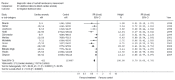
consistency – in women with positive attitudes, white and socioeconomically advantaged women were more likely to act in line with their attitudes (76% of white women had the test compared with 45% of South Asian women; P < 0.001, and 78% of socio-economically advantaged women had the test compared with 63% of socioeconomically disadvantaged women; P < 0.001); in women with a negative attitude, no difference was found between ethnic or social groups
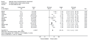
  • informed choice – the rates of informed choice were higher for white women (56% versus 20% of South Asian women; P < 0.001) and for socio-economically advantaged women (59% versus 14% for socio-economically disadvantaged women; P < 0.001).

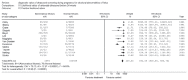
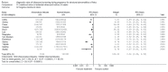
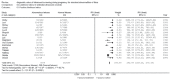
After controlling for confounding variables (ethnicity, age, socio-economic status and hospital attended), it was found that both South Asian women and socio-economically disadvantaged women with positive attitudes were less likely to act consistently with their attitudes compared with white and socio-economically advantaged women (OR 0.22, 95% CI 0.10 to 0.45 for South Asian versus white women; and OR 0.62, 95% CI 0.41 to 0.93 for social groups).

The study was not able to determine the cause of lower consistency between positive attitudes and behaviour of these women.

In the last study, 1127 women returned the questionnaire. A total of 75% of women selected first-trimester screening (option 1 or option 2) as their first choice, with 68.2 % preferring results within 1 hour (option 1) and 6.8% preferring a combined test. Twenty-four percent opted for the integrated test and just 1% opted for second-trimester testing as their first choice.

Evidence summary

There is high-quality evidence to indicate that pregnant women do not have sufficient knowledge to make the informed decisions that need to be made regarding Down’s syndrome screening and they find the concept of risk calculation particularly difficult to understand. Providing them with more information does not lead to an increase in their anxiety level.

Good evidence from a cohort study shows that women taking part in prenatal screening programme are inadequately informed regarding aspects of testing and the further pathway of management when an increased risk is identified.

Results from a cross-sectional study indicate that women undergoing a serum screening test for Down’s syndrome develop less attachment for the baby owing to the uncertainty surrounding interpretation of the test result.

Evidence from a review of literature shows that pregnant women from South Asia have a lower rate of uptake, acceptance and utilisation of screening tests.

For the screening tests in general, white women and women from socio-economically advantaged sections of society have a higher uptake, better knowledge, more consistency of actions related to positive attitude, and a higher rate of informed decision making when compared with women from South Asia and socio-economically disadvantaged sections of society.

9.2.7. Health economics evidence

Antenatal screening for Down’s syndrome

A systematic search of the literature was conducted to identify economic evaluations of screening for Down’s syndrome. The search identified 132 abstracts, of which 40 full papers were retrieved for further consideration. Six studies are included in the review.

One study801 was conducted to examine the performance of integrated Down’s syndrome screening (first- and second-trimester measurements integrated into a single screening test) when ratios of the levels of the same serum markers measured in both these trimesters (cross-trimester ratios) are added as new screening markers. The addition of cross-trimester ratios to an integrated test significantly improves the efficacy and safety of prenatal screening for Down’s syndrome. So, the addition of cross-trimester ratios is cost-effective and could be usefully introduced into screening programmes.

Another UK study802 was conducted to compare the effects, safety and cost-effectiveness of antenatal screening strategies. The main outcomes of the study were the number of liveborn babies with Down’s syndrome, miscarriages due to chorionic villus sampling or amniocentesis, healthcare costs of the screening programme, and additional costs and additional miscarriages per additional affected live birth prevented by adopting a more effective strategy. Compared with no screening, the additional cost per additional liveborn baby with Down’s syndrome prevented was £22,000 for measurement of NT. The cost of the integrated test was £51,000 compared with the measurement of NT. All other strategies were more costly and less effective, or cost more per additional affected baby prevented. Depending on the cost of the screening test, the first-trimester combined test and the quadruple test would also be cost-effective options. The main conclusions of the study were that the choice of screening strategy should be between the integrated test, first-trimester combined test, quadruple test or NT measurement depending on how much service providers are willing to pay, the total budget available and values on safety. Screening based on maternal age, the second-trimester double test and the first-trimester serum test was less effective, less safe and more costly than these four options.

One HTA study316 was conducted to identify the most effective, safe and cost-effective method of antenatal screening for Down’s syndrome using NT, maternal serum and urine markers in the first and second trimesters of pregnancy and maternal age in various combinations. The cost-effectiveness analysis showed that the screening using the integrated test is less costly than might be expected because the extra screening costs tend to be offset by savings in the cost of diagnosis arising from the low FPR. It was estimated that to achieve an 85% detection rate the cost to the UK NHS would be £15,300 per Down’s syndrome pregnancy detected. The corresponding cost of using the second-trimester quadruple test would be £16,800 and using the first-trimester combined test it would be £19,000.

Antenatal screening for Down’s syndrome + structural anomalies

One HTA297 study was conducted and one of the aims of this study was to refine and update a decision model of cost-effectiveness of options for routine scanning for fetal anomalies. The initial eight options considered were reduced to three dominating options: one second-trimester scan alone, one third-trimester scan alone and a combination of the one second-trimester scan followed by one third-trimester scan. More representative cost data are required before precise estimates of the additional costs and benefits of alternative options can be determined. Also, it is clear from the analysis that one second-trimester analysis scan emerged as a clear reference case, being one of the cheapest options yet still detecting a significant number of anomalies. When termination is acceptable and available, a third-trimester scan alone or the combination of one second- with one third-trimester scan, although comparable in economic terms, may be impractical because of the delay in identifying anomalies.

Another study803 was conducted to compare the cost-effectiveness of different programmes of routine antenatal ultrasound screening to detect four key fetal anomalies: serious cardiac anomalies, spina bifida, Down’s syndrome and lethal anomalies. The study showed that there was a substantial overlap between the cost ranges of each screening programme demonstrating considerable uncertainty about the relative economic efficiency of alternative screening programmes consisting of one second-trimester ultrasound scan. The cost per target anomaly detected (cost-effectiveness) for this programme was in the range £5,000–109,000, but in any 1000 women it would also fail to detect between 3.6 and 4.7 target anomalies. The model highlighted the weakness of the available evidence and demonstrated the need for more information both about current practice and costs.

Finally, a study804 was conducted in the UK to determine the most clinically and cost-effective policy of scanning and screening for fetal anomalies in early pregnancy. The number of anomalies detected and missed, the number of iatrogenic losses resulting from invasive tests, the total cost of strategies and the cost per abnormality detected were compared between strategies. First-trimester screening for chromosomal anomalies costs more than the second-trimester screening but results in fewer iatrogenic losses. Strategies which include a second-trimester ultrasound scan result in more anomalies being detected and have lower costs per anomaly detected.

GDG interpretation of evidence

Accuracy and effectiveness of screening

The integrated test was found to be cost-effective and resulted in the fewest losses of normal fetuses. However, there are concerns regarding the practicality of screening by this method. There is also evidence that women prefer a one-stage test to the integrated test.

Evidence shows that the combined test in the first trimester is also cost-effective and has good diagnostic value for detection of Down’s syndrome and other chromosomal anomalies.

Among the currently used second-trimester tests, the quadruple test seems to have the best screening performance but the measurement of inhibin A (the fourth analyte) is not generally available in the UK.

Although isolated ‘soft markers’ on second-trimester ultrasound (18–23 weeks) with the exception of thickened nuchal fold have limited effectiveness in screening for Down’s syndrome, two or more soft markers should prompt referral for fetal medicine opinion.

Other than the presence of increased nuchal fold thickening, isolated soft markers noted on the second-trimester scan should not be used to adjust the risk for Down’s syndrome which has been derived from an established, nationally approved screening programme.

Women’s views

Levels of knowledge among women are not currently adequate for informed decision making about whether or not to undergo screening.

The biggest gap in knowledge is in understanding risk.

Increasing pre-screen knowledge does not raise anxiety levels.

Fewer South Asian women than white women are offered screening and fewer of those who are offered it choose to go ahead with it. Some healthcare professionals appear to have misconceptions regarding the likely attitudes of South Asian women to screening and termination of pregnancy.

The knowledge of those women opting out of screening seems to be better than that of those who are screened (16–26% don’t know why they are being screened).

Serum screening can have a detrimental effect on women’s attachment to pregnancy even with a low-risk result, owing to the uncertainty created by the probabilistic nature of the way the result is presented.

Recommendations on screening for Down’s syndrome

All pregnant women should be offered screening for Down’s syndrome. Women should understand that it is their choice to embark on screening for Down’s syndrome.

Screening for Down’s syndrome should be performed by the end of the first trimester (13 weeks 6 days), but provision should be made to allow later screening (which could be as late as 20 weeks 0 days) for women booking later in pregnancy.

The ‘combined test’ (nuchal translucency, beta-human chorionic gonadotrophin, pregnancy-associated plasma protein-A) should be offered to screen for Down’s syndrome between 11 weeks 0 days and 13 weeks 6 days. For women who book later in pregnancy the most clinically and cost-effective serum screening test (triple or quadruple test) should be offered between 15 weeks 0 days and 20 weeks 0 days.

When it is not possible to measure nuchal translucency, owing to fetal position or raised body mass index, women should be offered serum screening (triple or quadruple test) between 15 weeks 0 days and 20 weeks 0 days.

Information about screening for Down’s syndrome should be given to pregnant women at the first contact with a healthcare professional. This will provide the opportunity for further discussion before embarking on screening. (Refer to Section 3.3 for more information about giving antenatal information). Specific information should include:

  • the screening pathway for both screen-positive and screen-negative results
  • the decisions that need to be made at each point along the pathway and their consequences
  • the fact that screening does not provide a definitive diagnosis and a full explanation of the risk score obtained following testing
  • information about chorionic villus sampling and amniocentesis
  • balanced and accurate information about Down’s syndrome.

If a woman receives a screen-positive result for Down’s syndrome, she should have rapid access to appropriate counselling by trained staff.

The routine anomaly scan (at 18 weeks 0 days to 20 weeks 6 days) should not be routinely used for Down’s syndrome screening using soft markers

The presence of an isolated soft marker, with an exception of increased nuchal fold, on the routine anomaly scan, should not be used to adjust the a priori risk for Down’s syndrome.

The presence of an increased nuchal fold (6 mm or above) or two or more soft markers on the routine anomaly scan should prompt the offer of a referral to a fetal medicine specialist or an appropriate healthcare professional with a special interest in fetal medicine.

Research recommendations on screening for Down’s syndrome

There should be multicentred studies to evaluate the practicality, cost-effectiveness and acceptability of a two-stage test for Down’s syndrome and other screening contingencies including the integrated test.

Further research should be undertaken into the views and understanding of women going through the screening process.

Copyright © 2008, National Collaborating Centre for Women’s and Children’s Health.

No part of this publication may be reproduced, stored or transmitted in any form or by any means, without the prior written permission of the publisher or, in the case of reprographic reproduction, in accordance with the terms of licences issued by the Copyright Licensing Agency in the UK [www.cla.co.uk]. Enquiries concerning reproduction outside the terms stated here should be sent to the publisher at the UK address printed on this page.

The use of registered names, trademarks, etc. in this publication does not imply, even in the absence of a specific statement, that such names are exempt from the relevant laws and regulations and therefore for general use.

Bookshelf ID: NBK51878

Views

Recent Activity

Your browsing activity is empty.

Activity recording is turned off.

Turn recording back on

See more...