From: Chapter 24, The Third Heart Sound

NCBI Bookshelf. A service of the National Library of Medicine, National Institutes of Health.

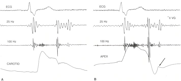
Four-channel phonocardiogram taken at a paper speed of 100 mm/sec. The top channel shows an electrocardiogram (EGG lead II); the second and third channels record from a single microphone placed near the cardiac apex. The cardiovascular sound is filtered so that the second channel records frequencies below 25 Hz and the third channel below 100 Hz. The fourth channel displays a carotid arterial pulse (left) and a tracing of the apex movement (right). The heart sounds are labeled I and II. A prominent ventricular gallop (VG) is recorded 0.16 second after the second heart sound. Note that it is recorded best on the low-frequency channel. A low-intensity, early to midsystolic murmur is also present (not labeled). The apex tracing shows a sustained systolic wave and a rapid early diastolic filling wave (arrow). The ventricular gallop occurs at the peak of the rapid filling wave. The examiner should simultaneously feel the carotid impulse and listen at the apex in order to time the heart sounds and gallop.
From: Chapter 24, The Third Heart Sound

NCBI Bookshelf. A service of the National Library of Medicine, National Institutes of Health.