From: Antigen recognition by T cells

NCBI Bookshelf. A service of the National Library of Medicine, National Institutes of Health.

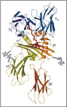
The structure of a T-cell receptor binding to an MHC class II molecule has been determined, and shows the T-cell receptor binding to an equivalent site, and in an equivalent orientation, to the way that TCRs bind to MHC class I molecules (see Fig. 3.27). The structure of the molecules is shown in a cartoon form, with the MHC class II α and β chains shown in light green and orange respectively. Only the Vα and Vβ domains of the T-cell receptor are shown, colored in blue. The peptide is colored red, while carbohydrate residues are indicated in gray. The TCR sits in a shallow saddle formed between the MHC class II α and β chain α-helical regions, at roughly 90° to the long axis of the MHC class II molecule and the bound peptide. Courtesy of E.L. Reinherz, reprinted with permission from Science 286:1913-1921. ©1999 American Association for the Advancement of Science.
From: Antigen recognition by T cells

NCBI Bookshelf. A service of the National Library of Medicine, National Institutes of Health.