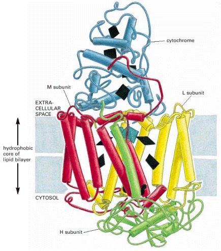
Figure 10-38. The three-dimensional structure of the photosynthetic reaction center of the bacterium Rhodopseudomonas viridis.

Figure 10-38The three-dimensional structure of the photosynthetic reaction center of the bacterium Rhodopseudomonas viridis.

The structure was determined by x-ray diffraction analysis of crystals of this transmembrane protein complex. The complex consists of four subunits L, M, H, and a cytochrome. The L and M subunits form the core of the reaction center, and each contains five α helices that span the lipid bilayer. The locations of the various electron carrier coenzymes are shown in black. Note that the coenzymes are arranged in the spaces between the helices. (Adapted from a drawing by J. Richardson based on data from J. Deisenhofer, O. Epp, K. Miki, R. Huber, and H. Michel, Nature 318:618–624, 1985.)

From: Membrane Proteins

Cover of Molecular Biology of the Cell
Molecular Biology of the Cell. 4th edition.
Alberts B, Johnson A, Lewis J, et al.
New York: Garland Science; 2002.
Copyright © 2002, Bruce Alberts, Alexander Johnson, Julian Lewis, Martin Raff, Keith Roberts, and Peter Walter; Copyright © 1983, 1989, 1994, Bruce Alberts, Dennis Bray, Julian Lewis, Martin Raff, Keith Roberts, and James D. Watson .

NCBI Bookshelf. A service of the National Library of Medicine, National Institutes of Health.