From: Cell Junctions

NCBI Bookshelf. A service of the National Library of Medicine, National Institutes of Health.

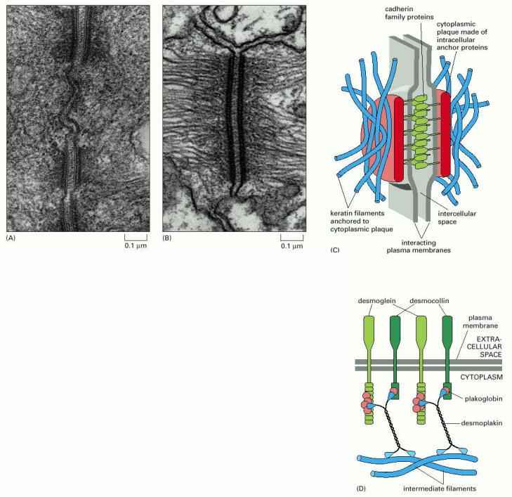
(A) An electron micrograph of three desmosomes between two epithelial cells in the intestine of a rat. (B) An electron micrograph of a single desmosome between two epidermal cells in a developing newt, showing clearly the attachment of intermediate filaments. (C) The structural components of a desmosome. On the cytoplasmic surface of each interacting plasma membrane is a dense plaque composed of a mixture of intracellular anchor proteins. A bundle of keratin intermediate filaments is attached to the surface of each plaque. Transmembrane adhesion proteins of the cadherin family bind to the plaques and interact through their extracellular domains to hold the adjacent membranes together by a Ca2+-dependent mechanism. (D) Some of the molecular components of a desmosome. Desmoglein and desmocollin are members of the cadherin family of adhesion proteins. Their cytoplasmic tails bind plakoglobin (γ-catenin), which in turn binds to desmoplakin. Desmoplakin also binds to the sides of intermediate filaments, thereby tying the desmosome to these filaments. (A, from N.B. Gilula, in Cell Communication [R.P. Cox, ed.], pp. 1–29. New York: Wiley, 1974. Reprinted by permission of John Wiley & Sons, Inc.; B, from D.E. Kelly, J. Cell Biol. 28:51–59, 1966. © The Rockefeller University Press.)
From: Cell Junctions

NCBI Bookshelf. A service of the National Library of Medicine, National Institutes of Health.