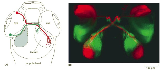
Figure 21-100. The neural map from eye to brain in a young zebrafish.

Figure 21-100The neural map from eye to brain in a young zebrafish

(A) Diagrammatic view, looking down on the top of the head. (B) Fluorescence micrograph. Fluorescent tracer dyes have been injected into each eye—red into the anterior part, green into the posterior part. The tracer molecules have been taken up by the neurons in the retina and carried along their axons, revealing the paths they take to the optic tectum in the brain and the map that they form there. (Courtesy of Chi-Bin Chien, from D.H. Sanes, T.A. Reh, and W.A. Harris, Development of the Nervous System. San Diego, CA: Academic Press, 2000.)

From: Neural Development

Cover of Molecular Biology of the Cell
Molecular Biology of the Cell. 4th edition.
Alberts B, Johnson A, Lewis J, et al.
New York: Garland Science; 2002.
Copyright © 2002, Bruce Alberts, Alexander Johnson, Julian Lewis, Martin Raff, Keith Roberts, and Peter Walter; Copyright © 1983, 1989, 1994, Bruce Alberts, Dennis Bray, Julian Lewis, Martin Raff, Keith Roberts, and James D. Watson .

NCBI Bookshelf. A service of the National Library of Medicine, National Institutes of Health.