

NCBI Bookshelf. A service of the National Library of Medicine, National Institutes of Health.
Frank SA. Immunology and Evolution of Infectious Disease. Princeton (NJ): Princeton University Press; 2002.
"The CTLs destroy host cells when their TCRs bind matching MHC-peptide complexes." This sort of jargon-filled sentence dominates discussions of the immune response to parasites. I had initially intended this book to avoid such jargon, so that any reasonably trained biologist could read any chapter without getting caught up in technical terms. I failed—the quoted sentence comes from a later section in this chapter.
The vertebrate immune system has many specialized cells and molecules that interact in particular ways. One has to talk about those cells and molecules, which means that they must be named. I could have tried a simpler or more logically organized naming system, but then I would have created a private language that does not match the rest of the literature. Thus, I use the standard technical terms.
In this chapter, I introduce the major features of immunity shared by vertebrates. I present enough about the key cells and molecules so that one can understand how immune recognition shapes the diversity of parasites. I have not attempted a complete introduction to immunology, because many excellent ones already exist. I recommend starting with Sompayrac's (1999) How the Immune System Works, which is a short, wonderfully written primer. One should keep a good textbook by one's side—I particularly like Janeway et al. (1999). Mims's texts also provide good background because they describe immunology in relation to parasite biology (Mims et al. 1998, 2001).
The first section of this chapter describes nonspecific components of immunity. Nonspecific recognition depends on generic signals of parasites such as common polysaccharides in bacterial cell walls. These signals trigger various killing mechanisms, including the complement system, which punches holes in the membranes of invading cells, and the phagocytes, which engulf invaders.
The second section introduces specific immunity, the recognition of small regions on particular parasite molecules. Specific recognition occurs when molecules of the host immune system bind to a molecular shape on the parasite that is not shared by other parasites. Sometimes all parasites of the same species share the specificity, and recognition differentiates between different kinds of parasites. Other times, different parasite genotypes vary in molecular shape, so that the host molecules that bind specifically to one parasite molecule do not bind another parasite molecule that differs by as little as one amino acid. A parasite molecule that stimulates specific recognition is called an antigen. The small region of the parasite molecule recognized by the host is called an epitope. Antigenic variation occurs when a specific immune response against one antigenic molecule fails to recognize a variant antigenic molecule.
The third section presents the B cells, which secrete antibodies. Antibodies are globular proteins that fight infection by binding to small regions (epitopes) on the surface molecules of parasites. Different antibodies bind to different epitopes. An individual can make billions of different antibodies, each with different binding specificity. Diverse antibodies provide recognition and defense against different kinds of parasites, and against particular parasites that vary genetically in the structure of their surface molecules. Antibodies bind to surface molecules and help to clear parasites outside of host cells.
The fourth section focuses on specific recognition by the T cells. Host cells continually break up intracellular proteins into small peptides. The hosts' major histocompatibility complex (MHC) molecules bind short peptides in the cell. The cell then transports the bound peptide-MHC pair to the cell surface for presentation to roving T cells. Each T cell has receptors that can bind only to particular peptide-MHC combinations presented on the surface of cells. Different T cell clones produce different receptors. When a T cell binds to a peptide-MHC complex on the cell surface and also receives stimulatory signals suggesting parasite invasion, the T cell can trigger the death of the infected cell. T cells bind to parasite peptides digested in infected cells and presented on the infected cell's surface, helping to clear intracellular infections.
The final section summarizes the roles of antibodies and T cells in specific immunity.
2.1. Nonspecific Immunity
Nonspecific immunity recognizes parasites by generic signs that indicate the parasite is an invader rather than a part of the host. The nonspecific complement system consists of different proteins that work together to punch holes in the surfaces of cells. Host cells have several surface molecules that shut off complement attack, causing complement to be directed only against invading cells. Common structural carbohydrates found on the surfaces of many parasites trigger complement attack, whereas the host cells' carbohydrate molecules do not trigger complement.
Phagocytic cells such as macrophages and neutrophils engulf invading parasite cells. Various signals indicate to the phagocytes that nearby cells are invaders. For example, certain lipopolysaccharides commonly occur in the outer walls of gram-negative bacteria such as E. coli. Mannose, which occurs in the cell walls of many invaders, also stimulates phagocytes. In addition, phagocytes respond to signs of tissue damage and inflammation.
Nonspecific defense by itself may not entirely clear an infection, and in some cases parasites can avoid nonspecific defense. For example, the protective capsules of staphylococci and the surface polysaccharide side chains of salmonellae protect those bacteria from attachment by nonspecific killing molecules (Mims et al. 1993, p. 12.2).
2.2. Specific Immunity: Antigens and Epitopes
Nonspecific immunity recognizes common, repetitive structural features that distinguish parasites from the host's cells. By contrast, specific immunity recognizes small regions of particular parasite molecules. Specific recognition may depend on just five or ten amino acids of a parasite protein. Such specificity means that different parasite species often differ at recognition sites. Indeed, different parasite genotypes may vary such that a host can recognize particular sites on one genotype but not on another.
This book is about parasite variation in regard to specific immunity, so it is important to get the jargon right. Specific host immunity recognizes and binds to an epitope, which is a small molecular site within a larger parasite molecule. An antigen is a parasite molecule that stimulates a specific immune response because it contains one or more epitopes. For example, if one injects a large foreign protein into a host, the host recognizes thousands of different epitopes on the surface of the protein antigen.
Antigenic variation occurs when a specific immune response against one antigenic molecule fails to recognize a variant antigenic molecule. The antigenic variants differ at one or more epitopes, the sites recognized by specific immunity.
2.3. B Cells and Antibodies
B cells mature in the bone marrow (bursa in birds). They then develop into lymphocytes, immune cells that circulate in the blood and lymph systems. B cells express globular proteins (immunoglobulins) on their cell surfaces. These immunoglobulins form the B cell receptors (BCRs). B cells also secrete those same immunoglobulins, which circulate as antibodies. In other words, antibodies are simply secreted BCRs. I will often use the word antibody for B cell immunoglobulin, but it is important to remember that the same immunoglobulins can be either BCRs or antibodies. Immunoglobulin is usually abbreviated as Ig.
The B cells generate alternative antibody specificities by specially controlled recombination and mutation processes (fig. 2.1). The host maintains a huge diversity of antibody specificities, each specificity in low abundance. Novel parasite epitopes often bind to at least one rare antibody specificity. Binding stimulates the B cells to divide, forming an expanded clonal lineage that increases production of the matching antibody.

Figure 2.1
The coding and assembly of antibody molecules. Randomly chosen alternatives of the variable (V), diversity (D), and joining (J) regions from different DNA modules combine to form an RNA transcript, which is then translated into a protein chain. Two heavy (more...)
Each antibody molecule has two kinds of amino acid chains, the heavy chains and the light chains (fig. 2.1). A heavy chain has three regions that affect recognition, variable (V), diversity (D), and joining (J). A light chain has only the V and J regions. In humans, there are approximately one hundred different V genes, twelve D genes, and four J genes (Janeway 1993).
Each progenitor of a B cell clone undergoes a special type of DNA recombination that brings together a V-D-J combination to form a heavy chain coding region. There are 100 × 12 × 4 = 4,800 V-D-J combinations. A separate recombination event creates a V-J combination for the light chain, of which there are 100 × 4 = 400 combinations. The independent formation of heavy and light chains creates the potential for 4,800 × 400 = 1,920,000 different antibodies. In addition, randomly chosen DNA bases are added between the segments that are brought together by recombination, greatly increasing the total number of antibody types.
Recombination creates a large number of different antibodies. Initially, each of these antibodies is rare. Upon infection a few of these rare types may match a parasite epitope, stimulating amplification of the B cell clones. The matching B cells increase their mutation rate, creating many slightly different antibodies that vary in their affinity to the invader (fig. 2.2). Those mutant cells that bind more tightly are stimulated to divide more rapidly. This evolutionary fine-tuning of the B cell population is called affinity maturation.
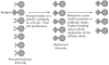
Figure 2.2
Clonal selection of B cells to produce antibodies that match an epitope of an invading antigen. Recombinational mechanisms produce a wide variety of different antibody molecules (fig. 2.1). All B cells of a particular clone are derived from a single ancestral (more...)
Naive B cells produce IgM immunoglobulins before stimulation and affinity maturation. After affinity maturation, B cells produce various types of immunoglobulins by changing the constant region (fig. 2.1). The most common are IgG in the circulatory system and IgA on mucosal surfaces.
On first encounter with a novel parasite, the rare, matching antibodies cannot control infection. While the host increases production of matching antibodies, the infection spreads. Eventually the host may produce sufficient antibody to clear parasites that carry the matching epitope. If the parasites, in turn, vary the matched epitope, the host must expand new antibody types to clear the variant parasites.
Once the host expands an antibody specificity against a matching epitope, it maintains a memory of that epitope. Upon later exposure to the same epitope, the host can quickly produce large numbers of matching antibodies. This memory allows the host to clear subsequent reinfection without noticeable symptoms.
Antibodies typically bind to surface epitopes of parasites. Thus, antibodies aid clearance of parasites circulating in the blood or otherwise exposed to direct attack. Once an intracellular parasite enters a host cell, the host must use other defenses such as T cells.
2.4. T Cells and MHC
Host cells continually break up intracellular proteins into small peptides. The host's major histocompatibility complex (MHC) molecules bind these short peptides within the cell. The cell then transports the bound peptide-MHC pair to the cell surface for presentation to roving T cells. T cells are lymphocytes that mature in the thymus.
T cell receptors (TCRs) vary in binding specificity. Each T cell receptor can bind only to particular peptide-MHC combinations presented on the surface of cells. Different T cell clones produce different TCRs. The TCR variability is generated by a process similar to the recombinational mechanisms that produce antibody diversity in B cells. However, T cells do not go through affinity maturation, so once the recombination process sets the TCR for a T cell lineage, the TCR does not change much for that lineage.
A parasite peptide is called an epitope when it binds to MHC and a TCR. In this case, an antigen is the protein from the which the epitope is digested.
There are two different kinds of MHC molecules and two main classes of T cells. Most cells of the body express the MHC class I molecules, presenting class I peptide-MHC complexes on their surface. The class I molecules bind a subset of T cells that have the cellular determinant protein CD8 on their surface, the CD8+ T cells. When the CD8+ T cells are stimulated by various signals of attack, they become armed with killing function and are known as cytotoxic T lymphocytes (CTLs). The CTLs destroy host cells when their TCRs bind matching peptide-MHC complexes. The CTLs play a central role in clearing intracellular infections.
Specialized antigen-presenting cells (APCs) take up external proteins including parasite proteins, digest those proteins into short peptides, and present the peptides bound to MHC class II molecules. T cells with the cellular determinant protein CD4 on their surface, the CD4+ T cells, can bind to class II peptide-MHC complexes presented on the surfaces of APCs if they have matching TCRs. The CD4+ cells are often called helper T cells because they frequently provide a helping signal needed to stimulate an antibody or CTL response.
Upon first exposure to a parasite, some of the parasite epitopes presented by MHC will match rare TCR specificities. TCR binding along with other stimulatory signals trigger rapid division of T cell clones with matching TCR specificities. The first infection by a parasite may spread widely in the host before matching T cells can be amplified. After amplification, eventual clearance of parasites with matching epitopes may end the infection or may favor the rise of variant epitopes, which must also be recognized and cleared. Once the host expands a TCR specificity against a matching epitope, it often maintains a memory of that epitope. Upon later exposure to the same epitope, the host produces large numbers of matching T cells more quickly than on first exposure.
T cells can recognize only those epitopes that bind to MHC for presentation. MHC class I binding specificity depends on short peptides of about 8–10 amino acids; class II binds to a sequence of about 13–17 amino acids (Janeway et al. 1999). The highly polymorphic MHC alleles vary between host individuals, causing each individual to have a particular spectrum of presentation efficiencies for different peptides. Thus, the strength of a host's response to a particular epitope depends on its MHC genotype.
2.5. Summary
I have greatly simplified the immune response. For example, different kinds of "helper" T cells regulate B cell stimulation, antibody affinity maturation, deployment and maintenance of CTLs, and other immune responses. Among antibodies, specialized types stimulate different inflammatory responses or killing mechanisms.
In spite of this complexity, antibodies do play a key role in clearing parasites located outside of cells, and MHC presentation to specific T cell receptors plays a key role in defense against parasites located within cells. B and T cell recognition is highly specific to particular epitopes, which are often small sets of amino acids. Parasites can escape that specific recognition by varying only one or two amino acids in an epitope. This recognition and escape provides the basis for antigenic variation.
- Vertebrate Immunity - Immunology and Evolution of Infectious DiseaseVertebrate Immunity - Immunology and Evolution of Infectious Disease
Your browsing activity is empty.
Activity recording is turned off.
See more...