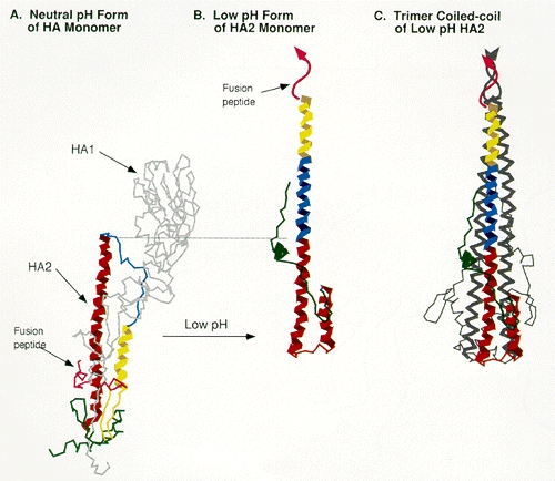
Figure 8. Ribbon diagram of the neutral and low pH forms of the influenza virus hemagglutinin.

Figure 8

Ribbon diagram of the neutral and low pH forms of the influenza virus hemagglutinin. (A) Neutral pH form of the hemagglutinin monomer crystal structure: HA1 is shown in gray, and HA2 in colors that correspond to the amino-terminal helix, the central extended chain, and the carboxy-terminal helix (Wilson et al. 1981). (B) Crystal structure of the low pH form of HA2 showing the formation of an extended helix (Bullough et al. 1994). (C) Trimer of the low pH form of HA2 showing the triple-stranded coiled-coil structure.

From: Retroviral Env Gene Products: Receptor Binding and Host-Range Determinants

Cover of Retroviruses
Retroviruses.
Coffin JM, Hughes SH, Varmus HE, editors.
Cold Spring Harbor (NY): Cold Spring Harbor Laboratory Press; 1997.
Copyright © 1997, Cold Spring Harbor Laboratory Press.

NCBI Bookshelf. A service of the National Library of Medicine, National Institutes of Health.