From: Osteogenesis: The Development of Bones
Copyright © 2000, Sinauer
Associates.
NCBI Bookshelf. A service of the National Library of Medicine, National Institutes of Health.
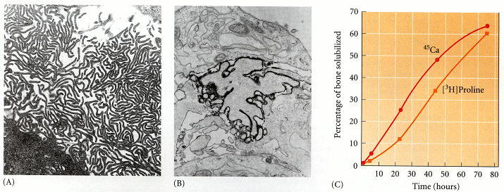
Osteoclast activity on the bone matrix. (A) Electron micrograph of the ruffled membrane of a chick osteoclast cultured on reconstituted bone matrix. (B) Section of ruffled membrane stained for the presence of an ATPase capable of transporting hydrogen ions from the cell. The ATPase is restricted to the membrane of the cell process. (C) Solubilization of inorganic and collagenous matrix components (as measured by the release of [45Ca] and [3H] proline, respectively) by 10,000 osteoclasts incubated on labeled bone fragments. (A and C from Blair et al. 1986; B from Baron et al. 1986, photograph courtesy of the authors.)
From: Osteogenesis: The Development of Bones
NCBI Bookshelf. A service of the National Library of Medicine, National Institutes of Health.