| NCBI National Center for Biotechnology Information |  |
 |
3MZS:
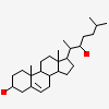
Crystal Structure of Cytochrome P450 CYP11A1 in complex with 22-hydroxy-cholesterol
| Biological unit 1: | monomeric |
| Source organism: | Bos taurus |
| Number of proteins: | 1 (Cholesterol side-chain cleavage enzyme) |
| Number of chemicals: | 3 ((3alpha,8alpha,22R)-cholest-5-ene-3,22-diol,PRO... ▼)
|
Similar Structures (973)
Showing 1 to 10 out of 973 selected structures
| PDB ID | Description | Taxonomy | Aligned Protein | RMSD | Aligned Residues | Sequence Identity | |||
|---|---|---|---|---|---|---|---|---|---|
| 1 | Full |
3NA1 | Crystal structure of human CYP11A1 in complex with 20-hydroxycholesterol |
Homo sapiens |
1 | 1.14Å | 470 | 74% | |
| 2 | Full |
3N9Y | Crystal structure of human CYP11A1 in complex with cholesterol |
Homo sapiens |
1 | 1.11Å | 469 | 74% | |
| 3 | Full |
3NA0 | Crystal structure of human CYP11A1 in complex with 20,22-dihydroxycholesterol |
Homo sapiens |
1 | 1.05Å | 467 | 74% | |
| 4 | Full |
3N9Z | Crystal structure of human CYP11A1 in complex with 22-hydroxycholesterol |
Homo sapiens |
1 | 1.05Å | 467 | 74% | |
| 5 | Full |
6XZ8 | Structure of aldosterone synthase (CYP11B2) in complex with N-[(1R)-1-[5-(6-chloro-1,1-dimethyl-3-oxo-isoindolin-2-yl)-3-pyridyl]ethyl]methanesulfonamide |
Homo sapiens |
1 | 2.18Å | 462 | 37% | |
| 6 | Full |
3K9V | Crystal structure of rat mitochondrial P450 24A1 S57D in complex with CHAPS |
Rattus norvegicus |
1 | 3.31Å | 462 | 28% | |
| 7 | Full |
4FDH | Structure of human aldosterone synthase, CYP11B2, in complex with fadrozole |
Homo sapiens |
1 | 2.20Å | 461 | 37% | |
| 8 | Full |
6XZ9 | Structure of aldosterone synthase (CYP11B2) in complex with 5-chloro-3,3-dimethyl-2-[5-[1-(1-methylpyrazole-4-carbonyl)azetidin-3-yl]oxy-3-pyridyl]isoindolin-1-one |
Homo sapiens |
1 | 2.21Å | 461 | 37% | |
| 9 | Full |
6M7X | Structure of human CYP11B1 in complex with fadrozole |
Homo sapiens |
1 | 2.39Å | 461 | 37% | |
| 10 | Full |
4DVQ | Structure of human aldosterone synthase, CYP11B2, in complex with deoxycorticosterone |
Homo sapiens |
1 | 2.20Å | 460 | 38% |
