?
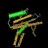 
Src homology 2 (SH2) domain found in Nck Nck proteins are adaptors that modulate actin cytoskeleton dynamics by linking proline-rich effector molecules to tyrosine kinases or phosphorylated signaling intermediates. There are two members known in this family: Nck1 (Nckalpha) and Nck2 (Nckbeta and Growth factor receptor-bound protein 4 (Grb4)). They are characterized by having 3 SH3 domains and a C-terminal SH2 domain. Nck1 and Nck2 have overlapping functions as determined by gene knockouts. Both bind receptor tyrosine kinases and other tyrosine-phosphorylated proteins through their SH2 domains. In addition they also bind distinct targets. Neuronal signaling proteins: EphrinB1, EphrinB2, and Disabled-1 (Dab-1) all bind to Nck-2 exclusively. And in the case of PDGFR, Tyr(P)751 binds to Nck1 while Tyr(P)1009 binds to Nck2. Nck1 and Nck2 have a role in the infection process of enteropathogenic Escherichia coli (EPEC). Their SH3 domains are involved in recruiting and activating the N-WASP/Arp2/3 complex inducing actin polymerization resulting in the production of pedestals, dynamic bacteria-presenting protrusions of the plasma membrane. A similar thing occurs in the vaccinia virus where motile plasma membrane projections are formed beneath the virus. Recently it has been shown that the SH2 domains of both Nck1 and Nck2 bind the G-protein coupled receptor kinase-interacting protein 1 (GIT1) in a phosphorylation-dependent manner. In general SH2 domains are involved in signal transduction. They typically bind pTyr-containing ligands via two surface pockets, a pTyr and hydrophobic binding pocket, allowing proteins with SH2 domains to localize to tyrosine phosphorylated sites. |
