All WormBook content, except where otherwise noted, is licensed under a Creative Commons Attribution License, which permits unrestricted use, distribution, and reproduction in any medium, provided the original work is properly cited.
NCBI Bookshelf. A service of the National Library of Medicine, National Institutes of Health.
WormBook: The Online Review of C. elegans Biology [Internet]. Pasadena (CA): WormBook; 2005-2018.
In mammals, flies, and worms, sex is determined by distinctive regulatory mechanisms that cause males (XO or XY) and females (XX) to differ in their dose of X chromosomes. In each species, an essential X chromosome-wide process called dosage compensation ensures that somatic cells of either sex express equal levels of X-linked gene products. The strategies used to achieve dosage compensation are diverse, but in all cases, specialized complexes are targeted specifically to the X chromosome(s) of only one sex to regulate transcript levels. In C. elegans, this sex-specific targeting of the dosage compensation complex (DCC) is controlled by the same developmental signal that establishes sex, the ratio of X chromosomes to sets of autosomes (X:A signal). Molecular components of this chromosome counting process have been defined. Following a common step of regulation, sex determination and dosage compensation are controlled by distinct genetic pathways. C. elegans dosage compensation is implemented by a protein complex that binds both X chromosomes of hermaphrodites to reduce transcript levels by one-half. The dosage compensation complex resembles the conserved 13S condensin complex required for both mitotic and meiotic chromosome resolution and condensation, implying the recruitment of ancient proteins to the new task of regulating gene expression. Within each C. elegans somatic cell, one of the DCC components also participates in the separate mitotic/meiotic condensin complex. Other DCC components play pivotal roles in regulating the number and distribution of crossovers during meiosis. The strategy by which C. elegans X chromosomes attract the condensin-like DCC is known. Small, well-dispersed X-recognition elements act as entry sites to recruit the dosage compensation complex and to nucleate spreading of the complex to X regions that lack recruitment sites. In this manner, a repressed chromatin state is spread in cis over short or long distances, thus establishing the global, epigenetic regulation of X chromosomes that is maintained throughout the lifetime of hermaphrodites.
1. Introduction
In numerous organisms, sex is determined by a chromosome counting mechanism that distinguishes one sex chromosome from two. In flies and worms, XX embryos become females (or hermaphrodites), while XO or XY embryos become males (Bridges, 1916; Madl and Herman, 1979; Nigon, 1951). Sex can also be specified by the presence of a particular sex chromosome, such as the Y chromosome of mammals: XY embryos are males, and XX embryos females (Gubbay et al., 1990; Koopman et al., 1991; Sinclair et al., 1990). These sex-determining mechanisms cause the two sexes to differ in their dose of X chromosomes, yet both sexes require equivalent levels of X-chromosome gene products. A chromosome-wide regulatory process called dosage compensation neutralizes the difference in X-linked gene dose between sexes by equalizing X-chromosome transcript levels.
The strategies for dosage compensation are diverse in different organisms (Figure 1), but in all known cases, specialized dosage compensation complexes are targeted exclusively to the X chromosome(s) of one sex to modulate gene expression in only that sex. This selective recruitment of the dosage compensation machinery establishes the epigenetic regulation of X chromosomes that is maintained throughout the lifetime of the animal. Female mammals randomly inactivate one X chromosome (Plath et al., 2002). Male flies double the transcription rate of their single X chromosome (Meller and Kuroda, 2002). Hermaphrodite worms keep both X chromosomes active, but repress transcript levels from each X chromosome by half (Meyer and Casson, 1986). In flies and mammals, this gene regulation utilizes non-coding RNAs that coat the regulated X (Meller and Kuroda, 2002; Plath et al., 2002), and the chromosome-wide regulation is accompanied by the sex-specific modification of histones on the dosage compensated X chromosomes (Bone et al., 1994; Heard et al., 2001; Hilfiker et al., 1997; Mermoud et al., 2002). In all three species, dosage compensation is essential, and failure to accomplish this global regulation causes either male- or female-specific lethality.
Fundamental questions are relevant to all forms of dosage compensation. First, what is the composition of the machinery that implements dosage compensation? Second, what are the sex-specific factors that activate the dosage compensation machinery in only one sex? Third, what are the cis-acting recruitment sites that target X chromosomes for regulation by the dosage compensation complex? Fourth, how is gene expression coordinately controlled along an entire X chromosome? Fifth, what is the molecular mechanism for fine-tuning X-linked gene expression by only two-fold? These basic questions have been addressed in C. elegans, using integrated genetic, biochemical, and cell biological approaches to dissect this regulatory process.
2. Genes encoding the dosage compensation machinery
In C. elegans, the processes of dosage compensation and sex determination are coordinately regulated by a group of genes that respond to the primary sex-determination signal. Following this common step of regulation, sex determination and dosage compensation are separately controlled by distinct genetic pathways.
The discovery of sex-specific lethal mutations that preferentially killed XX hermaphrodites was pivotal for the identification of dosage compensation genes (Hodgkin, 1983; Plenefisch et al., 1989). Mutations in eight genes-sdc-1, sdc-2, sdc-3, dpy-21, dpy-26, dpy-27, dpy-28, and dpy-30 reduced the viability of XX but not XO animals. All mutations disrupted dosage compensation, causing a two-fold elevation in X-linked transcript levels in XX but not XO animals (DeLong et al., 1993; Hsu and Meyer, 1994; Meyer and Casson, 1986; Nusbaum and Meyer, 1989; Villeneuve and Meyer, 1987). XX animals that escape lethality have a dumpy (Dpy) phenotype caused by the over expression of X-linked genes. The specific X-linked genes that contribute to this phenotype have not been determined. XO mutants appear wild type, except for dpy-30 XO animals, which are developmentally delayed and have numerous morphological and behavioral abnormalities. The sdc mutations disrupt sex determination as well as X-chromosome gene regulation, causing the rare, viable XX Dpy mutant embryos to develop as males. The sdc genes set the hermaphrodite mode of both sex determination and dosage compensation, acting early in the genetic regulatory hierarchy prior to the divergence of the sex determination and dosage compensation genes into two separate pathways (DeLong et al., 1993; Nusbaum and Meyer, 1989; Villeneuve and Meyer, 1987). The dosage compensation pathway includes the dpy genes listed above.
The hermaphrodite-specific elevation in X-linked transcript levels caused by all dosage compensation mutations was consistent with either of two mechanisms for dosage compensation: random inactivation of a single hermaphrodite X chromosome or repression of both hermaphrodite X chromosomes by half. X inactivation was unlikely since neither of the two genetic phenomena it would cause had been observed: first, hermaphrodites are not mosaic in phenotype for cell-autonomous X-linked markers; second, most X-linked loss-of-function mutations fail to behave as dominant alleles with variable penetrance and expressivity. The molecular architecture of the dosage compensation proteins and their localization to the two hermaphrodite X chromosomes (see below) confirmed that dosage compensation involves the repression of both X chromosomes by half.
3. Dosage compensation proteins assemble onto hermaphrodite X chromosomes to regulate gene expression and also function separately in chromosome segregation
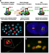
Molecular analysis of the dosage compensation genes showed the similarity of DPY-26, DPY-27, and DPY-28 proteins to components of 13S condensin, a complex essential for the resolution and compaction of mitotic and meiotic chromosomes from yeast to man (Figure 2A; Chan et al., 2004; Chuang et al., 1994; Hirano, 2002; Lieb et al., 1996; Swedlow and Hirano, 2003; C. Tsai and B. Meyer, in preparation). The prototypical condensin complex contains at least five subunits, including a pair of SMC (Structural Maintenance of Chromosomes) proteins (SMC2 and SMC4) and three non-SMC ancillary factors, which belong to the Chromosome-Associated Polypeptide CAP-D2, CAP-G, and CAP-H/Barren families (Hirano, 2002; Swedlow and Hirano, 2003). DPY-26 resembles CAP-H, DPY-28 resembles CAP-D2, and DPY-27 resembles SMC4. These three dosage compensation proteins form part of a complex that associates with both X chromosomes of hermaphrodites (Figure 2A–B).
Biochemical analysis of the dosage compensation complex identified an additional component, MIX-1, the C. elegans homolog of SMC2 (Lieb et al., 1996). Mutations in mix-1 disrupt not only dosage compensation, but also mitotic and meiotic chromosome resolution, condensation, and segregation, causing the death of both XX and XO animals (Chan et al., 2004; Hagstrom et al., 2002; Lieb et al., 1996). The role of MIX-1 in these vital cellular processes precluded its identification in genetic screens that exploited the hermaphrodite-specific action of dosage compensation genes.
MIX-1 binds to X chromosomes of hermaphrodites when it associates with the dosage compensation protein DPY-27. MIX-1 co-localizes with the centromeres of mitotic chromosomes in both sexes (Figure 2C) or with the meiotic chromosomes in diplotene/diakinesis (Figure 2E) when it associates with the mitotic/meiotic-specific SMC4 protein called SMC-4 (Chan et al., 2004; Hagstrom et al., 2002; Lieb et al., 1996). That is, MIX-1 partitions its roles in two separate biological process, gene expression and chromosome segregation, through its participation in two separate condensin-like complexes, a dosage compensation-specific complex and a mitotic/meiotic complex (Figure 2A) (Chan et al., 2004; Hagstrom et al., 2002). Together, these results suggest that the C. elegans dosage compensation process evolved by recruiting components used in other chromosome behaviors to the new task of fine-tuning gene expression. The similarity of the dosage compensation complex to condensin and the participation of MIX-1 in both complexes suggest a common mechanism for repressing X-chromosome gene expression during dosage compensation and for establishing chromosome resolution and higher order chromosome structure during mitotic and meiotic chromosome segregation.
Other components of the dosage compensation complex have also retained their roles in conserved cellular processes. DPY-28 figures prominently in meiosis, where it regulates the number and distribution of crossovers between homologous chromosomes (C. Tsai and B. Meyer, in preparation). DPY-30 not only binds X chromosomes to down regulate gene expression, it associates with the COMPASS complex to methylate histones (C. Hassig and B. Meyer, unpublished). DPY-30 is the C. elegans homolog of the S. cerevisiae COMPASS protein Sdc1p (Hsu and Meyer, 1994; Nagy et al., 2002).
Recruitment of the DPY and MIX proteins to X chromosomes requires the activities of SDC-2 (a novel protein), SDC-3 (a protein with two zinc fingers), and DPY-30 (Chuang et al., 1996; Davis and Meyer, 1997; Klein and Meyer, 1993; Lieb et al., 1998; Lieb et al., 1996; Yonker and Meyer, 2003). These three proteins recruit all other dosage compensation proteins to X, including DPY-21 (a novel protein) and SDC-1 (a protein with seven zinc fingers), through their own association with X (Chu et al., 2002; Davis and Meyer, 1997; Dawes et al., 1999; Yonker and Meyer, 2003). SDC-3 requires both SDC-2 and DPY-30 for its localization to X (Davis and Meyer, 1997), and DPY-30 requires both SDC-2 and SDC-3 to bind X (C. Hassig and B. Meyer, unpublished). SDC-2 is unique in that it can localize to X independent ly from all other dosage compensation proteins, suggesting that SDC-2 is pivotal for X chromosome recognition and confers chromosome specificity to dosage compensation (Dawes et al., 1999). Once the dosage compensation machinery assembles on X chromosomes, it appears to remain localized to X throughout the entire cell cycle, including mitosis (Figure 2D; Lieb et al., 1998).
4. Sex-specific targeting of the dosage compensation complex to hermaphrodite X chromosomes
SDC-2 not only confers chromosome-specificity to dosage compensation, it also confers hermaphrodite-specificity. All properties of SDC-2 are consistent with its role as the sex-specific switch that activates dosage compensation in hermaphrodites (Dawes et al., 1999). All other dosage compensation proteins are supplied maternally and are diffusely distributed throughout the nuclei of very young embryos (<30 cells) in both sexes and only later become localized to X chromosomes of XX but not XO animals. SDC-2 differs in four important ways. First, SDC-2 is not maternally contributed and not expressed in very young embryos. Its initial accumulation occurs around the 40-cell stage, the stage in which the dosage compensation machinery assembles on X. Second, SDC-2 localizes to hermaphrodite X chromosomes from the onset of its accumulation. Third, SDC-2 is not expressed in wild-type XO embryos, indicating that SDC-2, unlike other dosage compensation proteins, is sex-specifically regulated. sdc-2 is repressed in males by the male-specific gene xol-1, the master sex-determination switch gene and direct target of the primary sex-determination signal (Figure 3; Miller et al., 1988). When xol-1 is active (in XO embryos), male development ensues; when xol-1 is inactive (in XX embryos) hermaphrodite development ensues, including the activation of dosage compensation (Rhind et al., 1995). Finally, ectopic expression of sdc-2 in XO animals triggers assembly of the dosage compensation complex on the single X, causing death. Thus, SDC-2 is the hermaphrodite-specific switch that is both necessary and sufficient to activate dosage compensation.
The mechanism by which xol-1 controls sdc-2 remains a mystery. Whether it acts directly or indirectly is also not known. Unexpectedly, the crystal structure of XOL-1 revealed it to be a GHMP kinase family member, despite having sequence identity of less than 10% (Luz et al., 2003). GHMP kinases are small molecule kinases such as galactokinase and homoserine kinase. XOL-1 does not appear to bind ATP, suggesting that it may not have the enzymatic activity of a kinase. However, clues to XOL-1 function come from the identity of GHMP kinase family members as regulators of gene expression. S. cerevisiae GAL3p, a protein that is structurally similar to galactokinase and can be converted to a galactokinase by the substitution of two amino acids, is a galactose- and ATP-dependent transcriptional inducer of GAL genes (Platt et al., 2000). Moreover, GAL1 of Kluyveromyces lactis is a bifunctional protein that acts as both a galactokinase and a transcriptional inducer of GAL genes (Meyer et al., 1991). XOL-1 might also act with a small effector molecule to repress transcription of a downstream target gene such as sdc-2.
5. The X:A signal controls dosage compensation by regulating xol-1
xol-1, in turn, is regulated by the primary sex-determination signal, the X:A signal, the ratio of X chromosomes to sets of autosomes (the ploidy; (Figure 3; Madl and Herman, 1979; Miller et al., 1988; Nigon, 1951; Rhind et al., 1995). The two-fold difference in X-chromosome dose between males and hermaphrodites is translated into the ON/OFF state of xol-1. A set of X-linked genes called X Signal Elements (XSEs) communicate X-chromosome dose by repressing xol-1 in a dose-dependent manner. These XSEs include the nuclear receptor SEX-1 and the RNA binding protein FOX-1 (Akerib and Meyer, 1994; Carmi et al., 1998; Carmi and Meyer, 1999; Hodgkin et al., 1994; Nicoll et al., 1997; Skipper et al., 1999). XSEs act cumulatively to inhibit xol-1 by two different mechanisms, transcriptional repression and obstruction of proper pre-mRNA splicing.
Recent experiments showed the autosomal component of the X:A signal to include a set of dose-sensitive genes on autosomes called Autosomal-signal elements (ASE) that communicate the poidy. xol-1 is activated by this set of dose-sensitive ASEs, which include the T-box protein SEA-1 (Powell et al., 2005) and the zinc finger protein SEA-2 (P. Nix and B. Meyer, unpublished). In XX animals, the double dose of XSEs opposes the double dose of ASEs, causing xol-1 to be repressed. The mechanism by which this process occurs is not yet known. In XO animals, the single dose of XSEs cannot oppose the double dose of ASEs, allowing xol-1 to be active (Figure 3).
6. Recruitment of the dosage compensation complex for gene-specific versus chromosome-wide repression
Not only does SDC-2 activate dosage compensation, it also induces hermaphrodite sexual differentiation in concert with SDC-1 and SDC-3 (Chu et al., 2002; Dawes et al., 1999). Proof that SDC-2 acts as the pivotal sex-specific factor to initiate the hermaphrodite program of sexual development came in part from the observation that overexpression of SDC-2 and SDC-1 in XO animals, made viable by a dosage compensation mutation, caused XO animals to develop as hermaphrodites. SDC-2 activates hermaphrodite sexual development by binding directly to regulatory regions of the male sex-determination gene her-1, repressing its transcription 20-fold, thereby directing the sex determination pathway to the hermaphrodite mode (Dawes et al., 1999). SDC-2 thus acts both as a strong gene-specific repressor and a weaker chromosome-wide repressor. Unexpectedly, SDC-2, together with SDC-1 and SDC-3, recruits other dosage compensation components to her-1, directing this chromosome repression machinery to silence an individual, autosomal gene (Chu et al., 2002).
Functional dissection of her-1 in vivo revealed three DNA recognition elements required for SDC binding, recruitment of the DCC, and transcriptional repression (Chu et al., 2002; Li et al., 1999). These her-1 elements differed in location (promoter and 2nd intron), sequence, and strength of repression. Two her-1 binding sites share a 15-nucleotide repeat essential for repression, but the her-1 sites bear no obvious resemblance to any sequences on X, leading to the speculation that the chromosome-wide repression complex achieves different degrees of repression in part by associating with different DNA sequences. These results also imply that the dosage compensation complex regulates transcription along X chromosomes using diverse DNA recognition elements.
Further analysis revealed important molecular differences in the composition and targeting of the repression complex to her-1 versus X (Yonker and Meyer, 2003). The dosage compensation protein DPY-21, a novel protein, localizes to X but not to her-1. Furthermore, within the complex, different proteins play the lead role in recognizing DNA targets. SDC-2 recognizes X-chromosome targets, while SDC-3 recognizes her-1 targets. These results begin to explain how closely related complexes can achieve uniformly weak repression of many genes in one context and strong repression of a specific gene in another.
7. Recruitment and spreading of the dosage compensation complex along hermaphrodite X chromosomes
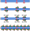
To define cis-acting X-chromosome sites that recruit the dosage compensation complex, a chromosome-wide search was conducted for regions of X sufficient to recruit the complex when detached from X (Csankovszki et al., 2004). Regions were analyzed in 32-ploid intestinal cell nuclei of XX hermaphrodite stains carrying either free or autosome-attached X-chromosome duplications (Figure 4). Provided that C. elegans X chromosomes contained discrete X-recognition elements (rex sites) that recruit the complex, four possibilities existed for how the complex might be targeted to X (Figure 5). First, a single site on X could recruit the complex and nucleate long-range spreading of the dosage compensation complex across the entire chromosome. Second, a limited number of recognition sites could recruit the complex, and some or all sites could nucleate short-range spreading. Third, a limited number of recognition sites could recruit the complex but the complex would not spread. It would only occupy sites that autonomously recruited it, influencing gene expression from a long distance, perhaps by altering chromosome structure. Fourth, a high density of X recognition sites could recruit the complex, but no spreading would occur, implying direct, short-range regulation by the complex.
The first possibility, the presence of a single recruitment site, was eliminated by analysis of hermaphrodites carrying one copy of the X duplication mnDp1 and two chromosomes deleted for the corresponding region (Figure 4A and Figure 6). The dosage compensation complex colocalized with both the duplication and the truncated X chromosomes, indicating that mnDp1 harbors at least one X-recognition element, as does the rest of X. The interpretation of multiple, independent X-recognition elements was greatly reinforced by the identification of many other non-overlapping, detached X regions that recruited the complex. Two classes of dosage compensation complex recruitment were found, strong recruitment typified by mnDp1 and limited, but reproducible recruitment typified by mnDp57 (Figure 4B and Figure 6).
The fourth possibility, a high density of recruitment sites, was eliminated by the discovery of large X segments incapable of recruiting the complex when detached from X (Figure 4C and Figure 6). Despite the inability to autonomously recruit the complex, these regions, typified by stDp2, harbor dosage compensated genes when present on the native X chromosome (Figure 6). These results indicated that the dosage compensation complex regulates gene expression in X regions that lack recruitment sites either by spreading into the regions from neighboring X-recognition elements (second possibility) or by acting over a distance (third possibility). The second possibility predicts binding of the dosage compensation complex to the region on the native X corresponding to stDp2, and the third possibility predicts no binding. Robust localization of the dosage compensation complex was found on the stDp2 region (Figure 4D), on other native X regions corresponding to duplications with no recruitment ability, and also on regions corresponding to duplications with limited, but reproducible binding. Thus, the C. elegans X chromosomes have discrete X-recognition elements that recruit the dosage compensation complex and nucleate spreading of the complex over short or long distances (Figure 7; Csankovszki et al., 2004). In contrast, mammals utilize a single recruitment site that nucleates long-range spreading of dosage compensation components along the regulated X. Flies were initially thought to have multiple recruitment sites that nucleate local spreading, but recent evidence suggests that flies have a high density of recruitment sites and perhaps little or no spreading (Fagegaltier and Baker, 2004; Oh et al., 2004).
8. Molecular identification of discrete X-recognition elements
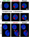
Small, discrete X-recognition elements responsible for recruitment activity were identified by a general strategy that assessed binding of the dosage compensation complex in vivo (Csankovszki et al., 2004). Transgenic nematode lines carrying extrachromosomal DNA arrays with tandem copies of cosmids from a recruitment region were made, and arrays were assayed for dosage compensation complex binding (Figure 8A). Arrays also carried lac operator repeats and lac repressor tagged with GFP. lac repressor bound to its operator DNA tagged arrays in interphase nuclei with GFP fluorescence. Co-localization of dosage compensation protein antibodies with arrays indicated the presence of an X-recognition element. This approach identified the element rex-1 from a region of X with limited recruitment ability (Figure 6 and Figure 8B; Csankovszki et al., 2004). Recruitment activity has subsequently been attributed to a 33 bp fragment (P. McDonel and B. Meyer, unpublished).
Multiple copies of the X-recognition element titrate the dosage compensation complexes from X and thereby impair dosage compensation (Figure 8B). Genetic assays indicated that dosage compensation was compromised in animals carrying arrays with X-recognition elements. Furthermore, X chromosomes could be detected easily by dosage compensation protein antibodies only in XX cells that lost arrays because of mitotic instability (Figure 8B; Csankovszki et al., 2004). Adjacent cells harboring arrays showed robust localization of the dosage compensation complex to the array but not the X chromosomes. These results indicate that X regions with limited recruitment ability harbor high-affinity recruitment sites capable of competing with other recruitment sites on X for the dosage compensation complex (Csankovszki et al., 2004).
A total of thirteen X-recognition elements have thus far been identified by this approach (J. Jans, P. McDonel, and B. Meyer, unpublished data). Further dissection of these sites will reveal the features of X-recognition elements essential for recruitment of the dosage compensation complex. Those features could be primary DNA sequence, DNA structure, or a combination of both.
In summary, the strategy by which C. elegans X chromosomes attract the condensin-like dosage compensation complex is now known. Discrete X-recognition elements serve as entry sites to recruit the dosage compensation complex and to nucleate spreading of the complex to X regions that lack recruitment sites (Figure 7). In this manner, a repressed chromatin state can be spread in cis over short or long distances, thus establishing the global regulation of X chromosomes that is maintained throughout the lifetime of hermaphrodites (Csankovszki et al., 2004).
9. Future endeavors
Future experiments will reveal whether the X-recognition protein SDC-2 binds X DNA directly or instead requires other X-bound cellular proteins to reach its target. Experiments will also determine the contributions, if any, of DNA structure or specific DNA motifs within X-recognition elements for the recruitment of the dosage compensation complex to X. Experiments will also define the mechanistic basis by which the dosage compensation complex spreads along the X chromosome and whether such spreading involves histone modifications and/or non-coding RNAs, as do other forms of epigenetic regulation across whole chromosomes or chromosomal domains. Ultimately, a detailed biochemical understanding of the two-fold X-chromosome repression will emerge.
10. References
- Akerib C.C. , Meyer B.J. Identification of X chromosome regions in Caenorhabditis elegans that contain sex-determination signal elements. Genetics. (1994);138:1105–1125. [PMC free article: PMC1206251] [PubMed: 7896094]
- Bone J.R. , Lavender J. , Richman R. , Palmer M.J. , Turner B.M. , Kuroda M.I. Acetylated histone H4 on the male X chromosome is associated with dosage compensation in Drosophila. Genes Dev. (1994);8:96–104. [PubMed: 8288132]
- Bridges C.B. Non-disjunction as proof of the chromosome theory of heredity. Genetics. (1916);1:1–51. [PMC free article: PMC1193653] [PubMed: 17245850]
- Carmi I. , Kopczynski J.B. , Meyer B.J. The nuclear hormone receptor SEX-1 is an X-chromosome signal that determines nematode sex. Nature. (1998);396:168–173. [PubMed: 9823896]
- Carmi I. , Meyer B.J. The primary sex determination signal of Caenorhabditis elegans. Genetics. (1999);152:999–1015. [PMC free article: PMC1460672] [PubMed: 10388819]
- Chan R.C. , Severson A.F. , Meyer B.J. Condensin restructures chromosomes in preparation for meiotic divisions. J. Cell Biol. (2004);167:613–625. [PMC free article: PMC2172564] [PubMed: 15557118]
- Chu D.S. , Dawes H.E. , Lieb J.D. , Chan R.C. , Kuo A.F. , Meyer B.J. A molecular link between gene-specific and chromosome-wide transcriptional repression. Genes Dev. (2002);16:796–805. [PMC free article: PMC186330] [PubMed: 11937488]
- Chuang P.T. , Albertson D.G. , Meyer B.J. DPY-27: a chromosome condensation protein homolog that regulates C. elegans dosage compensation through association with the X chromosome. Cell. (1994);79:459–474. [PubMed: 7954812]
- Chuang P.T. , Lieb J.D. , Meyer B.J. Sex-specific assembly of a dosage compensation complex on the nematode X chromosome. Science. (1996);274:1736–1739. [PubMed: 8939870]
- Csankovszki G. , McDonel P. , Meyer B.J. Recruitment and spreading of the C. elegans dosage compensation complex along X chromosomes. Science. (2004);303:1182–1185. [PubMed: 14976312]
- Davis T.L. , Meyer B.J. SDC-3 coordinates the assembly of a dosage compensation complex on the nematode X chromosome. Development. (1997);124:1019–1031. [PubMed: 9056777]
- Dawes H.E. , Berlin D.S. , Lapidus D.M. , Nusbaum C. , Davis T.L. , Meyer B.J. Dosage compensation proteins targeted to X chromosomes by a determinant of hermaphrodite fate. Science. (1999);284:1800–1804. [PubMed: 10364546]
- DeLong L. , Plenefisch J.D. , Klein R.D. , Meyer B.J. Feedback control of sex determination by dosage compensation revealed through Caenorhabditis elegans sdc-3 mutations. Genetics. (1993);133:875–896. [PMC free article: PMC1205407] [PubMed: 8462848]
- Fagegaltier D. , Baker B.S. X chromosome sites autonomously recruit the dosage compensation complex in Drosophila males. PLoS. Biol. (2004);2:e341. [PMC free article: PMC521175] [PubMed: 15502872]
- Gubbay J. , Collignon J. , Koopman P. , Capel B. , Economou A. , Munsterberg A. , Vivian N. , Goodfellow P. , Lovell-Badge R. A gene mapping to the sex-determining region of the mouse Y chromosome is a member of a novel family of embryonically expressed genes. Nature. (1990);346:245–250. [PubMed: 2374589]
- Hagstrom K.A. , Holmes V.F. , Cozzarelli N.R. , Meyer B.J. C. elegans condensin promotes mitotic chromosome architecture, centromere organization and sister chromatid segregation during mitosis and meiosis. Genes Dev. (2002);16:729–742. [PMC free article: PMC155363] [PubMed: 11914278]
- Heard E. , Rougeulle C. , Arnaud D. , Avner P. , Allis C.D. , Spector D.L. Methylation of histone H3 at Lys-9 is an early mark on the X chromosome during X inactivation. Cell. (2001);107:727–738. [PubMed: 11747809]
- Hilfiker A. , Hilfiker-Kleiner D. , Pannuti A. , Lucchesi J.C. mof, a putative acetyl transferase gene related to the Tip60 and MOZ human genes and to the SAS genes of yeast, is required for dosage compensation in Drosophila. EMBO J. (1997);16:2054–2060. [PMC free article: PMC1169808] [PubMed: 9155031]
- Hirano T. The ABCs of SMC proteins: two-armed ATPases for chromosome condensation, cohesion and repair. Genes Dev. (2002);16:399–414. [PubMed: 11850403]
- Hodgkin J. X chromosome dosage and gene expression in Caenorhabditis elegans: Two unusual dumpy genes. Mol. Gen. Genet. (1983);192:452.
- Hodgkin J. , Zellan J.D. , Albertson D.G. Identification of a candidate primary sex determination locus, fox-1, on the X chromosome of Caenorhabditis elegans. Development. (1994);120:3681–3689. [PubMed: 7821230]
- Hsu D.R. , Meyer B.J. The dpy-30 gene encodes an essential component of the Caenorhabditis elegans dosage compensation machinery. Genetics. (1994);137:999–1018. [PMC free article: PMC1206076] [PubMed: 7982580]
- Klein R.D. , Meyer B.J. Independent domains of the SDC-3 protein control sex determination and dosage compensation in C. elegans. Cell. (1993);72:349–364. [PubMed: 8431944]
- Koopman P. , Gubbay J. , Vivian N. , Goodfellow P. , Lovell-Badge R. Male development of chromosomally female mice transgenic for Sry. Nature. (1991);351:117–121. [PubMed: 2030730]
- Li W. , Streit A. , Robertson B. , Wood W.B. Evidence for multiple promoter elements orchestrating male-specific regulation of the her-1 gene in Caenorhabditis elegans. Genetics. (1999);152:237–248. [PMC free article: PMC1460597] [PubMed: 10224257]
- Lieb J.D. , Albrecht M.R. , Chuang P.T. , Meyer B.J. MIX-1: an essential component of the C. elegans mitotic machinery executes X chromosome dosage compensation. Cell. (1998);92:265–277. [PubMed: 9458050]
- Lieb J.D. , Capowski E.E. , Meneely P. , Meyer B.J. DPY-26, a link between dosage compensation and meiotic chromosome segregation in the nematode. Science. (1996);274:1732–1736. [PubMed: 8939869]
- Luz J.G. , Hassig C.A. , Pickle C. , Godzik A. , Meyer B.J. , Wilson I.A. XOL-1, primary determinant of sexual fate in C. elegans, is a GHMP kinase family member and a structural prototype for a class of developmental regulators. Genes Dev. (2003);17:977–990. [PMC free article: PMC196039] [PubMed: 12672694]
- Madl J.E. , Herman R.K. Polyploids and sex determination in Caenorhabditis elegans. Genetics. (1979);93:393–402. [PMC free article: PMC1214087] [PubMed: 295035]
- Meller V.H. , Kuroda M.I. Sex and the single chromosome. Adv. Genet. (2002);46:1–24. [PubMed: 11931222]
- Mermoud J.E. , Popova B. , Peters A.H. , Jenuwein T. , Brockdorff N. Histone H3 lysine 9 methylation occurs rapidly at the onset of random X chromosome inactivation. Curr. Biol. (2002);12:247–251. [PubMed: 11839280]
- Meyer B.J. , Casson L.P. Caenorhabditis elegans compensates for the difference in X chromosome dosage between the sexes by regulating transcript levels. Cell. (1986);47:871–881. [PubMed: 3779843]
- Meyer J. , Walker-Jonah A. , Hollenberg C.P. Galactokinase encoded by GAL1 is a bifunctional protein required for induction of the GAL genes in Kluyveromyces lactis and is able to suppress the gal3 phenotype in Saccharomyces cerevisiae. Mol. Cell Biol. (1991);11:5454–5461. [PMC free article: PMC361914] [PubMed: 1922058]
- Miller L.M. , Plenefisch J.D. , Casson L.P. , Meyer B.J. xol-1: a gene that controls the male modes of both sex determination and X chromosome dosage compensation in C. elegans. Cell. (1988);55:167–183. [PubMed: 3167975]
- Nagy P.L. , Griesenbeck J. , Kornberg R.D. , Cleary M.L. A trithorax-group complex purified from Saccharomyces cerevisiae is required for methylation of histone H3. Proc. Natl. Acad. Sci. USA. (2002);99:90–94. [PMC free article: PMC117519] [PubMed: 11752412]
- Nicoll M. , Akerib C.C. , Meyer B.J. X-chromosome-counting mechanisms that determine nematode sex. Nature. (1997);388:200–204. [PubMed: 9217163]
- Nigon V. Polyploidie experimentale chez un nematode libre, Rhabditis elegans maupas. Bull. Biol. Fr. Belg. (1951);85:187–255.
- Nusbaum C. , Meyer B.J. The Caenorhabditis elegans gene sdc-2 controls sex determination and dosage compensation in XX animals. Genetics. (1989);122:579–593. [PMC free article: PMC1203732] [PubMed: 2759421]
- Oh H. , Bone J.R. , Kuroda M.I. Multiple classes of MSL binding sites target dosage compensation to the X chromosome of Drosophila. Curr. Biol. (2004);14:481–487. [PubMed: 15043812]
- Plath K. , Mlynarczyk-Evans S. , Nusinow D.A. , Panning B. Xist RNA and the mechanism of X chromosome inactivation. Annu. Rev. Genet. (2002);36:233–278. [PubMed: 12429693]
- Platt A. , Ross H.C. , Hankin S. , Reece R.J. The insertion of two amino acids into a transcriptional inducer converts it into a galactokinase. Proc. Natl. Acad. Sci. USA. (2000);97:3154–3159. [PMC free article: PMC16208] [PubMed: 10737789]
- Plenefisch J.D. , DeLong L. , Meyer B.J. Genes that implement the hermaphrodite mode of dosage compensation in Caenorhabditis elegans. Genetics. (1989);121:57–76. [PMC free article: PMC1203606] [PubMed: 2917714]
- Powell, J.R., Jow, M.M., and Meyer, B.J. (2005). The T-box transcription factor SEA-1 is an autosomal element of the X:A signal that determines C. elegans sex. Dev. Cell, in press. [PMC free article: PMC2649673] [PubMed: 16139225]
- Rhind N.R. , Miller L.M. , Kopczynski J.B. , Meyer B.J. xol-1 acts as an early switch in the C. elegans male/hermaphrodite decision. Cell. (1995);80:71–82. [PubMed: 7813020]
- Sinclair A.H. , Berta P. , Palmer M.S. , Hawkins J.R. , Griffiths B.L. , Smith M.J. , Foster J.W. , Frischauf A.M. , Lovell-Badge R. , Goodfellow P.N. A gene from the human sex-determining region encodes a protein with homology to a conserved DNA-binding motif. Nature. (1990);346:240–244. [PubMed: 1695712]
- Skipper M. , Milne C.A. , Hodgkin J. Genetic and molecular analysis of fox-1, a numerator element involved in Caenorhabditis elegans primary sex determination. Genetics. (1999);151:617–631. [PMC free article: PMC1460491] [PubMed: 9927456]
- Swedlow J.R. , Hirano T. The making of the mitotic chromosome: modern insights into classical questions. Mol. Cell. (2003);11:557–569. [PubMed: 12667441]
- Villeneuve A.M. , Meyer B.J. sdc-1: a link between sex determination and dosage compensation in C. elegans. Cell. (1987);48:25–37. [PubMed: 3791412]
- Yonker S.A. , Meyer B.J. Recruitment of C. elegans dosage compensation proteins for gene-specific versus chromosome-wide repression. Development. (2003);130:6519–6532. [PubMed: 14660541]
Footnotes
- *
Edited by Lisa R. Girard. Last revised June 24, 2005. Published June 25, 2005. This chapter should be cited as: Meyer, B. J. X-Chromosome dosage compensation (June 25, 2005), WormBook, ed. The C. elegans Research Community, WormBook, doi/10.1895/wormbook.1.8.1, http://www
.wormbook.org [PMC free article: PMC4781388] [PubMed: 18050416].
- §
To whom correspondence should be addressed. E-mail: ude.yelekreb@reyemjb
- 1. Introduction
- 2. Genes encoding the dosage compensation machinery
- 3. Dosage compensation proteins assemble onto hermaphrodite X chromosomes to regulate gene expression and also function separately in chromosome segregation
- 4. Sex-specific targeting of the dosage compensation complex to hermaphrodite X chromosomes
- 5. The X:A signal controls dosage compensation by regulating xol-1
- 6. Recruitment of the dosage compensation complex for gene-specific versus chromosome-wide repression
- 7. Recruitment and spreading of the dosage compensation complex along hermaphrodite X chromosomes
- 8. Molecular identification of discrete X-recognition elements
- 9. Future endeavors
- 10. References
- Condensin-driven remodelling of X chromosome topology during dosage compensation.[Nature. 2015]Condensin-driven remodelling of X chromosome topology during dosage compensation.Crane E, Bian Q, McCord RP, Lajoie BR, Wheeler BS, Ralston EJ, Uzawa S, Dekker J, Meyer BJ. Nature. 2015 Jul 9; 523(7559):240-4. Epub 2015 Jun 1.
- Review Sex in the wormcounting and compensating X-chromosome dose.[Trends Genet. 2000]Review Sex in the wormcounting and compensating X-chromosome dose.Meyer BJ. Trends Genet. 2000 Jun; 16(6):247-53.
- The Caenorhabditis elegans dosage compensation machinery is recruited to X chromosome DNA attached to an autosome.[Genetics. 2000]The Caenorhabditis elegans dosage compensation machinery is recruited to X chromosome DNA attached to an autosome.Lieb JD, de Solorzano CO, Rodriguez EG, Jones A, Angelo M, Lockett S, Meyer BJ. Genetics. 2000 Dec; 156(4):1603-21.
- SUMOylation is essential for sex-specific assembly and function of the Caenorhabditis elegans dosage compensation complex on X chromosomes.[Proc Natl Acad Sci U S A. 2013]SUMOylation is essential for sex-specific assembly and function of the Caenorhabditis elegans dosage compensation complex on X chromosomes.Pferdehirt RR, Meyer BJ. Proc Natl Acad Sci U S A. 2013 Oct 1; 110(40):E3810-9. Epub 2013 Sep 16.
- Review Caenorhabditis elegans Dosage Compensation: Insights into Condensin-Mediated Gene Regulation.[Trends Genet. 2018]Review Caenorhabditis elegans Dosage Compensation: Insights into Condensin-Mediated Gene Regulation.Albritton SE, Ercan S. Trends Genet. 2018 Jan; 34(1):41-53. Epub 2017 Oct 13.
- X-Chromosome dosage compensation - WormBookX-Chromosome dosage compensation - WormBook
Your browsing activity is empty.
Activity recording is turned off.
See more...


