By agreement with the publisher, this book is accessible by the search feature, but cannot be browsed.
Copyright © 2001, Sinauer Associates, Inc.
Bookshelf ID: NBK10900

An official website of the United States government

NCBI Bookshelf. A service of the National Library of Medicine, National Institutes of Health.
Purves D, Augustine GJ, Fitzpatrick D, et al., editors. Neuroscience. 2nd edition. Sunderland (MA): Sinauer Associates; 2001.
The ultimate target of afferent auditory information is the auditory cortex. Although the auditory cortex has a number of subdivisions, a broad distinction can be made between a primary area and peripheral, or belt, areas. The primary auditory cortex (A1) is located on the superior temporal gyrus in the temporal lobe and receives point-to-point input from the ventral division of the medial geniculate complex; thus, it contains a precise tonotopic map. The belt areas of the auditory cortex receive more diffuse input from the belt areas of the medial geniculate complex and therefore are less precise in their tonotopic organization.
The primary auditory cortex (A1) has a topographical map of the cochlea (Figure 13.14), just as the primary visual cortex (V1) and the primary somatic sensory cortex (S1) have topographical maps of their respective sensory epithelia. Unlike the visual and somatic sensory systems, however, the cochlea has already decomposed the acoustical stimulus so that it is arrayed tonotopically along the length of the basilar membrane. Thus, A1 is said to comprise a tonotopic map, as do most of the ascending auditory structures between the cochlea and the cortex. Orthogonal to the frequency axis of the tonotopic map is a striped arrangement of binaural properties. The neurons in one stripe are excited by both ears (and are therefore called EE cells), while the neurons in the next stripe are excited by one ear and inhibited by the other ear (EI cells). The EE and EI stripes alternate, an arrangement that is reminiscent of the ocular dominance columns in V1 (see Chapter 12). The sorts of sensory processing that occur in the other divisions of the auditory cortex are not well understood, but they are likely to be important to higher-order processing of natural sounds, including those used for communication. It appears that some areas are specialized for processing combinations of frequencies, while others are specialized for processing modulations of amplitude or frequency.
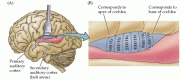
The human auditory cortex. (A) Diagram showing the brain in left lateral view, including the depths of the lateral sulcus, where part of the auditory cortex occupying the superior temporal gyrus normally lies hidden. The primary auditory cortex (A1) is (more...)
Sounds that are especially important for intraspecific communication often have a highly ordered temporal structure. In humans, the best example of such time-varying signals is speech, where different phonetic sequences are perceived as distinct syllables and words. Behavioral studies in cats and monkeys show that the auditory cortex is especially important for processing temporal sequences of sound. If the auditory cortex is ablated in these animals, they lose the ability to discriminate between two complex sounds that have the same frequency components but which differ in temporal sequence. Thus, without the auditory cortex, monkeys cannot discriminate one conspecific communication sound from another. Studies of human patients with bilateral damage to the auditory cortex also reveal severe problems in processing the temporal order of sounds. It seems likely, therefore, that specific regions of the human auditory cortex are specialized for processing elementary speech sounds, as well as other temporally complex acoustical signals, such as music. Indeed, Wernicke's area, which is critical to the comprehension of human language, lies within the secondary auditory area (Figure 13.15; see also Chapter 27).
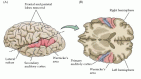
The human auditory cortical areas related to processing speech sounds. (A) Diagram of the brain in left lateral view, showing locations in the intact hemisphere. (B) An oblique section (plane of dashed line in A) shows the cortical areas on the superior (more...)
By agreement with the publisher, this book is accessible by the search feature, but cannot be browsed.
Your browsing activity is empty.
Activity recording is turned off.
See more...