U.S. flag

An official website of the United States government

NCBI Bookshelf. A service of the National Library of Medicine, National Institutes of Health.

Adam MP, Feldman J, Mirzaa GM, et al., editors. GeneReviews® [Internet]. Seattle (WA): University of Washington, Seattle; 1993-2024.

  • This publication is provided for historical reference only and the information may be out of date.

This publication is provided for historical reference only and the information may be out of date.

Cover of GeneReviews®

GeneReviews® [Internet].

Show details

Mucolipidosis III Alpha/Beta – RETIRED CHAPTER, FOR HISTORICAL REFERENCE ONLY

Synonyms: Mucolipidosis IIIA, Pseudo-Hurler Polydystrophy

, MD, PhD, , MD, FACMG, and , PhD.

Author Information and Affiliations

Initial Posting: ; Last Update: May 10, 2012.

Estimated reading time: 27 minutes

Summary

NOTE: THIS PUBLICATION HAS BEEN RETIRED. THIS ARCHIVAL VERSION IS FOR HISTORICAL REFERENCE ONLY, AND THE INFORMATION MAY BE OUT OF DATE.

Clinical characteristics.

Mucolipidosis alpha/beta (ML III alpha/beta; pseudo-Hurler polydystrophy), a slowly progressive disorder with clinical onset at approximately age three years, is characterized by slow growth rate and subnormal stature; radiographic evidence of mild to moderate dysostosis multiplex; joint stiffness and pain initially in the shoulders, hips, and fingers; gradual mild coarsening of facial features; and normal to mildly impaired cognitive development. If present, organomegaly is mild. Pain from osteoporosis that is clinically and radiologically apparent in childhood becomes more severe from adolescence. Cardiorespiratory complications (restrictive lung disease, thickening and insufficiency of the mitral and aortic valves, left and/or right ventricular hypertrophy) are common causes of death, typically in early to middle adulthood.

Diagnosis/testing.

In ML III alpha/beta the activity of nearly all lysosomal hydrolases is up to tenfold higher in plasma and other body fluids than in normal controls because of inadequate targeting to lysosomes. Urinary excretion of oligosaccharides (OSs), a nonspecific finding, is often excessive. Significant deficiency (1%-10% of normal) of the activity of the enzyme UDP-N-acetylglucosamine: lysosomal hydrolase N-acetylglucosamine-1-phosphotransferase (GNPTA), encoded by GNPTAB, confirms the diagnosis. Bidirectional sequencing of the entire GNPTAB coding region detects two pathogenic variants in more than 95% of individuals with ML III alpha/beta.

Management.

Treatment of manifestations: Low-impact physical therapy is usually well tolerated. Myringotomy tube placement may be indicated in the treatment of recurrent otitis media. Carpal tunnel signs may require tendon release. In late childhood or early adolescence symptomatic relief of hip pain may be initially accomplished with over-the-counter analgesics; in some older adolescents and adults with milder phenotypic variants, bilateral hip replacement has been successful. Later in the disease course management focuses on relief of general bone pain associated with osteoporosis, which has responded in a few individuals to scheduled intermittent IV administration of the bisphosphonate pamidronate.

Prevention of secondary complications: Because of concerns about airway management, surgical intervention should be undertaken only in tertiary care settings with pediatric anesthesiologists and intensivists.

Surveillance: Twice-yearly outpatient clinic visits for young children; annual routine follow-up visits after age six years unless bone pain, deteriorating ambulation, and/or cardiac and respiratory monitoring need more frequent attention.

Genetic counseling.

ML III alpha/beta is inherited in an autosomal recessive manner. At conception, each sib of an affected individual has a 25% chance of being affected, a 50% chance of being an asymptomatic carrier, and a 25% chance of being unaffected and not a carrier. Carrier testing for at-risk relatives and prenatal diagnosis for pregnancies at increased risk are possible if the pathogenic variants in the family are known.

Diagnosis

Clinical Diagnosis

The following clinical features contribute to early diagnosis of mucolipidosis III alpha/beta (ML III alpha/beta) [Cathey et al 2010] but are not by themselves diagnostic:

  • Average age at which features are recognized as distinctive: three years (range: late infancy to late childhood)
  • Slow growth rate that gradually decreases
  • Frequent upper respiratory infection and/or otitis media (variably present)
  • Joint stiffness initially in the shoulders, hips, and fingers
  • Joint pain that is exacerbated by strenuous exercise or physical therapy
  • Gradual mild coarsening of facial features
  • Slight corneal cloudiness, noticeable only by slit-lamp examination (variably present)
  • Absent to mild organomegaly
  • Inconsistently, mild to moderate kyphoscoliosis
  • Normal to mildly impaired cognitive development
  • Osteoporosis associated with pain; clinically and radiologically apparent in childhood and more adversely affecting gait and range of motion in large joints in older individuals

In infancy and early childhood skeletal radiographs reveal mild to moderate dysostosis multiplex [Spranger et al 2002]:

  • Long bones. Initially normal or slightly undertubulated; moderate to severe dysplasia of proximal femoral epiphyses
  • Hands and feet. Only mildly shortened diaphyses of metacarpals and phalanges; smaller than normal carpal bones
  • Ribs. Widening especially in lateral and frontal (costochondral junction) parts, but narrower than normal in dorsal parts
  • Spine. Mild generalized platyspondyly; irregular upper and lower end plates and dorsal scalloping; anterior inferior hook in lower thoracic and/or higher lumbar vertebrae; narrow intervertebral spaces
  • Pelvis. Dysplasia with hypoplastic iliac bones; flared iliac wings; elongation of pubic and ischial bones; shallow acetabula; coxa valga
  • Skull. Size proportionate to stature; normal sella turcica

In late childhood or adolescence skeletal radiographs reveal the following:

  • Long bones. Severe dysplasia of the proximal femoral epiphyses; often the epiphyses disappear altogether.
  • Hands. Radiographic abnormalities of the bones remain mild despite slowly progressive claw-like deformation of hands and fingers, mainly caused by hardening of soft tissue around the small joints.
  • Spine. Changes worsen; deficits of ossification remain visible; shape of individual vertebrae does not change significantly except when osteopenia is severe; a minority of affected individuals have severe kyphoscoliosis.
  • Skull. Calvarium thickens gradually; shape of sella turcica usually remains unaltered, anteroposterior elongation is rarely observed.
  • Bone density. Generalized osteopenia is consistent and slowly progressive.
  • Skeletal age. Delayed as evident in ossification of wrist bones and epiphyses of long bones

Testing

Biochemical Testing

Activity of lysosomal hydrolases. In ML III alpha/beta the activity of nearly all lysosomal hydrolases is up to tenfold higher in plasma and other body fluids than in normal controls because mannose-6-phosphate (M6P), which is essential to proper targeting of lysosomal acid hydrolases to lysosomes, cannot be added adequately to the hydrolases (see Molecular Pathogenesis).

The following lysosomal hydrolases are of most interest as their increased activity is relevant in the differential diagnosis of ML III and lysosomal storage disorders:

  • β-D-hexosaminidase (EC 3.2.1.52)
  • β-D-glucuronidase (EC 3.2.1.31)
  • β-D-galactosidase (EC 3.2.1.23)
  • α-D-mannosidase (EC 3.2.1.24)

Note: The acid hydrolases are improperly targeted to the lysosomes but not quantitatively deficient in leukocytes in ML III alpha/beta. In contrast to storage disorders resulting from deficiency of a single lysosomal enzyme, ML III alpha/beta cannot be diagnosed by assay of acid hydrolases in leukocytes.

Urinary excretion of oligosaccharides (OSs). This is a simple and inexpensive test. Excessive urinary excretion of OSs is a nonspecific finding that orients the clinician to consider one of the oligosaccharidoses. In ML III alpha/beta excessive urinary excretion of OSs is variably present; however, false negative results are rare.

Urinary excretion of glycosaminoglycans (GAGs) (i.e., acid mucopolysaccharides [AMPS]) is normal. This is a useful tool to distinguish ML III alpha/beta from the MPS disorders with onset beyond infancy.

UDP-N-acetylglucosamine: lysosomal hydrolase N-acetylglucosamine-1-phosphotransferase (GNPTAB) enzyme activity. Demonstration of a significant deficiency (1%-10% of normal) of the enzyme UDP-N-acetylglucosamine: lysosomal hydrolase N-acetylglucosamine-1-phosphotransferase (GNPTA) (EC 2.7.8.17), encoded by GNPTAB, in fibroblasts confirms the diagnosis of ML III alpha/beta [Kudo et al 2005, Kudo et al 2006]. Testing of GNPTAB enzyme activity is not routinely performed as part of clinical diagnostic evaluations.

Molecular Genetic Testing

Gene. GNPTAB is the only gene in which pathogenic variants are known to cause ML III alpha/beta.

Table 1.

Molecular Genetic Testing Used in Mucolipidosis III Alpha/Beta

Gene 1Test MethodPathogenic Variants Detected 2Variant Detection Frequency by Test Method 3
GNPTAB Sequence analysis 4Sequence variants>95% 5
Deletion/duplication analysis 6Partial- or whole-gene deletions or duplicationsUnknown; none reported 7
1.
2.

See Molecular Genetics for information on allelic variants.

3.

The ability of the test method used to detect a variant that is present in the indicated gene

4.

Sequence analysis detects variants that are benign, likely benign, of uncertain significance, likely pathogenic, or pathogenic. Pathogenic variants may include small intragenic deletions/insertions and missense, nonsense, and splice site variants; typically, exon or whole-gene deletions/duplications are not detected. For issues to consider in interpretation of sequence analysis results, click here.

5.

Bidirectional sequencing of the entire GNPTAB coding region detects two pathogenic variants in more than 95% of individuals with ML III alpha/beta.

6.

Testing that identifies deletions/duplications not readily detectable by sequence analysis of the coding and flanking intronic regions of genomic DNA. Methods used may include: quantitative PCR, long-range PCR, multiplex ligation-dependent probe amplification (MLPA), and chromosomal microarray (CMA) that includes this gene/chromosome segment.

7.

Variant detection rate is unknown and may be very low.

Interpretation of test results. In cases of apparent homozygosity for a single pathogenic variant, carrier status for each parent should be confirmed to determine if the child is a compound heterozygote for a deletion and the detected variant.

Testing Strategy

To confirm/establish the diagnosis in a proband requires a combination of clinical evaluation and laboratory testing. The following order of testing is recommended:

1.

Identification of characteristic clinical and radiographic findings

2.

Assay of oligosaccharides (OS) in urine

3.

Assay of several acid hydrolases in plasma; for example:

  • β-D-hexosaminidase (EC 3.2.1.52)
  • β-D-glucuronidase (EC 3.2.1.31)
  • β-D-galactosidase (EC 3.2.1.23)
  • α-D-mannosidase (EC 3.2.1.24)
  • Arylsulfatase A (EC 3.1.6.1)

Note: (1) In ML III, specific activity of lysosomal enzymes is elevated in plasma, deficient in fibroblasts, and normal in leukocytes. (2) The specific activity of lysosomal hydrolytic enzymes in leukocytes is useful in the differential diagnosis of other late-onset lysosomal disorders but is of no value in the diagnosis of ML III alpha/beta itself.

4.

Sequence analysis of GNPTAB

5.

Deletion/duplication analysis of GNPTAB; appropriate when:

  • Only one clearly pathogenic alteration can be identified by sequencing in a proband who has been clinically/biochemically diagnosed; OR
  • A proband appears homozygous for a pathogenic alteration but only one parent is identified to be a carrier of the alteration.

Carrier testing for at-risk relatives relies on molecular genetic testing. Prior identification of the pathogenic variants in the family is preferred; however, if the affected child is not available for testing, sequence analysis of the entire gene in both carrier parents can be performed to try to identify both pathogenic alleles.

Note: Assessment of enzyme activity cannot reliably identify heterozygous individuals.

Prognostication. Molecular genetic studies that reveal an obvious genotype-phenotype correlation support the clinical distinction between ML III alpha/beta and the allelic but clinically more severe disorder ML II. Pathogenic variants that completely inactivate the phosphotransferase primordial enzyme consistently result in ML II irrespective of their location within the gene. Pathogenic variants with less adverse effect on this enzyme activity usually result in ML III alpha/beta or more rarely in intermediate phenotypes that are not yet fully defined [Paik et al 2005, Steet et al 2005, Tiede et al 2005, Bargal et al 2006, Kudo et al 2006, Otomo et al 2009, Tappino et al 2009, Cathey et al 2010, David-Vizcarra et al 2010].

Clinical Characteristics

Clinical Description

Mucolipidosis III alpha/beta (ML III alpha/beta; pseudo-Hurler polydystrophy) is a slowly progressive inborn error of metabolism with clinical onset at approximately age three years and fatal outcome in early to middle adulthood [Leroy 2007, Cathey et al 2010]. Comprehensive data on life expectancy are still lacking.

Growth. Weight and length at birth are within normal limits. Gradual slowing of growth rate begins in late infancy to early childhood. Concerns about small stature rarely arise before age three years, when worsening shoulder, hip, and knee contractures adversely affect stature. ML III alpha/beta does not cause frank dwarfism as does ML II (see Figure 1); however, stature from early childhood is often below the third centile on standard growth curves (see Figure 2A, Figure 2B). Final stature is well below expected for an individual’s average family stature.

Figure 1. . Girl (age 9.

Figure 1.

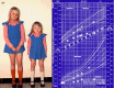
Girl (age 9.5 yrs) on the right has mucolipidosis type III alpha/beta. Boy (age 3 yrs) on the left has mucolipidosis II. Hands in the two children are significantly different: short, broad with claw-like in ML II and rather long in ML III alpha/beta. (more...)

Figure 2. A.

Figure 2

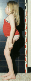
A. Deficient linear growth in ML III alpha/beta is illustrated by the difference in stature in dizygotic twin girls. The affected twin is shown on the right. B. Growth of the affected twin (solid circles) and her healthy twin (open circles) plotted on (more...)

Craniofacial. True macrocephaly does not occur. Dysmorphic facial features are absent or minimal in younger children. Coarsening of facial features is gradual and more apparent in profile, including full cheeks, depressed nasal bridge, and prominent mouth. Gingival hypertrophy is mild and does not usually interfere with tooth eruption.

Ophthalmologic. Epicanthal folds persist longer than normal. Proptosis, often observed in ML II, is rare. The corneas are clear by routine clinical inspection, but opacities may be appreciated by slit-lamp examination.

Audiologic. Episodes of otitis media occur in individuals with ML III alpha/beta more frequently than in the general population. Conductive hearing loss, documented in some affected individuals, has not been studied systematically. Sensorineural hearing loss is not a typical feature of ML III.

Respiratory. Mild hoarseness of the voice is an inconsistent finding. Upper-respiratory infections are more frequent than expected in some (but not all) children. From late childhood bronchitis and bronchopneumonia are the most consistent clinical complications.

Adults exhibit restrictive lung disease caused by stiffening of the thoracic cage, slowly progressive sclerosis of bronchi, and hardening and thickening of the interstitial tissue (extracellular matrix) in lung parenchyma.

Cardiovascular. Individuals with ML III alpha/beta are at risk for cardiac involvement. Gradual thickening and subsequent insufficiency of the mitral valve and the aortic valve are common from late childhood onward [Steet et al 2005].

Left and/or right ventricular hypertrophy is often documented on echocardiography in older individuals. Pulmonary hypertension may occur in some older individuals, but at present is still insufficiently documented.

Rapid progression of cardiac disease is rarely observed in ML III alpha/beta.

Pneumonia may compound mild cardiac insufficiency. Death in early adulthood is often from cardiopulmonary causes, even without complicating factors such as pneumonia.

Gastrointestinal. Prominence of the abdomen especially upon standing upright is caused in part by lumbar hyperlordosis, compensation for hip and knee flexion contractures, and hypotonia of the abdominal wall musculature. Diastasis of the medial recti and small umbilical hernias may also be present. In general, individuals with ML III alpha/beta do not present with organomegaly.

Skeletal/ soft connective tissue. Stiffness of all large and small joints is a cardinal feature. Limited range of motion in the shoulders is frequently the initial evidence of ML III alpha/beta and is mainly of soft tissue origin.

Limited range of motion in the hips and knees explains the slow gait and inability of children to run effectively. Flexion contractures in the hips and knees cause the squatting standing posture, most apparent in lateral view (see Figure 3).

Figure 3. . Same patient with ML III alpha/beta as in Figure 2 at age 12 years.

Figure 3.

Same patient with ML III alpha/beta as in Figure 2 at age 12 years. Profile view shows posture adversely affected by flexion contractures and stiffness in the hips and knees with compensatory dorsal hyperlordosis and sacral hyperkyphosis. Hands and fingers (more...)

Secondary but severe arthritic changes in the hips that can lead to destruction of the proximal femoral epiphyses make walking increasingly difficult and painful. Significant hardening of the surrounding soft tissues contributes to hip dysfunction. Many affected individuals become wheelchair bound before or during early adulthood.

Range of motion in the wrists and ankles is less adversely affected, than in the other large joints. Dupuytren-type palmar contractures may appear from late childhood onward and exacerbate the moderate to severe claw-like flexion deformity of the fingers associated with recurrent swelling and progressive stiffness. Neuropathic carpal tunnel signs can become severe in some individuals.

In ML III alpha/beta the hands and fingers are usually of near-normal length in contrast to the severely affected hands in ML II.

Before the appropriate diagnosis is made, many individuals with ML III alpha/beta have been evaluated for a rheumatologic disorder.

Osteoporosis affects the entire skeleton. Bone pain becomes the most distressing symptom in Ml III alpha/beta, even in individuals with limited ambulation. Osteolytic bone lesions also are associated with significant bone pain in those who are non-ambulatory.

Neuromotor development and intellect are the most variable features in ML III alpha/beta, ranging from normal to mild or moderate developmental delay in reaching motor milestones. Onset and development of receptive and expressive language skills occur at the expected age. Stuttering has not been observed in individuals with ML III alpha/beta. Although psychometric tests often reveal an IQ within normal limits, approximately half of the affected children require school assistance, often because of their physical limitations.

Other. The neck is short. Thickening of the skin is inconsistent and mild.

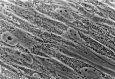
Previously used diagnostic testing. Phase-contrast or electron microscopic (EM) demonstration of large amounts of dense cytoplasmic inclusions (I-cells) in cultured fibroblasts was previously used to help confirm the diagnosis of ML II and ML III alpha/beta (see Figure 4).

Figure 4. . Living culture of skin fibroblasts derived from a person with ML III alpha/beta viewed by the contrast light microscope.

Figure 4.

Living culture of skin fibroblasts derived from a person with ML III alpha/beta viewed by the contrast light microscope. The cytoplasm is filled with dense granular inclusions that consistently spare a juxtanuclear zone that represents the endoplasmic (more...)

Note: On electron microscopy (EM) the mesenchymal cells in any tissue reveal large numbers of cytoplasmic vacuoles comprising swollen lysosomes bound by a unit membrane. The contents are pleomorphic, but not dense. This phenomenon is specific to ML II and ML III alpha/beta and is not observed in any lysosomal storage disorder.

The activity of lysosomal enzymes is severely reduced in I-cells, but significantly increased in the corresponding culture media.

The cytologic and enzymatic findings in cell culture cannot distinguish between ML II (I-cell disease) and ML III alpha/beta (see ML II).

Genotype-Phenotype Correlations

GNPTAB sequencing has been available only since 2005; however, the overall results of several studies have confirmed that homozygous and compound heterozygous genotypes that produce no or nearly no functional GlcNAc-1-phosphotransferase activity (caused by premature translation termination and/or frameshift effects) result in the ML II phenotype. The combination of less "morbid" pathogenic variants (e.g., missense and most of the splice site variants that result in up to 10% of residual GlcNAc-1-phosphotransferase activity) often yield the more slowly evolving ML III alpha/beta phenotype [Tiede et al 2005, Paik et al 2005, Bargal et al 2006, Kudo et al 2006, Encarnação et al 2009, Otomo et al 2009, Tappino et al 2009, Cathey et al 2010, David-Vizcarra et al 2010, Cury et al 2011].

Clearly, some children have phenotypes clinically intermediate between the reference phenotypes delineated ML II and ML III alpha/beta [Cathey et al 2010, David-Vizcarra et al 2010]. Astute clinicians often label the mutated GNPTAB-based disorder in such individuals as cases of ML II/III. Some of the intermediate phenotypes consistently correlate with a specific mutated genotype, whereas others have a homozygous mutated genotype.

At present it is not known whether the interesting but rarely available post-mortem pathology studies in ML III alpha/beta [Kerr et al 2011, Kobayashi et al 2011] will enhance genotype-phenotype correlation when compared with similar studies on ML II published several decades ago. Such studies clearly contribute to various aspects of pathogenesis.

Nomenclature

Pseudo-Hurler-polydystrophy (PHP) was the term used in 1966 by Maroteaux and Lamy when they first clinically and radiologically delineated the multisystem disorder that had only progressive stiffening of the large and small joints in common with Hurler disease or mucopolysaccharidosis I (MPS I)

Mucolipidosis (ML). The term PHP has been largely replaced by the term mucolipidosis III, introduced in 1970 by Spranger and Wiedemann, who provided the first clinical classification of the group of metabolic disorders clinically intermediate between the lipidoses and the MPSs (storage disorders of glycosaminoglycans). Their hypothesis that some of these disorders could be pathogenetically and genetically related was confirmed in 1973 when the alignment of ML III (PHP) and ML II (I-cell disease) was shown by the discovery of the "in vitro" cytologic and biochemical I-cell phenomenon in PHP fibroblasts.

Although mucolipidosis is a clinically useful name, biochemists consider it a misnomer because "mucolipids" do not exist in nature. The term mucolipidosis has been used in four different inborn errors of metabolism; only ML II and ML III alpha/beta are GNPTAB related. Mucolipidosis I (also called sialidosis type II) and mucolipidosis IV, are genetically distinct disorders.

Oligosaccharidoses (OSs). During the 1970s, excessive urinary excretion of OSs was documented in most of the mucolipidoses; therefore, the term "oligosaccharidoses" and later the term "glycoproteinoses" have been substituted for the term mucolipidoses.

Mucolipidosis II, mucolipidosis III alpha/beta, and mucolipidosis III gamma. Because even the trivial name of the causal enzyme defect UDP-N-acetylglucosamine: lysosomal hydrolase N-acetylglucosamine 1-phosphotransferase is long, the current naming of ML II and ML III alpha/beta as "UPDGlcNAc 1-P-transferase deficiency disorders" is cumbersome; it is, however, strictly the most correct one [Leroy 2007].

The enzyme GNPT is the product of two genes, one encoding the alpha and beta subunits, and the other encoding the gamma subunit [Bao et al 1996]:

  • Pathogenic variants in GNPTAB cause ML III alpha/beta and the allelic disorder ML II.
  • Pathogenic variants in GNPTG cause the variant ML III, designated ML III gamma [Cathey et al 2008].

Prevalence

Estimates of the prevalence of ML III alpha/beta based on objective data are not available. It is, however, likely that the prevalence is of the same order of magnitude as that of ML II and hence estimated to range between 2.5x10⁻⁶ and 1.10-5.

Consequently, the carrier rate is estimated at between 1:158 and 1:316 (see ML II).

Differential Diagnosis

Mucolipidosis III gamma. The clinical features of mucolipidosis III gamma (also known as variant ML III) are similar to but milder than those observed in individuals with mucolipidosis III alpha/beta (ML III alpha/beta). In most of the published case reports of ML III gamma, affected individuals are of Middle Eastern descent [Raas-Rothschild et al 2000, Raas-Rothschild et al 2004, Cathey et al 2008]. If the diagnosis of ML III is strongly suspected clinically and molecular analysis of GNPTAB does not reveal pathogenic variants, analysis of GNPTG should be performed.

See Nomenclature.

Lysosomal storage disease. Clinical findings in ML III alpha/beta overlap those observed in nearly all late-onset mild forms of the delineated entities among the MPSs including the following:

  • Attenuated MPS I (formerly called Hurler-Scheie syndrome or Scheie syndrome)
  • Attenuated MPS II (Hunter syndrome)
  • Morquio disease type B (MPS IV B)
  • Maroteaux-Lamy disease type B (MPS VI B)
  • Sly disease type B (MPS VII B)

While sharing several clinical aspects of dysostosis multiplex, the entities mentioned are associated with evidence of more severe storage on physical examination. In all the MPSs, the size of the head is enlarged, a finding not present in ML III alpha/beta. Biochemical testing distinguishes the MPSs.

Among the group of OSs, the more challenging differential diagnoses include: alpha-mannosidosis, late infantile and juvenile galactosialidosis, and childhood dysmorphic sialidosis (ML I) [Leroy 2007].

Disorders clinically allied to the oligosaccharidoses relevant in the differential diagnosis include late infantile sialic acid storage disorder or Salla disease (see Free Sialic Acid Storage Disorders) and multiple sulfatase deficiency (mucosulfatidosis). In both disorders the neurodegenerative aspects are much more prominent. In free sialic acid storage disorders, dysostosis multiplex is absent or minimal and urinary excretion of free sialic acid (not of OSs) is excessive. In multiple sulfatase deficiency both urinary AMPS and sulfatides are excessive.

Rheumatologic disorders are often suspected in persons with ML III alpha/beta because of slowly decreasing range of motion in large and small joints and increasing pain in the hips. Rheumatoid arthritis has clinical and laboratory signs of inflammation and specific antibodies; activity of the lysosomal enzymes in leukocytes is normal. Dysostosis multiplex is absent.

Osteochondrodysplasias with clinical similarities to ML III alpha/beta but without the radiologic signs of dysostosis multiplex [Spranger et al 2002] include the following:

Management

Evaluations Following Initial Diagnosis

To establish the extent of disease in an individual diagnosed with mucolipidosis III alpha/beta (ML III alpha/beta), the following evaluations are recommended:

  • Radiographic skeletal survey if either not performed or incomplete in the diagnostic evaluation
  • Baseline evaluations with an orthopedic surgeon and a metabolic bone specialist to better determine if/when surgical interventions or bisphosphonate therapy may be initiated (see Treatment of Manifestations)
  • Cardiac evaluation with echocardiography to assess valve thickening and ventricular size and function
  • Baseline ophthalmologic examination
  • Hearing screen
  • Developmental assessment to help establish appropriate expectations for the child’s developmental progress
  • Consultation with a clinical geneticist and/or genetic counselor

Treatment of Manifestations

Supportive and symptomatic management is indicated.

Skeletal. No measures are effective in treating the progressive limitation of motion in large and small joints. The classic physiotherapeutic early intervention programs that are often beneficial in children with developmental delay, neuromotor delay, or cerebral palsy cannot be recommended unequivocally in ML III alpha/beta for the following reasons:

  • Stretching exercises are ineffective and painful.
  • The unknowing therapist may inflict damage to the surrounding joint capsule and adjacent tendons and cause subsequent soft tissue calcification.

Therapies that are "low impact" in regard to joint and tendon strain, including short sessions of aqua therapy, are usually well tolerated.

Management of pain in the hips during and following walking requires attention from late childhood or early adolescence.

Carpal tunnel signs may require tendon release procedures for temporary relief.

Later in the disease course more general bone pain of variable intensity is present.

Encouraging results have been obtained in several individuals with ML III alpha/beta with monthly IV administration of pamidronate, a biphosphonate. The recommended dose is 1 mg/kg monthly. The protocol under development is different from that applied to individuals with osteogenesis imperfecta. Bone density needs to be monitored closely. At present, information as to when in the disease course or at what age to initiate such treatment is insufficient. Bone pain in the two individuals about whom information has been published was reduced within a few months of initiating therapy. In some wheelchair-bound individuals ambulation has been transiently restored for more than one year. Bone densitometry is improved [Robinson et al 2002].

Several remarks need to be made regarding this symptomatic treatment:

  • Parents and affected individuals must keep in mind that this treatment does not cure the disorder. It neither represses the slow process of bone resorption nor alters its course.
  • The long-term effect(s) are unknown.
  • The end point to the treatment regimen remains incompletely defined [Robinson et al 2002; Sillence, personal communication].
  • Not all affected individuals benefit from bisphosphonate treatment. The use of bisphosphonates in ML III alpha/beta and other bone diseases is an area of active clinical research worldwide.

In older adolescents and adults with milder phenotypic variants of ML III alpha/beta, bilateral hip replacement has been successful.

Audiologic. Recurrent otitis media occurs more often in ML III alpha/beta than in a control population. The prevalence decreases with age. Myringotomy tube placement may be considered necessary as a preventive measure of conductive hearing deficiency but should not be considered a "routine" procedure in this condition because of the unique airway issues and hence the anesthesia risks involved (see Prevention of Secondary Complications).

Prevention of Secondary Complications

Because of concerns about airway management, surgical intervention should be avoided as much as possible and undertaken only in tertiary care settings with pediatric anesthesiologists and intensivists. Individuals with ML III alpha/beta are small and have a narrow airway, reduced tracheal suppleness from stiff connective tissue, and progressive narrowing of the airway from mucosal thickening. The use of a much smaller endotracheal tube than for age- and size-matched controls is necessary. Fiberoptic intubation must be available.

Poor compliance of the thoracic cage and the progressively sclerotic lung parenchyma further complicate airway management, especially in older individuals. Functional decline of lung parenchyma is likely due at least in part to slowly progressive degeneration of soft connective tissue in the extracellular matrix, a phenomenon insufficiently studied but concomitant to the osteopenia in bone. As subclinical cardiac failure may become overt during anesthesia, any surgical intervention should be preceded by a thorough cardiologic evaluation Extubation may also be a challenge.

Surveillance

Young children with ML III alpha/beta and their families benefit from outpatient clinic visits about twice a year.

From age six years similar follow-up visits are recommended on a yearly basis unless bone pain and deteriorating ambulation become major handicaps and/or cardiac and respiratory monitoring need more frequent attention.

Evaluation of Relatives at Risk

See Genetic Counseling for issues related to testing of at-risk relatives for genetic counseling purposes.

Therapies Under Investigation

Preliminary results suggest that IV bisphosphonate may alleviate bone pain in some affected individuals; however, such treatment is still considered investigational.

Search ClinicalTrials.gov in the US and www.ClinicalTrialsRegister.eu in Europe for access to information on clinical studies for a wide range of diseases and conditions.

Genetic Counseling

Genetic counseling is the process of providing individuals and families with information on the nature, mode(s) of inheritance, and implications of genetic disorders to help them make informed medical and personal decisions. The following section deals with genetic risk assessment and the use of family history and genetic testing to clarify genetic status for family members; it is not meant to address all personal, cultural, or ethical issues that may arise or to substitute for consultation with a genetics professional. —ED.

Mode of Inheritance

Mucolipidosis III alpha/beta (ML III alpha/beta) is inherited in an autosomal recessive manner.

Risk to Family Members

Parents of a proband

  • The parents of an affected child are obligate heterozygotes and therefore carry one mutated allele.
  • Heterozygotes (carriers) are asymptomatic.

Sibs of a proband

  • At conception, each sib of an affected individual has a 25% chance of being affected, a 50% chance of being an asymptomatic carrier, and a 25% chance of being unaffected and not a carrier.
  • Once an at-risk sib is known to be unaffected, the risk of his/her being a carrier is 2/3.
  • Heterozygotes (carriers) are asymptomatic.

Offspring of a proband. Individuals with ML III alpha/beta do not commonly reproduce. The offspring of an individual with ML III alpha/beta are obligate heterozygotes (carriers) for a pathogenic variant in GNPTAB.

Other family members of a proband. Each sib of the proband’s parents is at a 50% risk of being a carrier.

Carrier (Heterozygote) Detection

Carrier testing for at-risk family members is possible once the pathogenic variants have been identified in the family.

Related Genetic Counseling Issues

See Management, Evaluation of Relatives at Risk for information on testing at-risk relatives for the purpose of early diagnosis and treatment

Family planning

  • The optimal time for determination of genetic risk, clarification of carrier status, and discussion of the availability of prenatal testing is before pregnancy.
  • It is appropriate to offer genetic counseling (including discussion of potential risks to offspring and reproductive options) to young adults who are affected, are carriers, or are at risk of being carriers.

DNA banking is the storage of DNA (typically extracted from white blood cells) for possible future use. Because it is likely that testing methodology and our understanding of genes,allelic variants, and diseases will improve in the future, consideration should be given to banking DNA of affected individuals.

Prenatal Testing and Preimplantation Genetic Diagnosis

Once the GNPTAB pathogenic variants have been identified in an affected family member, prenatal diagnosis for a pregnancy at increased risk and preimplantation genetic diagnosis are possible.

Resources

GeneReviews staff has selected the following disease-specific and/or umbrella support organizations and/or registries for the benefit of individuals with this disorder and their families. GeneReviews is not responsible for the information provided by other organizations. For information on selection criteria, click here.

  • International Advocate for Glycoprotein Storage Diseases (ISMRD)
    3921 Country Club Drive
    Lakewood CA 90712
    Email: info@ismrd.org
  • National MPS Society
    PO Box 14686
    Durham NC 27709-4686
    Phone: 877-677-1001 (toll-free); 919-806-0101
    Fax: 919-806-2055
    Email: info@mpssociety.org
  • Society for Mucopolysaccharide Diseases (MPS)
    MPS House Repton Place
    White Lion Road
    Amersham Buckinghamshire HP7 9LP
    United Kingdom
    Phone: 0345 389 9901
    Email: mps@mpssociety.co.uk

Molecular Genetics

Information in the Molecular Genetics and OMIM tables may differ from that elsewhere in the GeneReview: tables may contain more recent information. —ED.

Table A.

Mucolipidosis III Alpha/Beta: Genes and Databases

Data are compiled from the following standard references: gene from HGNC; chromosome locus from OMIM; protein from UniProt. For a description of databases (Locus Specific, HGMD, ClinVar) to which links are provided, click here.

Table B.

OMIM Entries for Mucolipidosis III Alpha/Beta (View All in OMIM)

252600MUCOLIPIDOSIS III ALPHA/BETA
607840N-ACETYLGLUCOSAMINE-1-PHOSPHOTRANSFERASE, ALPHA/BETA SUBUNITS; GNPTAB

Molecular Pathogenesis

The partial inactivation of UDP-N-acetylglucosamine: lysosomal hydrolase N-acetylglucosamine 1-phosphotransferase (encoded by GNPTAB and GNPTG [see Mucolipidosis III Gamma]) may result from pathogenic variants that allow reduced or residual protein production (missense or some of the splice-site variants). Hence synthesis of the common mannose-6-phosphate (M6P recognition marker) moiety to lysosomal acid hydrolases is reduced significantly though not completely abolished. Binding to specific M6P receptors in the trans-Golgi network is highly inadequate and results in very ineffective receptor-mediated transport of lysosomal enzymes to the lysosomal intracellular compartment. The mutant hydrolases leave the cells and appear in excessive amounts in body fluids as well as in vitro in the cell culture media. Once outside, the enzymes cannot reenter normal fibroblasts (and thus are often referred to as "low-uptake" lysosomal enzymes). In contrast, normal mature acid hydrolases, which are normally structured phosphoglycoproteins ("high-uptake" lysosomal enzymes), can enter any type of cultured fibroblast (including "I-cells") by pinocytosis [Kornfeld & Sly 2001]. Quantitative differences between low-uptake enzymes in body fluids and tissue cell culture media of individuals with ML III alpha/beta and those with ML III gamma are insignificant and have not been studied in the in vivo intercellular matrix.

N-linked glycosylation of lysosomal hydrolases occurs in the endocytoplasmic reticulum, which is also the site of the preceding stepwise build-up of OSs and of their "en bloc" transfer from the dolicholpyrophosphoryl-OS-precursor carrier to some of the asparagine residues in the nascent hydrolase proteins.

As the newly formed glycoproteins traverse the Golgi cisterns, sequential enzymatic modification of the N-linked OSs occurs along two different pathways: one pathway modifies the N-linked OSs into complex type glycan side chains, whereas the other, quantitatively the more important pathway at least in mesenchymal tissues, converts the precursor glycans into oligomannosyl-type OS side chains. Specific phosphorylation alone is adversely affected by biallelic inactivating GNPTAB pathogenic variants at a late step in this synthetic pathway (see next paragraph). A significant decrease of this specific phosphorylation manifests clinically as ML III alpha/beta; complete lack of phosphorylation of the oligomannosyl glycans causes ML II. Formation of the M6P recognition marker in lysosomal hydrolases is significantly reduced in ML III alpha/beta, and nearly or totally absent in ML II.

Normal formation of the M6P recognition marker is a two-step process. The first step is catalyzed by UDP-N-acetylglucosamine: lysosomal hydrolase N-acetylglucosamine-1-phosphotransferase (trivial name GlcNAc-phosphotransferase GNPTAB) (EC 2.7.8.17). This enzyme, also known as N-acetylglucosamine (GlcNAc)-1-phosphotransferase, comprises the subunits alpha and beta, the amino- and the carboxyl end, respectively, of the native protein encoded by GNPTAB. Inactivity or deficiency of this enzyme causes ML II and ML III alpha/beta, respectively. The second step, which is not affected in individuals with ML II or III alpha/beta, involves the action of N-acetylglucosamine-1-phosphodiester alpha-N-acetylglucosaminidase, which removes the blocking N-acetylglucosamine (GlcNAc) residue from the phosphorylated oligomannosyl type ORs, thereby exposing the M6P recognition marker. To date, pathogenic variants that inactivate the N-acetylglucosamine-1-phosphodiester alpha-N-acetylglucosaminidase have not been reported in either ML II or ML III alpha/beta.

Gene structure. GNPTAB, located on chromosome 12q23.3, has 21 exons and spans 85 kb of genomic DNA. The GNPTAB encodes the alpha and beta subunits of the oligomeric human GNPTAB in a single 6.2-kb alpha/beta transcript (see Molecular Pathogenesis). See Table A, Gene for a detailed summary of gene and protein information.

Pathogenic variants. Several dozen pathogenic variants are known. Types of pathogenic variants include: (a) missense, nonsense, and splice-site variants; and (b) small insertions and deletions that result in a shift of the proper reading frame (see Table 2 [pdf]). Any of the latter type of variants combined with a splice-site or missense variant can be found in individuals with MLII. Most individuals with ML III alpha/beta are homozygous or compound heterozygous for missense or splice-site variants or combinations of either. Some mild phenotypes with signs and symptoms probably characteristic of long survival have been correlated with the mutated genotypes detected [Paik et al 2005, Steet et al 2005, Tiede et al 2005, Bargal et al 2006, Kudo et al 2006, Encarnação et al 2009, Otomo et al 2009, Tappino et al 2009, Cathey et al 2010]. To date, no larger-scale rearrangements have been reported in individuals with ML III alpha/beta.

Normal gene product. GNPTAB encodes the alpha and beta subunits of the oligomeric human GNPTAB in a single 6.2-kb alpha/beta transcript. The subunit structure of the human GNPTAB enzyme is a 540-kd complex of disulfide-linked homodimers. Each is composed of a 166-kd alpha subunit (encoded by the GNPTAB precursor gene) and a 51-kd gamma subunit (encoded by GNPTG). Each of these subcomplexes is non-covalently associated with a 56-kd beta subunit (encoded by the GNPTAB precursor gene). Therefore, GNPTAB is a hexameric enzyme complex that may be symbolized by α2β2γ2 [Kudo et al 2005, Tiede et al 2005, Kudo et al 2006].

Following its translation this alpha/beta precursor polypeptide undergoes proteolytic cleavage at the lysine (residue 928) - asparagine (residue 929) peptide bond. This bond is enzymatically released by site-1 protease (S1P). Cells deficient in S1P failed to activate the alpha/beta precursor and exhibited the I-cell phenotype in vitro [Marschner et al 2011]. The N-terminal alpha subunit, the larger of the two, consists of 928 amino acids. The beta subunit (the C-terminal part of the precursor) consists of 328 amino acids. The 1256-amino acid precursor protein has a predicted molecular mass of 144 kd, two transmembrane domains, and 19 potential glycosylation sites [Kudo et al 2005, Tiede et al 2005, Kudo et al 2006]. In a recent study of Ml II and ML III alpha/beta fibroblast (I-cell) strains, the use of anti-peptide antibodies against the alpha and beta subunits showed that pathogenic variants in the gamma subunit adversely affected the assembly and intracellular distribution of the former subunits [Zarghooni & Dittakavi 2009].

Abnormal gene product. See Molecular Pathogenesis.

References

Literature Cited

  • Bargal R, Zeigler M, Abu-Libdeh B, Zuri V, Mandel H, Ben Neriah Z, Stewart F, Elcioglu N, Hindi T, Le Merrer M, Bach G, Raas-Rothschild A. When mucolipidosis III meets mucolipidosis II: GNPTA gene mutations in 24 patients. Mol Genet Metab. 2006;88:359–63. [PubMed: 16630736]
  • Bao M, Booth JL, Elmendorf BJ, Canfield WM. Bovine UDP-N-acetylglucosamine:lysosomal-enzyme N-acetylglucosamine-1-phosphotransferase. I. Purification and subunit structure. J Biol Chem. 1996;271:31437–45. [PubMed: 8940155]
  • Cathey SS, Kudo M, Tiede S, Raas-Rothschild A, Braulke T, Beck M, Taylor HA, Canfield WM, Leroy JG, Neufeld EF, McKusick VA. Molecular order in mucolipidosis II and III nomenclature. Am J Med Genet. 2008;146A:512–3. [PubMed: 18203164]
  • Cathey SS, Leroy JG, Wood T, Eaves K, Simensen RJ, Kudo M, Stevenson RE, Friez MJ. Phenotype and genotype in mucolipidoses II and III alpha/beta: a study of 61 probands. J Med Genet. 2010;47:38–48. [PMC free article: PMC3712854] [PubMed: 19617216]
  • Cury GK, Velho RV, Alegra T, Matte U, Artigalas O, Burin M, Ribeiro EM, Lourenço CM, Kim CA, Valadares ER, Miguel DSCG, Acosta AX, Galera MF, Schwartz IVD. Mucolipidoses type II and III: molecular analysis of the GNPTAB and GNPTG genes in 15 Brazilian patients. J Inherit Metab Dis. 2011;34:S206.
  • David-Vizcarra G, Briody J, Ault J, Fietz M, Fletcher J, Savarirayan R, Wilson M, McGill J, Edwards M, Munns C, Alcausin M, Cathey S, Sillence D. The natural history and osteodystrophy of mucolipidosis types II and III. J Paediatr Child Health. 2010;46:316–22. [PMC free article: PMC4188554] [PubMed: 20367762]
  • Encarnação M, Lacerda L, Costa R, Prata MJ, Coutinho MF, Ribeiro H, Lopes L, Pineda M, Ignatius J, Galvez H, Mustonen A, Vieira P, Lima MR, Alves S. Molecular analysis of the GNPTAB and GNPTG genes in 13 patients with mucolipidosis type II or type III - identification of eight novel mutations. Clin Genet. 2009;76:76–84. [PubMed: 19659762]
  • Kerr DA, Memoli VA, Cathey SS, Harris BT. Mucolipidosis type III α/β: the first characterization of this rare disease by autopsy. Arch Pathol Lab Med. 2011;135:503–10. [PMC free article: PMC4188553] [PubMed: 21466370]
  • Kobayashi H, Takahashi-Fujigasaki J, Fukuda T, Sakurai K, Shimada Y, Nomura K, Ariga M, Ohashi T, Eto Y, Otomo T, Sakai N, Ida H. Pathology of the first autopsy case diagnosed as mucolipidosis type III α/β suggesting autophagic dysfunction. Mol Genet Metab. 2011;102:170–5. [PubMed: 21051253]
  • Kornfeld S, Sly WS. I-Cell disease and pseudo-Hurler polydystrophy: disorders of lysosomal enzyme phosphorylation and localization. In: Scriver CR, Beaudet AL, Sly WS, Valle D, Vogelstein B, eds. The Metabolic and Molecular Bases of Inherited Disease. 8 ed. New York, NY: McGraw-Hill; 2001:3469-82.
  • Kudo M, Bao M, D’Sousa A, Ying F, Pan H, Roe BA, Canfield WM. The α- and β-subunits of the human UDP-N-acetylglucosamine: lysosomal enzyme N-acetylglucosamine-1-phosphotransferase [corrected] are encoded by a single cDNA. J Biol Chem. 2005;280:36141–9. [PubMed: 16120602]
  • Kudo M, Brem MS, Canfield WM. Mucolipidosis II (I-cell disease) and Mucolipidosis III (classical pseudo-Hurler polydystrophy) are caused by mutations in the GlcNAc-phosphotransferase alpha / beta -subunits precursor gene. Am J Hum Genet. 2006;78:451–63. [PMC free article: PMC1380288] [PubMed: 16465621]
  • Leroy JG. Oligosaccharidoses, disorders allied to the oligosaccharides. In: Rimoin DL, Connor JM, Pyeritz RE, Korf BR, eds. Emery and Rimoin’s Principles and Practice of Medical Genetics. 5 ed. Philadelphia, PA: Churchill Livingstone; 2007:2413-48.
  • Marschner K, Kollmann K, Schweizer M, Braulke T, Pohl S. A key enzyme in the biogenesis of lysosomes is a protease that regulates cholesterol metabolism. Science. 2011;333:87–90. [PubMed: 21719679]
  • Otomo T, Muramatsu T, Yorifuji T, Okuyama T, Nakabayashi H, Fukao T, Ohura T, Yoshino M, Tanaka A, Okamoto N, Inui K, Ozono K, Sakai N. Mucolipidosis II and III alpha/beta: mutation analysis of 40 Japanese patients showed genotype-phenotype correlation. J Hum Genet. 2009;54:145–51. [PubMed: 19197337]
  • Paik KH, Song SM, Ki CS, Yu HW, Kim JS, Min KH, Chang SH, Yoo EJ, Lee IJ, Kwan EK, Han SJ, Jin DK. Identification of mutations in the GNPTA (MFGC4170) gene coding for GlcNAc-phosphotransferase alpha/beta subunits in Korean patients with mucolipidosis type II and type IIIA. Hum Mut. 2005;26:308–14. [PubMed: 16116615]
  • Raas-Rothschild A, Bargal R, Goldman O, Ben-Asher E, Groener JE, Toutain A, Stemmer E, Ben-Neriah Z, Flusser H, Beemer FA, Penttinen M, Olender T, Rein AJ, Bach G, Zeigler M. Genomic organization of the UDP-N-acetylglucosamine-1-phosphotransferase gamma subunit (GNPTAG) and its mutations in mucolipidosis III. J Med Genet. 2004;41:e52. [PMC free article: PMC1735719] [PubMed: 15060128]
  • Raas-Rothschild A, Cormier-Daire V, Bao M, Genin E, Salomon R, Brewer K, Zeigler M, Mandel H, Toth S, Roe B, Munnich A, Canfield WM. Molecular basis of variant pseudo-hurler polydystrophy (mucolipidosis IIIC). J Clin Invest. 2000;105:673–81. [PMC free article: PMC289169] [PubMed: 10712439]
  • Robinson C, Baker N, Noble J, King A, David G, Sillence D, Hofman P, Cundy T. The osteodystrophy of mucolipidosis type III and the effects of intravenous pamidronate treatment. J Inherit Metab Dis. 2002;25:681–93. [PubMed: 12705498]
  • Spranger JW, Brill PW, Poznanski A. Mucolipidosis III. In: Bone Dysplasias: Atlas of Genetic Disorders of Skeletal Development. 2 ed. New York, NY: Oxford University Press; 2002:295-9.
  • Steet RA, Hullin R, Kudo M, Martinelli M, Bosshard NU, Schaffner T, Kornfeld S, Steinmann B. A splicing mutation in the alpha/beta GlcNAc-1-phosphotransferase gene results in an adult onset form of mucolipidosis III associated with sensory neuropathy and cardiomyopathy. Am J Med Genet A. 2005;132A:369–75. [PubMed: 15633164]
  • Tappino B, Chuzhanova NA, Regis S, Dardis A, Corsolini F, Stroppiano M, Tonoli E, Beccari T, Rosano C, Mucha J, Blanco M, Szlago M, Di Rocco M, Cooper DN, Filocamo M. Molecular characterization of 22 novel UDP-N-acetylglucosamine-1-phosphate transferase alpha- and beta-subunit (GNPTAB) gene mutations causing mucolipidosis types IIalpha/beta and IIIalpha/beta in 46 patients. Hum Mutat. 2009;30:E956–73. [PubMed: 19634183]
  • Tiede S, Muschol N, Reutter G, Cantz M, Ullrich K, Braulke T. Missense mutations in N-acetylglucosamine-1-phosphotransferase alpha/beta subunit gene in a patient with mucolipidosis III and a mild clinical phenotype. Am J Med Genet A. 2005;137A:235–40. [PubMed: 16094673]
  • Zarghooni M, Dittakavi SS. Molecular analysis of cell lines from patients with mucolipidosis II and mucolipidosis III. Am J Med Genet A. 2009;149A:2753–61. [PubMed: 19938078]

Chapter Notes

Revision History

  • 29 August 2019 (ma) Chapter retired: Covered in GNPTAB-Related Disorders
  • 10 May 2012 (me) Comprehensive update posted live
  • 7 July 2009 (cd) Revision: deletion/duplication analysis available clinically for GNPTAB
  • 26 August 2008 (cg) Review posted live
  • 16 June 2008 (jgl) Original submission
Copyright © 1993-2024, University of Washington, Seattle. GeneReviews is a registered trademark of the University of Washington, Seattle. All rights reserved.

GeneReviews® chapters are owned by the University of Washington. Permission is hereby granted to reproduce, distribute, and translate copies of content materials for noncommercial research purposes only, provided that (i) credit for source (http://www.genereviews.org/) and copyright (© 1993-2024 University of Washington) are included with each copy; (ii) a link to the original material is provided whenever the material is published elsewhere on the Web; and (iii) reproducers, distributors, and/or translators comply with the GeneReviews® Copyright Notice and Usage Disclaimer. No further modifications are allowed. For clarity, excerpts of GeneReviews chapters for use in lab reports and clinic notes are a permitted use.

For more information, see the GeneReviews® Copyright Notice and Usage Disclaimer.

For questions regarding permissions or whether a specified use is allowed, contact: ude.wu@tssamda.

Bookshelf ID: NBK1875PMID: 20301730

Views

Tests in GTR by Gene

Similar articles in PubMed

See reviews...See all...

Recent Activity

Your browsing activity is empty.

Activity recording is turned off.

Turn recording back on

See more...