

NCBI Bookshelf. A service of the National Library of Medicine, National Institutes of Health.
Molecular Imaging and Contrast Agent Database (MICAD) [Internet]. Bethesda (MD): National Center for Biotechnology Information (US); 2004-2013.

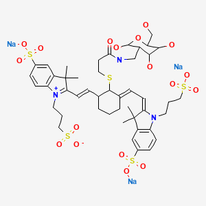
| Chemical name: | Indocyanine green derivative 02-2-deoxy-d-glucose |
|
| Abbreviated name: | ICG-Der-02-2DG | |
| Synonym: | ||
| Agent category: | Compound | |
| Target: | Glucose transporters, hexokinases | |
| Target category: | Transporter, enzyme | |
| Method of detection: | Optical, near-infrared fluorescence (NIR) imaging | |
| Source of signal: | ICG-Der-02 | |
| Activation: | No | |
| Studies: |
| No structure is available in PubChem. |
 In vitro
Background
[PubMed]
Optical fluorescence imaging is increasingly used to monitor biological functions of specific targets in small animals (1-3). However, the intrinsic fluorescence of biomolecules poses a problem when fluorophores that absorb visible light (350–650 nm) are used. Near-infrared (NIR) fluorescence (650–900 nm) detection avoids the natural background fluorescence interference of biomolecules, providing a high contrast between target and background tissues. NIR fluorophores have wider dynamic range and minimal background fluorescence as a result of reduced scattering compared with visible fluorescence detection. They also have high sensitivity, resulting from low background fluorescence, and high extinction coefficients, which provide high quantum yields. The NIR region is also compatible with solid-state optical components, such as diode lasers and silicon detectors. NIR fluorescence imaging is a noninvasive complement to radionuclide imaging in small animals or with probes in close proximity to the target in humans (4). Among the various optical imaging agents, only indocyanine green (ICG), with NIR fluorescence absorption at 780 nm and emission at 820 nm, is approved by the United States Food and Drug Administration for clinical applications in angiography, blood flow evaluation, and liver function assessment (5-8). It is also under evaluation in several clinical trials for other applications, such as optical imaging and mapping of both the lymphatic vessels and lymph nodes in cancer patients for surgical dissection of tumor cells and endoscopic imaging of the pancreas and colon.
The phosphorylation of glucose, an initial and important step in cellular metabolism, is catalyzed by hexokinases (HKs) (9). There are four HKs in mammalian tissues (HKI–HKIV). HKI, HKII, and HKIII have molecular weights of ~100,000 each; HKI is found mainly in the brain, and HKII is insulin-sensitive and is found in adipose and muscle cells. HKIII is found mainly in the liver and lung. HKIV, also known as glucokinase, has a molecular weight of ~50,000 and is specific to the liver and pancreas. Most brain HK is bound to mitochondria, enabling coordination between glucose consumption and oxidation. Tumor cells are known to be highly glycolytic because of increased expression of glycolytic enzymes and HK activity (10), which was detected in tumors from patients with lung, gastrointestinal, and breast cancers. The HKs, by converting glucose to glucose-6-phosphate, help maintain the downhill gradient that results in the transport of glucose into cells through the facilitative glucose transporters (GLUT1–GLUT13) (11). GLUT1 is considered to be the main transporter of glucose uptake. GLUT4 and HKII are the major transporter and HK isoform in skeletal muscle, heart, and adipose tissue, wherein insulin promotes glucose utilization. HKIV is associated with GLUT2 in liver and pancreatic β cells.
2-Deoxy-d-glucose (2DG) was first developed to inhibit glucose utilization by cancer cells (12). HKs phosphorylate 2DG to 2DG-6-phosphate, which inhibits phosphorylation of glucose. 2-[18F]Fluoro-2-deoxy-d-glucose ([18F]FDG) was later developed for use in molecular imaging studies (13). FDG is moved into cells by glucose transporters and is then phosphorylated by HK to FDG-6-phosphate. FDG-6-phosphate cannot be metabolized further in the glycolytic pathway and remains in the cells. Tumor cells do not contain a sufficient amount of glucose-6-phosphatase to reverse the phosphorylation. The elevated rates of glycolysis and glucose transport in many types of tumor cells and activated cells enhance the uptake of FDG in these cells relative to normal cells. Positron emission tomography (PET) with [18F]FDG has been used to assess alterations in glucose metabolism in brain, cancers, cardiovascular diseases, Alzheimer’s disease and other central nervous system disorders, and infectious, autoimmune, and inflammatory diseases (14-19). Various NIR dyes (such as Cypate, Cy5.5, and IRDye800CW) were conjugated to 2DG (20-22) as optical imaging agents for in vivo imaging of glucose utilization in tumors in mice. ICG derivative 02 (ICG-Der-02) contains one carboxyl functional group for covalent conjugation to the amino group of biomolecules. ICG-Der-02 is a hydrophilic dye. Guo et al. (23) evaluated ICG-Der-02-2DG for in vivo NIR optical imaging in tumor-bearing mice.
Related Resource Links:
- Gene information in NCBI (hexokinase, GLUT1)
- Articles in Online Mendelian Inheritance in Man (OMIM) (hexokinase, GLUT1)
Synthesis
[PubMed]
ICG-Der-02-N-hydroxysuccinimide ester (0.0128 mmol) was reacted with 2-amino-2-DG (0.064 mmol) for 18 h at room temperature in sodium phosphate buffer (pH 9) (23). ICG-Der-02-2DG was purified with high-performance liquid chromatography and verified with mass spectroscopy. There is one dye molecule per ICG-Der-02-2DG molecule. The yield of ICG-Der-02-2DG was ~32%, with 92% purity. ICG-Der-02-2DG displayed spectral properties similar to those of ICG-Der-02, with maximum absorption at 783 nm and maximum emission at 811 nm.
In Vitro Studies: Testing in Cells and Tissues
[PubMed]
In vitro uptake studies of rhodamine-2DG (RhB-2DG) were performed with MCF-7/estradiol, U87MG, and MCF-7 tumor cells in culture, showing that high, medium, and low fluorescence intensity correlated respectively with the GLUT1 expression levels (23). Fluorescence confocal microscopy showed that RhB-2DG accumulated in the cytoplasm of the tumor cells. Excess 2DG was able to block the fluorescence signal in the cytoplasm. Rhodamine was used because the investigators do not have confocal fluorescence microcopy with NIR capability.
Animal Studies
Rodents
[PubMed]
Guo et al. (23) performed in vivo NIR fluorescence imaging studies in nude mice (n = 5) bearing GLUT1-expressing MCF-7/estradiol xenografts at 0.5–48 h after intravenous injection of ICG-Der-02-2DG (10 nmol per mouse). ICG-Der-02-2DG accumulated mainly in the tumors, with a peak tumor/muscle ratio of 8.87 ± 0.31 at 4 h. The tumor/muscle ratio gradually decreased to ~7 at 48 h. Among the normal tissues, only kidneys exhibited strong fluorescence signal, indicative of renal excretion of ICG-Der-02-2DG. Ex vivo NIR fluorescence imaging showed that the tumors exhibited the highest fluorescence intensity (tissue/background ratio, ~17), followed by the kidney (~12), liver (~5), lung (~4), spleen (~3), intestine (~2), and heart (~2). No blocking experiment was performed.
References
- 1.
- Achilefu S. Lighting up tumors with receptor-specific optical molecular probes. Technol Cancer Res Treat. 2004;3(4):393–409. [PubMed: 15270591]
- 2.
- Becker A., Hessenius C., Licha K., Ebert B., Sukowski U., Semmler W., Wiedenmann B., Grotzinger C. Receptor-targeted optical imaging of tumors with near-infrared fluorescent ligands. Nat Biotechnol. 2001;19(4):327–31. [PubMed: 11283589]
- 3.
- Ntziachristos V., Bremer C., Weissleder R. Fluorescence imaging with near-infrared light: new technological advances that enable in vivo molecular imaging. Eur Radiol. 2003;13(1):195–208. [PubMed: 12541130]
- 4.
- Tung C.H. Fluorescent peptide probes for in vivo diagnostic imaging. Biopolymers. 2004;76(5):391–403. [PubMed: 15389488]
- 5.
- Yannuzzi, L.A., Indocyanine green angiography: a perspective on use in the clinical setting. Am J Ophthalmol, 2011151(5): p. 745-751 e1. [PubMed: 21501704]
- 6.
- Yamamoto M., Sasaguri S., Sato T. Assessing intraoperative blood flow in cardiovascular surgery. Surg Today. 2011;41(11):1467–74. [PubMed: 21969147]
- 7.
- Schaafsma B.E., Mieog J.S., Hutteman M., van der Vorst J.R., Kuppen P.J., Lowik C.W., Frangioni J.V., van de Velde C.J., Vahrmeijer A.L. The clinical use of indocyanine green as a near-infrared fluorescent contrast agent for image-guided oncologic surgery. J Surg Oncol. 2011;104(3):323–32. [PMC free article: PMC3144993] [PubMed: 21495033]
- 8.
- Manizate F., Hiotis S.P., Labow D., Roayaie S., Schwartz M. Liver functional reserve estimation: state of the art and relevance for local treatments: the Western perspective. J Hepatobiliary Pancreat Sci. 2010;17(4):385–8. [PubMed: 19936599]
- 9.
- Smith T.A. Mammalian hexokinases and their abnormal expression in cancer. Br J Biomed Sci. 2000;57(2):170–8. [PubMed: 10912295]
- 10.
- Suolinna E.M., Haaparanta M., Paul R., Harkonen P., Solin O., Sipila H. Metabolism of 2-[18F]fluoro-2-deoxyglucose in tumor-bearing rats: chromatographic and enzymatic studies. Int J Rad Appl Instrum B. 1986;13(5):577–81. [PubMed: 3818323]
- 11.
- Wood I.S., Trayhurn P. Glucose transporters (GLUT and SGLT): expanded families of sugar transport proteins. Br J Nutr. 2003;89(1):3–9. [PubMed: 12568659]
- 12.
- Laszlo J., Humphreys S.R., Goldin A. Effects of glucose analogues (2-deoxy-D-glucose, 2-deoxy-D-galactose) on experimental tumors. J Natl Cancer Inst. 1960;24:267–81. [PubMed: 14414406]
- 13.
- Fowler J.S., Ido T. Initial and subsequent approach for the synthesis of 18FDG. Semin Nucl Med. 2002;32(1):6–12. [PubMed: 11839070]
- 14.
- Phelps M.E. PET: the merging of biology and imaging into molecular imaging. J Nucl Med. 2000;41(4):661–81. [PubMed: 10768568]
- 15.
- Phelps M.E., Mazziotta J.C. Positron emission tomography: human brain function and biochemistry. Science. 1985;228(4701):799–809. [PubMed: 2860723]
- 16.
- Phelps M.E., Mazziotta J.C., Huang S.C. Study of cerebral function with positron computed tomography. J Cereb Blood Flow Metab. 1982;2(2):113–62. [PubMed: 6210701]
- 17.
- Rohren E.M., Turkington T.G., Coleman R.E. Clinical applications of PET in oncology. Radiology. 2004;231(2):305–32. [PubMed: 15044750]
- 18.
- Sokoloff L. Basic principles in imaging of regional cerebral metabolic rates. Res Publ Assoc Res Nerv Ment Dis. 1985;63:21–49. [PubMed: 2992057]
- 19.
- Spence A.M., Mankoff D.A., Muzi M. Positron emission tomography imaging of brain tumors. Neuroimaging Clin N Am. 2003;13(4):717–39. [PubMed: 15024957]
- 20.
- Kovar J.L., Volcheck W., Sevick-Muraca E., Simpson M.A., Olive D.M. Characterization and performance of a near-infrared 2-deoxyglucose optical imaging agent for mouse cancer models. Anal Biochem. 2009;384(2):254–62. [PMC free article: PMC2720560] [PubMed: 18938129]
- 21.
- Cheng Z., Levi J., Xiong Z., Gheysens O., Keren S., Chen X., Gambhir S.S. Near-infrared fluorescent deoxyglucose analogue for tumor optical imaging in cell culture and living mice. Bioconjug Chem. 2006;17(3):662–9. [PMC free article: PMC3191878] [PubMed: 16704203]
- 22.
- Chen Y., Zheng G., Zhang Z.H., Blessington D., Zhang M., Li H., Liu Q., Zhou L., Intes X., Achilefu S., Chance B. Metabolism-enhanced tumor localization by fluorescence imaging: in vivo animal studies. Opt Lett. 2003;28(21):2070–2. [PubMed: 14587818]
- 23.
- Guo J., Du C., Shan L., Zhu H., Xue B., Qian Z., Achilefu S., Gu Y. Comparison of near-infrared fluorescent deoxyglucose probes with different dyes for tumor diagnosis in vivo. Contrast Media Mol Imaging. 2012;7(3):289–301. [PubMed: 22539399]
- PMCPubMed Central citations
- PubChem SubstanceRelated PubChem Substances
- PubMedLinks to PubMed
- Review Cypate-2-Deoxy-d-glucose.[Molecular Imaging and Contrast...]Review Cypate-2-Deoxy-d-glucose.Leung K. Molecular Imaging and Contrast Agent Database (MICAD). 2004
- Review IRDye800-2-Deoxy-D-glucose.[Molecular Imaging and Contrast...]Review IRDye800-2-Deoxy-D-glucose.Leung K. Molecular Imaging and Contrast Agent Database (MICAD). 2004
- Review (99m)Tc-Ethylenedicysteine-deoxyglucose.[Molecular Imaging and Contrast...]Review (99m)Tc-Ethylenedicysteine-deoxyglucose.Leung K. Molecular Imaging and Contrast Agent Database (MICAD). 2004
- Review 6-Deoxy-6-[(18)F]fluoro-D-fructose.[Molecular Imaging and Contrast...]Review 6-Deoxy-6-[(18)F]fluoro-D-fructose.Leung K. Molecular Imaging and Contrast Agent Database (MICAD). 2004
- Review [(18)F]Fluoro-2-deoxy-2-D-glucose.[Molecular Imaging and Contrast...]Review [(18)F]Fluoro-2-deoxy-2-D-glucose.Leung K. Molecular Imaging and Contrast Agent Database (MICAD). 2004
- Indocyanine green derivative 02-2-deoxy-d-glucose - Molecular Imaging and Contra...Indocyanine green derivative 02-2-deoxy-d-glucose - Molecular Imaging and Contrast Agent Database (MICAD)
Your browsing activity is empty.
Activity recording is turned off.
See more...