U.S. flag

An official website of the United States government

NCBI Bookshelf. A service of the National Library of Medicine, National Institutes of Health.

Adam MP, Feldman J, Mirzaa GM, et al., editors. GeneReviews® [Internet]. Seattle (WA): University of Washington, Seattle; 1993-2024.

Cover of GeneReviews®

GeneReviews® [Internet].

Show details

HYAL2 Deficiency

Synonym: Orofacial Clefting and Cor Triatriatum Sinister Syndrome

, BSc, MBChB, PhD, , MD, , BSc,
PhD, and , BSc, MBBS, MRCPCH, PhD.

Author Information and Affiliations

Initial Posting: .

Estimated reading time: 19 minutes

Summary

Clinical characteristics.

HYAL2 deficiency is characterized by cardiac anomalies, cleft lip and palate (unilateral or bilateral), ophthalmic findings (including mild-to-severe myopia up to −16.75 diopters and increased risk of retinal detachment), hearing loss (typically conductive), and skeletal findings (including pectus excavatum and digital anomalies). Intellect is typically normal. To date, 17 individuals from seven families have been reported with HYAL2 deficiency.

Diagnosis/testing.

The diagnosis of HYAL2 deficiency is established in a proband with suggestive findings and biallelic pathogenic variants in HYAL2 identified by molecular genetic testing.

Management.

Treatment of manifestations: There is no cure for HYAL2 deficiency. Supportive care to improve quality of life, maximize function, and reduce complications is recommended. This can include care by specialists in pediatric cardiology and cardiac surgery and a multidisciplinary craniofacial team to coordinate timing and type of surgical interventions, manage issues with feeding and nutrition, and work with speech-language pathologists (regarding speech and communication issues) and audiologists (regarding hearing loss).

Surveillance: Members of the multidisciplinary care team will recommend intervals to monitor existing manifestations and the individual's response to supportive care.

Genetic counseling.

HYAL2 deficiency is inherited in an autosomal recessive manner. If both parents are known to be heterozygous for an HYAL2 pathogenic variant, each sib of an affected individual has at conception a 25% chance of being affected, a 50% chance of being an asymptomatic carrier, and a 25% chance of being unaffected and not a carrier. Once the HYAL2 pathogenic variants have been identified in an affected family member, carrier testing for relatives at risk and prenatal and preimplantation genetic testing are possible.

Diagnosis

No consensus clinical diagnostic criteria for HYAL2 deficiency have been published.

Suggestive Findings

HYAL2 deficiency should be suspected in a proband with one or more of the following clinical findings in the absence of intellectual disability and a family history consistent with autosomal recessive inheritance.

Clinical findings

  • Characteristic facial features (See Figure 1.)
  • Congenital cardiac anomalies (including coarctation of the aorta, mitral/pulmonary valve atresia, hypoplastic left ventricle, tetralogy of Fallot, double outlet right ventricle, hypoplastic pulmonary and aortopulmonary arteries with agenesis of the ductus venosus)
  • Cleft lip and palate (CLP), either unilateral or bilateral
  • Ophthalmologic features including:
    • Mild-to-severe myopia (up to −16.75 diopters), which can be asymmetric. When myopia is severe, complications include myopic macular degeneration and increased risk of retinal detachment.
    • Cataracts (posterior subcapsular cataract and wedge-shaped cortical cataract), suggesting that the high myopia may be part of a hereditary vitreoretinal degeneration phenotype
    • Ptosis (mild to moderate, bilateral). It is not currently known whether this is stationary or progressive.
  • Hearing loss. Typically conductive, diagnosed on newborn hearing screening or soon after birth and associated with CLP. Sensorineural hearing loss is described but rare.
  • Skeletal findings (including pectus excavatum and digital anomalies such as broad halluces and thumbs)
Figure 1. . Photographs of individuals with HYAL2 deficiency.

Figure 1.

Photographs of individuals with HYAL2 deficiency. Key features include frontal bossing, hypertelorism, broad and flattened nasal tip, and auricular anomalies (cupped ears, overfolded helices, preauricular pit) in almost all individuals. Additional variable (more...)

Family history is consistent with autosomal recessive inheritance (e.g., affected sibs and/or parental consanguinity). Absence of a known family history does not preclude the diagnosis.

Establishing the Diagnosis

The diagnosis of HYAL2 deficiency is established in a proband with suggestive findings and biallelic pathogenic (or likely pathogenic) variants in HYAL2 identified by molecular genetic testing (see Table 1).

Note: (1) Per ACMG/AMP variant interpretation guidelines, the terms "pathogenic variant" and "likely pathogenic variant" are synonymous in a clinical setting, meaning that both are considered diagnostic and can be used for clinical decision making [Richards et al 2015]. Reference to "pathogenic variants" in this section is understood to include likely pathogenic variants. (2) Identification of biallelic HYAL2 variants of uncertain significance (or of one known HYAL2 pathogenic variant and one HYAL2 variant of uncertain significance) does not establish or rule out the diagnosis.

Molecular genetic testing approaches can include a combination of gene-targeted testing (single-gene testing, multigene panel) and comprehensive genomic testing (exome sequencing, genome sequencing) depending on the phenotype.

Gene-targeted testing requires that the clinician determine which gene(s) are likely involved (see Option 1), whereas comprehensive genomic testing does not (see Option 2).

Option 1

Single-gene testing. Sequence analysis of HYAL2 is performed first to detect small intragenic deletions/insertions and missense, nonsense, and splice site variants. Typically, if only one or no variant is detected by the sequencing method used, the next step is to perform gene-targeted deletion/duplication analysis to detect exon and whole-gene deletions or duplications; however, to date such variants have not been identified as a cause of this disorder.

Note: Targeted analysis for the HYAL2 variant c.443A>G (p.Lys148Arg) can be performed first in individuals of Amish ancestry (see Table 7).

A multigene panel that includes HYAL2 and other genes of interest (see Differential Diagnosis) is most likely to identify the genetic cause of the condition while limiting identification of variants of uncertain significance and pathogenic variants in genes that do not explain the underlying phenotype. Note: (1) The genes included in the panel and the diagnostic sensitivity of the testing used for each gene vary by laboratory and are likely to change over time. (2) Some multigene panels may include genes not associated with the condition discussed in this GeneReview. (3) In some laboratories, panel options may include a custom laboratory-designed panel and/or custom phenotype-focused exome analysis that includes genes specified by the clinician. (4) Methods used in a panel may include sequence analysis, deletion/duplication analysis, and/or other non-sequencing-based tests.

For an introduction to multigene panels click here. More detailed information for clinicians ordering genetic tests can be found here.

Option 2

Comprehensive genomic testing does not require the clinician to determine which gene is likely involved. Exome sequencing is most commonly used; genome sequencing is also possible.

For an introduction to comprehensive genomic testing click here. More detailed information for clinicians ordering genomic testing can be found here.

Table 1.

Molecular Genetic Testing Used in HYAL2 Deficiency

Gene 1MethodProportion of Pathogenic Variants 2 Detectable by Method
HYAL2 Sequence analysis 3100% 4
Gene-targeted deletion/duplication analysis 5None reported to date 4
1.
2.

See Molecular Genetics for information on variants detected in this gene.

3.

Sequence analysis detects variants that are benign, likely benign, of uncertain significance, likely pathogenic, or pathogenic. Variants may include small intragenic deletions/insertions and missense, nonsense, and splice site variants; typically, exon or whole-gene deletions/duplications are not detected. For issues to consider in interpretation of sequence analysis results, click here.

4.

Data derived from the subscription-based professional view of Human Gene Mutation Database [Stenson et al 2020]

5.

Gene-targeted deletion/duplication analysis detects intragenic deletions or duplications. Methods used may include a range of techniques including quantitative PCR, long-range PCR, multiplex ligation-dependent probe amplification (MLPA), and a gene-targeted microarray designed to detect single-exon deletions or duplications.

Clinical Characteristics

Clinical Description

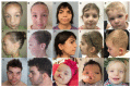
HYAL2 deficiency is characterized by congenital cardiac anomalies, cleft lip and palate that can be unilateral or bilateral, distinctive ophthalmic findings (including mild-to-severe myopia up to −16.75 diopters and increased risk of retinal detachment), hearing loss that is typically conductive, and skeletal findings (including pectus excavatum and digital anomalies). Intellect is typically normal.

To date, 17 individuals from seven families have been identified with HYAL2 deficiency [Muggenthaler et al 2017, Fasham et al 2022]. The following description of the phenotypic features associated with this condition is based on these reports.

Table 2.

HYAL2 Deficiency: Frequency of Selected Features

FeatureProportion of Persons w/Feature 1
Congenital cardiac anomalies12/17
Cleft lip & palate (CLP)Bilateral6/17
Unilateral4/17
OphthalmologicMyopia11/11
Ptosis5/13
Cataract3/14
Myopic maculopathy &/or retinal detachment4 persons w/myopia, 2 of whom had retinal detachment
SkeletalPectus excavatum7/16
Broad thumbs/halluces6/17
Ears/hearingExternal ear anomalies11/14
Conductive hearing loss5/15
Sensorineural hearing loss2/15
Duodenal web2 persons 2
1.

Limited clinical details are available for some of the reported individuals included in this table. The denominator of each fraction represents the total number of individuals in whom the corresponding finding was reported.

2.

Denominator unknown

Congenital cardiac anomalies range from the asymptomatic (left cor triatrium) to those likely to require surgical intervention in the first year of life. Many affected individuals have complex congenital cardiac malformations combining a number of the individual anomalies listed below.

  • Ventricular septal defect (4 individuals)
  • Tetralogy of Fallot (2 individuals)
  • Aortic regurgitation (2 individuals)
  • Hypoplastic left ventricle
  • Coarctation of the aorta
  • Mitral valve atresia
  • Pulmonary valve atresia
  • Aortic stenosis
  • Pulmonary hypertension
  • Double outlet right ventricle
  • Persistent left superior vena cava
  • Left cor triatrium

Cleft lip and palate. See Table 2.

Myopia, identified in all 11 reported individuals who had a formal eye examination, may be particularly severe (up to −16.75 diopters). It may be complicated by myopic macular degeneration (four individuals) with retinal detachment (two of the four). Additional ocular findings include two individuals with cataracts, suggesting that high myopia may be part of a hereditary vitreoretinal degeneration phenotype.

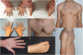
Pectus excavatum. See examples included in Figure 2.

Figure 2.

Figure 2.

Digital anomalies and pectus excavatum in individuals with HYAL2 deficiency A, B. Broad, proximally placed thumbs in an eight-year-old Polish female

Digital anomalies, common as a group, are individually variable (see Figure 2) and include the following:

  • Broad thumbs and/or halluces (6/17)
  • Syndactyly of fingers or toes (5/17), most commonly bilateral toe 2-3 syndactyly
  • Finger webbing
  • Hypoplastic nails
  • Fifth finger clinodactyly

Duodenal web has been observed in two individuals presenting with features of obstruction. It is not clear at this point if this is an infrequent feature of HYAL2 deficiency or coincidental.

Hearing loss. As would be expected in association with cleft lip and palate, hearing loss is usually conductive, can be unilateral or bilateral, and is of variable severity (mild to severe) and age of onset. In two individuals prelingual-onset static sensorineural hearing loss was reported; in one person this was bilateral, mild to severe, and mixed in one ear. In the other person this was bilateral, profound, and treated with cochlear implant [Fasham et al 2022].

Growth, including stature and head size, has to date been normal [Fasham et al 2022].

Other congenital anomalies. Bilateral extrarenal pelvises, congenital diaphragmatic hernia, and glabellar capillary nevus were described in one individual (Individual 4 in Fasham et al [2022]).

Bilateral cryptorchidism was described once [Fasham et al 2022].

Prognosis. The presence and severity of congenital cardiac anomalies primarily determine prognosis. Two sibs with severe complex congenital cardiac lesions died at age less than one year [Fasham et al 2022]. One, who died at age 10 days, had mitral valve atresia, hypoplastic left ventricle, double outlet right ventricle with pulmonary valve atresia, hypoplastic pulmonary and aortopulmonary arteries, and agenesis of the ductus venosus. The other, who died at age 10 months, had ventricular septal defect, atrial septal defect, persistent patent ductus arteriosus, pulmonary hypertension, and diaphragmatic hernia.

When congenital heart disease is absent or amenable to surgical treatment, survival does not seem impacted. The oldest reported individuals are ages 19 and 20 years.

Genotype-Phenotype Correlations

Although no genotype-phenotype correlations have been conclusively identified, the phenotypes reported in three individuals with a nonsense variant in trans with a missense variant suggests that nonsense variants may be associated with more severe congenital cardiac anomalies and severe myopia. These include the two sibs described in Clinical Description, Prognosis, who died during the first year of life due to severe cardiac anomalies, who were compound heterozygous for the HYAL2 variants p.Ser65Ter and p.Phe425Val, and a third individual who was compound heterozygous for the HYAL2 variants p.Arg277Cys and p.Arg295Ter and had coarctation of the aorta, ventricular septal defect, and severe myopic maculopathy [Fasham et al 2022].

Prevalence

To date 17 individuals from seven families with HYAL2 deficiency have been identified [Muggenthaler et al 2017, Fasham et al 2022]. Given that HYAL2 deficiency is a recently described condition, the exact prevalence is unknown. However, low prevalence of the phenotype and low numbers identified in cohorts of individuals with cleft lip and palate with or without other syndromic features suggests that HYAL2 deficiency is rare.

The HYAL2 variant c.443A>G (p.Lys148Arg) is a founder variant in individuals primarily of Indiana Amish ancestry. Seven of 266 individuals of Amish ancestry were found to be heterozygous for this variant [Muggenthaler et al 2017]. This variant was also found to have an allele frequency of 0.6% in the Anabaptist community (based on studies of more than 10,000 exomes from Amish and Mennonite individuals) [Fasham et al 2022].

Differential Diagnosis

The differential diagnosis of HYAL2 deficiency includes other syndromic clefting disorders; see Table 3 for selected examples.

Table 3.

Selected Genes of Interest in the Differential Diagnosis of HYAL2 Deficiency

Gene(s)DisorderMOIKey Features of This Disorder
Overlapping w/HYAL2 DeficiencyDistinguishing from HYAL2 Deficiency
ALX1
ALX3
ALX4
Frontonasal dysplasia (OMIM PS136760)AR
  • Hypertelorism, nasal anomalies, midline orofacial clefts
  • Congenital cardiac defects incl tetralogy of Fallot
  • Abnormalities of hair incl V-shaped "widow's peak" anterior hairline
  • Severe myopia absent
  • Pectus excavatum absent
COL2A1
COL11A1
COL11A2
COL9A1
COL9A2
COL9A3
Stickler syndrome AD
AR 1

Cleft palate, myopia, retinal detachment, auditory issues

  • Characteristic facial features
  • Skeletal abnormalities
EFNB1 Craniofrontonasal syndrome (OMIM 304110)XLHypertelorism, nasal anomalies, orofacial clefts
  • Craniosynostosis
  • Presentation in males is usually mild.
1.

Stickler syndrome caused by pathogenic variants COL2A1, COL11A1, or COL11A2 is inherited in an autosomal dominant manner; Stickler syndrome caused by pathogenic variants in COL9A1, COL9A2, or COL9A3 is inherited in an autosomal recessive manner.

Management

Clinical advice and practice guidelines for HYAL2 deficiency have been proposed [Fasham et al 2022]. Children should be under the care of a pediatrician to monitor their general health and development. Clinical management strategies should be directed to the early diagnosis and treatment of congenital cardiac anomalies and ophthalmologic findings (which may lead to visual impairment if untreated).

Evaluations Following Initial Diagnosis

To establish the extent of disease and needs in an individual diagnosed with HYAL2 deficiency, the evaluations summarized in Table 4 (if not performed as part of the evaluation that led to the diagnosis) are recommended.

Treatment of Manifestations

There is no cure for HYAL2 deficiency.

Supportive care to improve quality of life, maximize function, and reduce complications is recommended. This can include multidisciplinary care by specialists (see Table 5).

Table 5.

HYAL2 Deficiency: Treatment of Manifestations

Manifestation/ConcernTreatmentConsiderations/Other
Congenital cardiac anomalies Mgmt by pediatric cardiologist &/or pediatric cardiac surgeon to address specific lesion(s) identified
Timing/coordination of surgical, dental, & orthodontic mgmt of CLP Determined by multidisciplinary craniofacial teamSurgical repair timing & type of procedure determined by team
Feeding/Nutrition Multidisciplinary craniofacial teamEquipment & techniques for feeding infants w/cleft palate &/or cardiac anomalies (e.g., high-calorie foods/formulas &/or supplementation via nasogastric or enteral feeding as needed) to maintain adequate caloric intake, growth, & weight gain 1
Speech issues Assessment by speech-language pathologist as part of multidisciplinary craniofacial teamInterventions depend on etiology of speech issues & may incl speech therapy &/or use of assistive communication devices.
Ophthalmologic involvement Pediatric ophthalmologistMgmt of refractive errors, strabismus
Ophthalmologic subspecialistMore complex findings (e.g., cataract, myopic maculopathy &/or retinal detachment)
Low vision services
  • Children: through early intervention programs &/or school district
  • Adults: low vision clinic &/or community vision services / OT / mobility services
Hearing loss Consider otolaryngology eval as required for mgmt of middle ear effusions
  • Timely treatment of otitis media secondary to eustachian tube dysfunction due to cleft palate to prevent secondary hearing loss
  • Some persons may require placement of pressure-equalizing tubes.
Family/Community
  • Ensure appropriate social work involvement to connect families w/local resources, respite, & support.
  • Coordinate care to manage multiple subspecialty appointments, equipment, medications, & supplies.

CLP = cleft lip and palate; OT = occupational therapy

1.

Enteral feeding tubes may be needed if there is concern for aspiration or if the affected individual is unable to take in adequate calories for growth. Some children with more significant respiratory issues may require surgical feeding tubes and/or procedures to protect their lungs from microaspiration.

Surveillance

HYAL2 deficiency is primarily a developmental disorder and as such progressive complications are not expected. However, because the complications of congenital anomalies may become apparent with time, the evaluations summarized in Table 6 are recommended.

Evaluation of Relatives at Risk

See Genetic Counseling for issues related to testing of at-risk relatives for genetic counseling purposes.

Therapies Under Investigation

Search ClinicalTrials.gov in the US and EU Clinical Trials Register in Europe for access to information on clinical studies for a wide range of diseases and conditions. Note: There may not be clinical trials for this disorder.

Genetic Counseling

Genetic counseling is the process of providing individuals and families with information on the nature, mode(s) of inheritance, and implications of genetic disorders to help them make informed medical and personal decisions. The following section deals with genetic risk assessment and the use of family history and genetic testing to clarify genetic status for family members; it is not meant to address all personal, cultural, or ethical issues that may arise or to substitute for consultation with a genetics professional. —ED.

Mode of Inheritance

HYAL2 deficiency is inherited in an autosomal recessive manner.

Risk to Family Members

Parents of a proband

  • The parents of an affected child are presumed to be heterozygous for an HYAL2 pathogenic variant.
  • Molecular genetic testing is recommended for the parents of a proband to confirm that both parents are heterozygous for an HYAL2 pathogenic variant and to allow reliable recurrence risk assessment.
  • If a pathogenic variant is detected in only one parent and parental identity testing has confirmed biological maternity and paternity, it is possible that one of the pathogenic variants identified in the proband occurred as a de novo event in the proband or as a postzygotic de novo event in a mosaic parent [Jónsson et al 2017]. If the proband appears to have homozygous pathogenic variants (i.e., the same two pathogenic variants), additional possibilities to consider include:
  • Heterozygotes (carriers) are asymptomatic and are not at risk of developing the disorder.

Sibs of a proband

  • If both parents are known to be heterozygous for an HYAL2 pathogenic variant, each sib of an affected individual has at conception a 25% chance of being affected, a 50% chance of being an asymptomatic carrier, and a 25% chance of being unaffected and not a carrier.
  • Intrafamilial clinical variability has been observed among related individuals with HYAL2 deficiency (e.g., cleft palate and/or cardiac anomalies in some but not all affected family members) [Muggenthaler et al 2017, Fasham et al 2022].
  • Heterozygotes (carriers) are asymptomatic and are not at risk of developing the disorder.

Offspring of a proband. The offspring of an individual with HYAL2 deficiency are obligate heterozygotes (carriers) for a pathogenic variant in HYAL2.

Other family members. Each sib of the proband's parents is at a 50% risk of being a carrier of an HYAL2 pathogenic variant.

Carrier Detection

Carrier testing for at-risk relatives requires prior identification of the HYAL2 pathogenic variants in the family.

Related Genetic Counseling Issues

Family planning

  • The optimal time for determination of genetic risk and discussion of the availability of prenatal/preimplantation genetic testing is before pregnancy.
  • It is appropriate to offer genetic counseling (including discussion of potential risks to offspring and reproductive options) to young adults who are affected, are carriers, or are at risk of being carriers.
  • Carrier testing for reproductive partners of known carriers could be considered, particularly if both partners are of the same ethnic background. An HYAL2 founder variant has been identified in individuals of Amish ancestry (see Table 7).

Prenatal Testing and Preimplantation Genetic Testing

Once the HYAL2 pathogenic variants have been identified in an affected family member, prenatal and preimplantation genetic testing are possible.

Differences in perspective may exist among medical professionals and within families regarding the use of prenatal testing. While most centers would consider use of prenatal testing to be a personal decision, discussion of these issues may be helpful.

Resources

GeneReviews staff has selected the following disease-specific and/or umbrella support organizations and/or registries for the benefit of individuals with this disorder and their families. GeneReviews is not responsible for the information provided by other organizations. For information on selection criteria, click here.

Molecular Genetics

Information in the Molecular Genetics and OMIM tables may differ from that elsewhere in the GeneReview: tables may contain more recent information. —ED.

Table A.

HYAL2 Deficiency: Genes and Databases

GeneChromosome LocusProteinHGMDClinVar
HYAL2 3p21​.31 Hyaluronidase-2 HYAL2 HYAL2

Data are compiled from the following standard references: gene from HGNC; chromosome locus from OMIM; protein from UniProt. For a description of databases (Locus Specific, HGMD, ClinVar) to which links are provided, click here.

Table B.

OMIM Entries for HYAL2 Deficiency (View All in OMIM)

603551HYALURONOGLUCOSAMINIDASE 2; HYAL2

Molecular Pathogenesis

Hyaluronidase-2 (HYAL2) is proposed to act as a hyaluronidase enzyme, catalyzing the degradation of the high-molecular-weight glycosaminoglycan hyaluronan. HYAL2 undergoes complex post-translational modification before achieving its final topology as a mature GPI-anchored cell surface glycoprotein. The translational steps required to generate mature HYAL2 begin in the endoplasmic reticulum (ER), where even conservative amino acid substitutions may lead to ER-associated degradation and HYAL2 deficiency [Fasham et al 2022].

In silico three-dimensional homology modeling of HYAL2 missense variants using a crystal structure of HYAL1(2PE4) was undertaken by Fasham et al [2022] and predicted destabilization of protein folding in almost all instances. Analysis of immunoblotting, immunofluorescence, and glycosylation of variant / wild type human HYAL2 expressed in mouse embryonic fibroblasts was consistent with protein instability and a concomitant reduction or absence of mature HYAL2 at the cell surface.

Mechanism of disease causation. HYAL2 pathogenic variants may cause disease by one of the following mechanisms:

  • Nonsense-mediated mRNA decay and loss of functional protein
  • Missense variants that either:
    • Result in protein instability resulting in low total cell HYAL2 levels;
    • Disrupt the localization of HYAL2 resulting in low cell surface HYAL2 levels.

Table 7.

HYAL2 Pathogenic Variants

Reference SequencesDNA Nucleotide ChangePredicted Protein ChangeComment [Reference]
NM_003773​.5
NP_003764​.3
c.194C>Gp.Ser65TerNonsense variant seen in trans w/missense variant p.Phe425Val in 2 sibs who died during 1st yr of life due to severe cardiac anomalies [Fasham et al 2022] (See Genotype-Phenotype Correlations.)
c.443A>Gp.Lys148ArgFounder variant common in persons of Indiana Amish ancestry [Muggenthaler et al 2017, Fasham et al 2022]
c.829C>Tp.Arg277CysMissense variant seen in trans w/nonsense variant p.Arg295Ter in a person w/coarctation of aorta, ventricular septal defect, & severe myopic maculopathy [Fasham et al 2022] (See Genotype-Phenotype Correlations.)
c.883C>Tp.Arg295TerNonsense variant seen in trans w/missense variant p.Arg277Cys in a person w/coarctation of aorta, ventricular septal defect, & severe myopic maculopathy [Fasham et al 2022] (See Genotype-Phenotype Correlations.)
c.1273T>Gp.Phe425ValMissense variant seen in trans w/nonsense variant p.Ser65Ter in 2 sibs who died during 1st yr of life due to severe cardiac anomalies [Fasham et al 2022] (See Genotype-Phenotype Correlations.)

Variants listed in the table have been provided by the authors. GeneReviews staff have not independently verified the classification of variants.

GeneReviews follows the standard naming conventions of the Human Genome Variation Society (varnomen​.hgvs.org). See Quick Reference for an explanation of nomenclature.

Chapter Notes

Author Notes

Further information on our work with the Amish community can be found at Windows of Hope Project.

Acknowledgments

First and foremost, the authors would like to thank the patients and their families as well as the collaborators who have been involved in defining this emerging multisystem disorder. We would particularly like to acknowledge Professor Barbara Triggs-Raine (University of Manitoba) and her group, especially Ms Promita Ghosh, without whom we would have been unable to begin to define the pathomechanism of the condition.

This work was in part supported by the Medical Research Council, Medical Research Foundation, Newlife Foundation for Disabled Children, GW4-CAT Wellcome Trust, Fondazione Bambino Gesù, Vite Coraggiose, the Natural Science and Engineering Research Council of Canada, Genomic Answers for Kids, National Center for Advancing Translational Sciences of the National Institutes of Health, Institute for Translational Medicine and Therapeutics of the Perelman School of Medicine at the University of Pennsylvania, the National Human Genome Research Institute / National Heart, Lung and Blood Institute, Baylor-Hopkins Center for Mendelian Genomics, and Solve-RD.

Revision History

  • 21 September 2023 (bp) Review posted live
  • 19 December 2022 (eb) Original submission

References

Literature Cited

  • Fasham J, Lin S, Ghosh P, Radio FC, Farrow EG, Thiffault I, Kussman J, Zhou D, Hemming R, Zahka K, Chioza BA, Rawlins LE, Wenger OK, Gunning AC, Pizzi S, Onesimo R, Zampino G, Barker E, Osawa N, Rodriguez MC, Neuhann TM, Zackai EH, Keena B, Capasso J, Levin AV, Bhoj E, Li D, Hakonarson H, Wentzensen IM, Jackson A, Chandler KE, Coban-Akdemir ZH, Posey JE, Banka S, Lupski JR, Sheppard SE, Tartaglia M, Triggs-Raine B, Crosby AH, Baple EL. Elucidating the clinical spectrum and molecular basis of HYAL2 deficiency. Genet Med. 2022;24:631-44. [PMC free article: PMC9933146] [PubMed: 34906488]
  • Jónsson H, Sulem P, Kehr B, Kristmundsdottir S, Zink F, Hjartarson E, Hardarson MT, Hjorleifsson KE, Eggertsson HP, Gudjonsson SA, Ward LD, Arnadottir GA, Helgason EA, Helgason H, Gylfason A, Jonasdottir A, Jonasdottir A, Rafnar T, Frigge M, Stacey SN, Th Magnusson O, Thorsteinsdottir U, Masson G, Kong A, Halldorsson BV, Helgason A, Gudbjartsson DF, Stefansson K. Parental influence on human germline de novo mutations in 1,548 trios from Iceland. Nature. 2017;549:519-22. [PubMed: 28959963]
  • Muggenthaler MM, Chowdhury B, Hasan SN, Cross HE, Mark B, Harlalka GV, Patton MA, Ishida M, Behr ER, Sharma S, Zahka K, Faqeih E, Blakley B, Jackson M, Lees M, Dolinsky V, Cross L, Stanier P, Salter C, Baple EL, Alkuraya FS, Crosby AH, Triggs-Raine B, Chioza BA. Mutations in HYAL2, encoding hyaluronidase 2, cause a syndrome of orofacial clefting and cor triatriatum sinister in humans and mice. PLoS Genet. 2017;13:e1006470. [PMC free article: PMC5230738] [PubMed: 28081210]
  • Richards S, Aziz N, Bale S, Bick D, Das S, Gastier-Foster J, Grody WW, Hegde M, Lyon E, Spector E, Voelkerding K, Rehm HL, et al. Standards and guidelines for the interpretation of sequence variants: a joint consensus recommendation of the American College of Medical Genetics and Genomics and the Association for Molecular Pathology. Genet Med. 2015;17:405-24. [PMC free article: PMC4544753] [PubMed: 25741868]
  • Stenson PD, Mort M, Ball EV, Chapman M, Evans K, Azevedo L, Hayden M, Heywood S, Millar DS, Phillips AD, Cooper DN. The Human Gene Mutation Database (HGMD®): optimizing its use in a clinical diagnostic or research setting. Hum Genet. 2020;139:1197-207. [PMC free article: PMC7497289] [PubMed: 32596782]
Copyright © 1993-2024, University of Washington, Seattle. GeneReviews is a registered trademark of the University of Washington, Seattle. All rights reserved.

GeneReviews® chapters are owned by the University of Washington. Permission is hereby granted to reproduce, distribute, and translate copies of content materials for noncommercial research purposes only, provided that (i) credit for source (http://www.genereviews.org/) and copyright (© 1993-2024 University of Washington) are included with each copy; (ii) a link to the original material is provided whenever the material is published elsewhere on the Web; and (iii) reproducers, distributors, and/or translators comply with the GeneReviews® Copyright Notice and Usage Disclaimer. No further modifications are allowed. For clarity, excerpts of GeneReviews chapters for use in lab reports and clinic notes are a permitted use.

For more information, see the GeneReviews® Copyright Notice and Usage Disclaimer.

For questions regarding permissions or whether a specified use is allowed, contact: ude.wu@tssamda.

Bookshelf ID: NBK594824PMID: 37733894

Views

Related information

  • OMIM
    Related OMIM records
  • PMC
    PubMed Central citations
  • PubMed
    Links to PubMed
  • Gene
    Locus Links

Similar articles in PubMed

See reviews...See all...

Recent Activity

Your browsing activity is empty.

Activity recording is turned off.

Turn recording back on

See more...