Definition
Examination of the peripheral blood smear should be considered, along with review of the results of peripheral blood counts and red blood cell indices, an essential component of the initial evaluation of all patients with hematologic disorders. The examination of blood films stained with Wright's stain frequently provides important clues in the diagnosis of anemias and various disorders of leukocytes and platelets.
Normal human red blood cells are biconcave disks (diskocytes) with a mean diameter of about 7.5 μm. Erythrocytes are slightly smaller than small lymphocytes. The hemoglobin of red cells is located peripherally, leaving an area of central pallor equal to approximately 30 to 45% of the diameter of the cells. Cells of normal size and hemoglobin content (color) are termed normocytic and normochromic. Larger than normal erythrocytes are macrocytes (diameter greater than 9 μm); small red cells are microcytes (diameter less than 6 μm); and those with central pallor greater than 50% of the diameter are hypochromic. Abnormal variability in size is termed anisocytosis; unusual variation in shape is called poikilocytosis; and significant differences among erythrocytes in the amount of central pallor is referred to as anisochromia. Polychromatophilia means the erythrocytes have a blue-gray hue to the color of their cytoplasm.
From a diagnostic standpoint, poikilocytosis has no specificity, but the recognition of specific forms of poikilocytes (irregularly shaped cells) often points to specific disorders. Spherocytes are round, densely staining red cells that lack central pallor and have a smaller than normal diameter. In stomatocytes, the area of central pallor is elliptical rather than round, giving the cell the appearance of the opening of a mouth (stoma). Target cells (codocytes) have a centrally located disk of hemoglobin surrounded by an area of pallor with an outer rim of hemoglobin adjacent to the cell membrane giving the cell the appearance of a target. Leptocytes (or wafer cells) are thin, flat cells with the hemoglobin at the periphery of the cell. Sickle cells (drepanocytes) are elongated, sometimes crescent-shaped, erythrocytes with pointed ends. Elliptocytes (ovalocytes) range from slightly oval to elongated cigar-shaped forms. Teardrop erythrocytes (dacryocytes) are red cells with one end round and the other end more pointed. Acanthocytes have several (usually 3 to 7) irregularly spaced blunted projections from the margin of the cells. Echinocytes are also cells with cytoplasmic projections, but in contrast to acanthocytes, the projections are typically evenly spaced on the cell surface, more numerous (often 10 to 15), and frequently have sharper points. Schizocytes (schistocytes) are fragmented erythrocytes appearing in a variety of morphologic forms such as small triangular erythrocytes, helmet cells, and normal-size erythrocytes with 2 to 3 pointed surface projections (keratocytes, or "horn cells"). Round erythrocytes with a single, elliptical or round surface defect are termed bite cells. Rouleaux formation is a phrase denoting the stacking of erythrocytes, generally in a curving pattern.
Morphologic identification of inclusion bodies within erythrocytes can be helpful clinically. Howell–Jolly bodies are purple spheres, usually about 0.5 μm in diameter, presenting singly, or rarely multiply, in the cytoplasm. Basophilic stippling of erythrocytes refers to numerous very small coarse or fine blue granules within the cytoplasm. When the stippled particles are due to iron granules (demonstrable by the Prussian blue stain), they are termed Pappenheimer bodies. Malaria parasites may appear as cytoplasmic inclusion bodies within erythrocytes. Platelets overlying erythrocytes may be mistaken for erythrocyte inclusions.
There are a number of important morphologic abnormalities of mature granulocytes. Cytoplasmic vacuoles may be recognized. Toxic granulation refers to small, dark blue-staining granules. Döhle bodies are light blue cytoplasmic inclusions, 1 to 2 μm in diameter. The Pelger–Huët anomaly, a disorder characterized by impaired nuclear segmentation of mature neutrophilic granulocytes, appears morphologically as cells with bilobed nuclei (dumbbell or eyeglass shapes) or with round or oval nuclei (Stodtmeister cells). Hypersegmented neutrophils are cells in which there are six or more nuclear lobes.
Reactive lymphocytes are usually larger than small lymphocytes, may have cytoplasmic vacuolization, sometimes have deep blue staining of the periphery of the cytoplasm, and contain nuclei that may be kidney bean or monocytoid in shape.
Most platelets in the peripheral blood have diameters between 1 and 3 μm. Platelets greater than 3 μm in diameter are "large" (megathrombocytes). In a normal person usually less than 5% of the platelets appear large.
Figure 155.1 shows examples of morphologically normal and abnormal erythrocytes.
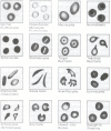
Figure 155.1
Morphologically abnormal erythrocytes.
Technique
Morphologic abnormalities of peripheral blood cells are discovered by microscopic examination with the oil immersion lens of well-prepared films of peripheral blood stained with Wright's stain. For appropriate interpretation of the morphology of erythrocytes, one concentrates on areas of the slide where the red cells appear singly and have central pallor. Examination of erythrocytes far out on the feathered edge discloses erythrocytes lacking central pallor, whereas in thick areas of the slide the morphology of the erythrocytes is distorted by contact between cells.
Artifactual changes of erythrocytes occur commonly on peripheral blood films. Cytoplasmic vacuolization of red cells is an artifact. Echinocytes (crenated red cells) are frequently caused by hypertonicity or alkalinity of the staining solution. Stomatocytes may form when the staining solution is too acidic. When target cells appear in one area of the slide and not in another, they are artifacts because naturally occurring target cells will be distributed evenly throughout the slide.
Neutrophils and monocytes often congregate at the feathered end and at the edges of the blood film. Granulocytes of blood anticoagulated with EDTA may acquire cytoplasmic vacuoles. Leukocytes may rupture during the preparation of the blood film, leaving amorphous nuclear material ("basket" cells). The lymphocytes of chronic lymphocytic leukemia are particularly prone to rupture.
Although platelets usually occur singly on the blood film, in some cases there is considerable aggregation of platelets, making estimation of the number of platelets more difficult.
Basic Science
Morphologic abnormalities of erythrocytes may be due either to production of abnormal erythrocytes in the bone marrow or to pathologic processes to which the erythrocytes are exposed in the circulation. Macrocytes usually reflect abnormal erythropoiesis in which there is a reduced number of cell divisions during maturation of erythroid precursors. Hypochromia generally arises because of impaired hemoglobinization of erythroid cells in the marrow. Spherocytes can be due to an inherited membrane abnormality of erythrocytes (hereditary spherocytosis) or can result from the action of phagocytes on erythrocytes sensitized with antibodies wherein the phagocytes remove portions of the red cell membrane, creating spherocytes. In addition, both spherocytes and schizocytes result from the action of abnormal physical forces in the circulation (particularly shear stress) that cause fragmentation of normal erythrocytes. Stomatocytes are overhydrated cells, which in three-dimensional views are bowl shaped; they may appear as an intermediate form in the transformation of diskocytes to spherocytes. The target cell is a bell-shaped cell with a relative excess of membrane; in patients with obstructive liver disease a significant increase in the total membrane content of cholesterol leads to the increase in cell surface area. The spur cells (acanthocytes) of chronic alcoholic liver disease have increased cholesterol but, in contrast to target cells, normal content of phospholipids. The echinocytes of pyruvate kinase deficiency form because of decreased ATP generation resulting in loss of water and potassium from the red cells. Sickle cells result from aggregation in the deoxygenated state of molecules of hemoglobin S, which have the substitution of valine for glutamic acid of the sixth amino acid position of the beta chain of hemoglobin. In hereditary elliptocytosis, the elliptical shape of the cell is due to membrane protein abnormalities. Infiltrative disorders of the bone marrow, with disruption of the vasculature of the marrow, are associated with the formation of elliptocytes and teardrop erythrocytes. Bite cells apparently arise when a phagocyte removes a portion of the red cell along with a Heinz body.
Howell–Jolly bodies are remnants of DNA; they are ordinarily removed from red cells by the spleen. Basophilic stippling occurs in conditions in which the biosynthesis of hemoglobin is impaired; the stippled particles are aggregates of ribosomes or, in the case of Pappenheimer bodies, aggregates of ferritin, lysosomes, ribosomes, and degenerating mitochondria.
Pelger–Hüet cells occur in a hereditary disorder in which the granulocytes function normally and no hematologic illness exists. As an acquired disorder, Pelger–Hüet cells generally reflect neutrophilic granulocytic dysplasia.
Clinical Significance
Two abnormalities of erythrocytes can be recognized by low-power microscopic examination of the blood. Rouleaux of erythrocytes is related to very high serum protein concentrations, generally due to multiple myeloma or to macroglobulinemia. Agglutination of red cells on the slide is usually due to cold agglutinins.
Macrocytes (frequently oval) in substantial numbers are observed in patients with megaloblastic anemias (vitamin B12 or folic acid deficiency) often with considerable anisocytosis (with some microcytes present as well). In addition, macrocytes may be prominent in individuals with erythroleukemia, myelodysplastic disorders, acquired sideroblastic anemia, and with antimetabolite or androgen drug therapy. A lesser degree of macrocytosis is seen commonly in alcoholic patients. Polychromatophilic macrocytes usually indicate a high reticulocyte count.
A predominance of hypochromic microcytic cells is found in iron deficiency anemia, thalassemia, and hereditary sideroblastic anemia, and in some patients with the anemia of chronic disorders and with lead intoxication. For individuals with mild anemia, the degree of microcytosis is usually substantially greater in patients with thalassemia minor than those with iron deficiency. Anisochromia with the presence of a dimorphic red cell population (hypochromic and normochromic) is observed in acquired sideroblastic anemia, patients with thalassemia minor after transfusions, and persons with iron deficiency following transfusions or treatment with iron.
Examination of peripheral blood films of normal persons reveals small numbers of poikilocytes, usually less than 2%. In the assessment of the significance of poikilocytosis, one must identify the predominant abnormal morphologic form and exclude artifactual alterations of the red cells.
Spherocytes are the predominant morphologic abnormality in patients with hereditary spherocytosis, autoimmune hemolytic anemia, and hemolytic transfusion reactions, and are common, along with schizocytes, in patients with red cell fragmentation disorders. Spherocytes may also be observed in less common hemolytic states such as the Heinz body hemolytic anemias and clostridial sepsis. Stomatocytes are seen in large numbers in alcoholics and in the rare disorder of hereditary stomatocytosis, and in small numbers in normal persons. There are four major circumstances in which target cells appear as the major morphologic abnormality: thalassemia, hepatic disease with jaundice, hemoglobin C disorders, and the postsplenectomy state. Lesser numbers of target cells are found in sickle cell anemia, iron deficiency, and lead intoxication. Leptocytes are seen in thalassemic disorders and with obstructive liver disease. Sickle cells and dense, deformed poikilocytes ("irreversibly sickled cells") are characteristic of sickle cell anemia, hemoglobin SC disease, hemoglobin S-thalassemia, and hemoglobin C-Harlem, but are not observed in sickle cell trait.
A large number of elliptocytes (25 to 75% of the red cells) usually indicates hereditary elliptocytosis. Moderate numbers of elliptocytes are seen in thalassemia and myelofibrosis, and lesser numbers in iron deficiency and hypersplenic states. Teardrop erythrocytes (usually with elliptocytes) are particularly prominent in patients with myelofibrosis with myeloid metaplasia and occur frequently in patients with other infiltrative disorders of the bone marrow such as leukemia and metastatic carcinoma. Acanthocytes are the principal morphologic abnormality in abetalipoproteinemia and in the "spur cell anemia" associated with severe alcoholic liver disease. Acanthocytes are found along with other poikilocytes after splenectomy. Conditions associated with the appearance of echinocytes are pyruvate kinase deficiency of erythrocytes, uremia, carcinomas, and immediately after the transfusion of aged or metabolically depleted blood (echinocytes form during storage of the blood). Correction of the metabolic abnormalities of uremia results in disappearance of echinocytes. Schizocytes are the morphologic hallmark of the hemolytic anemias associated with red cell fragmentation (i.e., the microangiopathic hemolytic anemias and those hemolytic anemias associated with malfunctioning cardiac prostheses). Schizocytes also may form during disseminated intravascular coagulation. Bite cells are principally related to the various Heinz body hemolytic anemias, such as glucose-6-phosphate dehydrogenase deficiency.
Howell–Jolly bodies are found in patients who have had splenectomies or are hyposplenic (e.g., sickle cell anemia) and rarely in megaloblastic anemias. The three disorders particularly associated with coarse basophilic stippling are lead poisoning, sideroblastic anemia, and thalassemia.
Nucleated erythrocytes, usually in small numbers (in adults), may appear in the blood when the marrow is under intense stimulation due to severe hemolysis, hemorrhage, or hypoxia. In addition, nucleated red cells and immature myeloid cells may be recognized with infiltrative disorders of the marrow such as myelofibrosis, leukemia, and metastatic carcinoma. In patients with megaloblastic anemias the nucleated erythrocytes in the blood have megaloblastic nuclear features.
Cytoplasmic vacuolization of granulocytes is observed in patients with bacteremia or other severe infections. Toxic granulation, a rather nonspecific finding, is found in a variety of disorders including infections and metabolic derangements. Döhle bodies are seen in patients with infections and burns, during pregnancy, after cytotoxic chemotherapy (particularly with cyclophosphamide), and with the May–Heggelin anomaly. Pelger-Hüet cells, on an acquired rather than hereditary basis, are particularly associated with myelodysplastic and myeloproliferative disorders. Hypersegmented neutrophils usually are an important clue to the presence of vitamin B12 or folic acid deficiency but are occasionally found in patients with myelodysplasia or myeloproliferative disorders.
The diagnosis of leukemia is commonly obvious by recognition of abnormal numbers and stages of development of myeloid or lymphoid cells in the blood. Immature monocytes suggest either leukemia or myelodysplasia. A significant increase in the number of basophils usually indicates a myeloproliferative disorder.
A high percentage of reactive lymphocytes may be seen in viral illnesses such as infectious mononucleosis, viral hepatitis, cytomegalovirus infection, HIV infection and rubella, or with reactions to drugs such as phenytoin and para-aminosalicylic acid. Lymphocytes with convoluted nuclei may be found in T cell lymphomas and in the Sezary syndrome.
An increased number of large platelets is observed in thrombocytopenia with immune-mediated hyperdestruction, disseminated intravascular coagulation, myeloproliferative disorders (particularly myelofibrosis), megaloblastic anemias, the Bernard–Soulier syndrome, and the May–Heggelin anomaly. Platelet size is normal in hypersplenic states. Microthrombocytes are found in the Wiskott–Aldrich syndrome. Hypogranular platelets are seen in the myeloproliferative disorders.
References
- Bessis M. Red cell shapes. An illustrated classification and its rationale. Nouv Rev Fr Hematol. 1972;12:721–46. [PubMed: 4268780]
- Bessis M, Lessin LS, Beutler E. Morphology of the erythron. In: Williams WJ, Beutler E, Ersler AJ, Lichtman MA, eds. Hematology. 3rd ed. New York: McGraw-Hill, 1983;257–79.
- Garg SK, Lackner H, Karpatkin S. The increased percentage of megathrombocytes in various clinical disorders. Ann Intern Med. 1972;77:361–69. [PubMed: 5066195]
- Lessin LS, Klug PP, Jensen WN. Clinical implications of red cell shape. In: Stollerman GH, ed. Advances in internal medicine. Chicago: Year Book Medical Publishers, 1976;21:451–99. [PubMed: 766592]
- Schwartz SO, Stansbury F. Significance of nucleated red blood cells in peripheral blood. Analysis of 1,496 cases. JAMA. 1954;154:1339–40. [PubMed: 13151849]
- Zeigler Z, Murphy S, Gardner FH. Microscopic platelet size and morphology in various hematologic disorders. Blood. 1978;51:479–86. [PubMed: 414804]
Publication Details
Author Information and Affiliations
Authors
Edward C. Lynch.Copyright
Publisher
Butterworths, Boston
NLM Citation
Lynch EC. Peripheral Blood Smear. In: Walker HK, Hall WD, Hurst JW, editors. Clinical Methods: The History, Physical, and Laboratory Examinations. 3rd edition. Boston: Butterworths; 1990. Chapter 155.
