

NCBI Bookshelf. A service of the National Library of Medicine, National Institutes of Health.
Mittal RK. Motor Function of the Pharynx, Esophagus, and its Sphincters. San Rafael (CA): Morgan & Claypool Life Sciences; 2011.
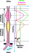
Intraluminal pressure measurements, used extensively to record esophageal motor activity, monitor contraction of the circular muscles of the esophagus which comprises only 50% or less of the mass of muscularis propria. Longitudinal muscle contraction causes esophageal shortening. Primary and secondary peristalses associated esophageal shortening have been known to radiologists since 1950s. Using radio-opaque markers implanted on the esophageal wall, cineradiography studies show peristalsis in the longitudinal muscle layer of the esophagus [63,114–116]. Strain gauze recording of the opossum esophagus shows coordination between circular and longitudinal muscle layers [117,118]. These studies found that at a given site in the esophagus longitudinal muscles contract prior to and outlasts circular muscle contraction by 2–4 seconds. Ultrasound imaging and manometry studies show that, actually, the two layers of the esophagus contract in a precisely coordinated way; the onset, peak, and termination of contraction of the two muscle layers are precisely coordinated [103,119]. Similarly, contraction proximal to the site of balloon is also seen in the longitudinal muscle layer [120]. Caudal to the distension site, the two layers of esophagus demonstrate relaxation. Similar to circular muscles, deglutitive inhibition also occurs in the longitudinal muscles as demonstrated by the changes in distance of the radio-opaque markers implanted along the length of the esophagus (to measure longitudinal muscle contraction) [121]. All of the above observations suggest that ascending contraction and descending relaxation “law of intestine” described by Bayless and Starling [122] in 1899 holds true for the circular and longitudinal muscle layers of the esophagus (Figure 15), as is also the case with the colon [123,124]. There is strong correlation between the amplitude of circular and longitudinal muscle contraction during peristalsis [103,125].
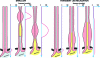
If it is the milking action of circular muscle that is responsible for bolus propulsion, what is the function of longitudinal muscle layer of the esophagus? There are several advantages for the two layers to contract together during peristalsis? First, contraction of longitudinal muscle layers brings together the rings of circular muscles at the site of contraction which increases muscle mass and efficiency of circular muscle contraction [58,101]. Second, increase in muscle thickness at the site of contraction reduces esophageal wall stress that prevents outward bulging or “aneurysm like effect.” Discoordination with regards to the timing of contraction between the two muscle layers occurs in patients with nutcracker esophagus (hypertensive esophageal peristalsis) [126]. Interestingly, these patients also have a high incidence of diverticular (out pouch) formation. Discoordination between the two layers is related to a hypercholinergic state because cholinestrase inhibitor (edrophonium) induces discoordination between the two muscle layers in the normal subjects and an anticholinergic (atropine) ameliorates temporal discoordination between the two muscle layers in patients with nutcracker esophagus [127].
Retrograde transport and transient LES relaxation is associated with a unique and distinct profile of contraction in the two muscle layers [64,127]. Contraction of longitudinal muscle of the distal esophagus starts right before the onset of transient LES relaxation, and it gets getter stronger and traverses in an antiperistaltic fashion toward the proximal esophagus during the entire duration of transient LES relaxation. Circular muscles do not contract during the entire period of TLESR (Figure 16). LES and crural diaphragm remain relaxed during the entire period of longitudinal muscle contraction, and with cessation of longitudinal muscle contraction, there is return of LES basal tone and crural diaphragm activity. Circular and longitudinal muscle layers of the esophagus contract together during peristalsis [119], and longitudinal muscle contracts independent of circular muscle during transient LES relaxation [127]. Since transient LES relaxation and swallow-mediated peristalsis are mediated via vagus nerve and brain stem, it is likely that the central program generator (CPG) can initiate two distinct motor programs, program 1, which is responsible for aboral transport with swallowing, and program 2, which is responsible for the retrograde transport of which transient LES relaxation is the key component. As will be discussed later, longitudinal muscle contraction of the distal esophagus is likely a key event that induces LES relaxation through the activation of stretch sensitive motor neurons of the LES.
- Peristalsis in the Circular and Longitudinal Muscles of the Esophagus - Motor Fu...Peristalsis in the Circular and Longitudinal Muscles of the Esophagus - Motor Function of the Pharynx, Esophagus, and its Sphincters
Your browsing activity is empty.
Activity recording is turned off.
See more...