

NCBI Bookshelf. A service of the National Library of Medicine, National Institutes of Health.
Baron S, editor. Medical Microbiology. 4th edition. Galveston (TX): University of Texas Medical Branch at Galveston; 1996.
General Concepts
Clinical Manifestations
A great variety of infections including abscesses, gangrene, cellulitis, bacteremia, pneumonia, peritonitis, bite wounds, and pelvic inflammatory disease.
Structure
Gram-positive and gram-negative cocci.
Classification and Antigenic Types
Diverse group of genera. Strictly anaerobic as well as aerotolerant species. P eptostreptococcus, Gemella, and Streptococcus are gram-positive genera of primary clinical importance. Veillonella is the gram-negative genus isolated most frequently from clinical specimens.
Pathogenesis
Infection usually results from invasion of damaged tissue by normal microbial flora. Most infections are polymicrobic. However, approximately 10%–15% of all clinical isolates come from pure culture infections.
Host Defenses
Undefined.
Epidemiology
Normal flora of skin, mouth, intestinal tract, and genitourinary tract.
Diagnosis
Laboratory isolation of organism from infected site with careful consideration of organism's role as a possible member of normal microbial flora of that particular site.
Control
Antibiotic therapy (e.g., penicillin, clindamycin), abscess drainage, debridement of necrotic tissue.
Clinical Manifestations
Anaerobic cocci are not involved in any single specific disease process; rather, they may be present in a great variety of infections involving all areas of the human body (Fig. 19-1). These infections may range in severity from mild skin abscesses, which disappear spontaneously after incision and drainage, to more serious and life-threatening infections such as brain abscess, bacteremia, necrotizing pneumonia, and septic abortion. Infection by anaerobic cocci (and by anaerobes in general) usually involves invasion of devitalized tissue by organisms that are part of the normal flora of the affected tissue or of the surrounding areas.
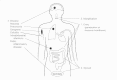
Figure 19-1
Pathogenesis of anaerobic cocci.
Brain abscess, with a mortality rate of 40%, is one of the more serious infections involving anaerobic cocci. Anaerobes, rather than facultative or aerobic organisms, are a major cause; anaerobic cocci, Bacteroides and Fusobacterium, respectively, are the predominant groups isolated. Anaerobic cocci often have been isolated in pure culture from brain abscesses. Chronic otitis media or mastoiditis frequently is the primary source of the organisms and may result as a direct extension of the infection into the brain. Pleuropulmonary infection, sinusitis, congenital heart defects, and bacterial endocarditis are other conditions predisposing individuals to brain abscess by blood-borne metastases.
Pleuropulmonary infections in which anaerobic cocci may be etiologic agents are lung abscesses, necrotizing pneumonia, aspiration pneumonitis, and empyema. The incidence of anaerobes in these infections is 50–90%; anaerobic cocci account for about 40% of the anaerobic isolates. F nucleatum and P melaninogenica are often isolated concomitantly. These organisms are part of the normal microbial flora of the mouth and enter the lower respiratory tract as the result of aspiration, usually in association with altered consciousness.
Anaerobic pleuropulmonary infections frequently develop slowly and often are chronic. The mortality rate is about 15%.
Anaerobic cocci are involved in several skin and soft tissue infections that may be confused with clostridial myonecrosis (gas gangrene). These infections are anaerobic streptococcal myonecrosis, progressive bacterial synergistic gangrene, necrotizing fasciitis, crepitant cellulitis, chronic burrowing ulcer, and synergistic necrotizing cellulitis. These are severe infections, and the mortality rates may be as high as 75%. These conditions may be characterized by a purulent exudate, by varying degrees of tissue necrosis involving the skin, fascia, and/or underlying muscles, and sometimes by systemic toxicity. The infecting organisms often produce gas. Anaerobic cocci often are isolated with other organisms in these infections. They are characteristically found with Staphylococcus aureus and Streptococcus pyogenes in progressive bacterial synergistic gangrene, and also are found with gram-negative aerobic or facultative bacilli or Bacteroides or both in synergistic nonclostridial myonecrosis and synergistic necrotizing cellulitis. Diabetes mellitus and vascular insufficiency (often associated with trauma) are predisposing factors. Decubitus ulcers and postoperative wound infections are other soft-tissue infections from which anaerobic cocci have been isolated.
Anaerobic cocci have been recognized as significant pathogens in puerperal fever and septic abortion since the early 1900s. Other infections of the female genital tract in which anaerobic cocci have been implicated are pyometra, tuboovarian abscesses, postoperative wound infections following gynecologic surgery, and pelvic inflammatory disease, often in association with gonococci. Anaerobic cocci (P prevotii, P anaerobius and S intermedius) and B fragilis are the most frequently isolated anaerobes from these infections. Like the anaerobic cocci in other infections, these organisms are part of the normal flora of the affected area or of the surrounding tissues—in this case, the vagina.
Periodontal disease, peritonitis, intraabdominal abscesses, and abscesses of the liver, spleen, and pancreas are types of intraabdominal infections from which anaerobic cocci have been isolated. Again, these are polymicrobic infections; concomitant isolates may be Bacteroides sp., E. coli and Streptococcus sp.
Structure, Classification and Antigenic Types
The anaerobic cocci are a physiologically diverse group that has recently undergone significant taxonomic changes. Anaerobic gram-positive cocci of clinical significance are found in three gram-positive genera (Peptostreptococcus, Gemella, and Streptococcus) and one gram-negative genus (Veillonella). There are other genera of anaerobic cocci, but they are rarely isolated from clinical specimens. Not all anaerobic cocci require stringent anaerobic conditions; for example, strains of Streptococcus intermedius are quite aerotolerant and may grow under reduced oxygen tension. Anaerobic cocci may be proteolytic or saccharolytic or both. They produce a variety of short-chain volatile fatty acids (i.e., acetic, propionic, butyric, caproic, and lactic acids) from the fermentation of simple sugars and amino acids. Both P magnus and P anaerobius possess species-specific cell wall antigens; in other anaerobic cocci, species-specific antigens have not yet been identified. Peptostreptococcus and Streptococcus are the most clinically important genera, with P magnus as the most frequent clinical isolate.
The anaerobic gram-positive cocci are difficult to speciate, but a few biochemical tests can be helpful. P anaerobius is the only species susceptible to sodium polyanethol sulfonate (SPS). P asaccharolyticus and P hydrogenalis are both indole positive, but alkaline phosphatase negative and positive, respectively. Of the indole-negative butyric acid producers, P tetradius is strongly saccharolytic and urease-positive, while P prevotii is weakly saccharolytic and usually urease-negative. P magnus and P micros are similar biochemically and are distinguished primarily on the basis of cell size and alkaline phosphatase reaction. The three prominent species of anaerobic cocci that are strongly saccharolytic and produce large amounts of lactic acid include S intermedius, S constellatus, G morbillorum. These latter species are either aerotolerant or become aerotolerant upon passage on laboratory media. Obligate anaerobic species in the genus Streptococcus are only rarely isolated from clinical specimens, but may be found in human feces, as can other genera of anaerobic gram-positive cocci.
Three genera of anaerobic gram-negative cocci can be found in human fecal flora: Veillonella, Acidominococcus, and Megosphora. Veillonella is considered the only clinically significant genus and V parvula is the species most frequently isolated from clinical specimens. Veillonella can be presumptively identified by the red fluorescence of colonies under ultraviolet light. This fluorescence is lost rapidly on exposure to oxygen.
Pathogenesis
Anaerobic cocci are opportunistic pathogens that cause a multitude of infections. They are part of the normal microbial flora of a healthy individual, but they can and do cause infections involving traumatized tissue or infections in the compromised host. They are isolated most often from a wide variety of polymicrobic infections (usually along with Bacteroides sp. or with facultative organisms or both), indicating a synergistic role in these infections. Approximately 10–15% of all isolates of anaerobic cocci come from pure culture infections, thus indicating that these organisms can be significant pathogens rather than innocuous commensals. In one series of 20 patients with anaerobic bacteremia, Peptostreptococcus species accounted for 21% of the 29 anaerobic isolates. The anaerobic cocci represent 25–30% of all anaerobic clinical isolates. Among anaerobes, they are second only to the gram-negative anaerobic bacilli in frequency of isolation from clinical specimens. The anaerobic cocci have received relatively little attention from microbiologists and clinicians. It is not known if anaerobic cocci produce toxins, capsules, or have other pathogenic attributes.
Host Defenses
P magnus infection (in pure culture) of hip prostheses has produced a serum antibody response; however, in most cases, specific immune responses to anaerobic cocci have not been investigated.
Epidemiology
Anaerobic cocci are part of the normal flora of the skin, the mouth, and the intestinal and genitourinary tracts of healthy individuals. Recently, with increasing study of the anaerobic cocci as pathogens, certain species are being associated with specific types of infection. As noted above, P prevotii and P anaerobius are associated with female genital tract and intraabdominal infections. P magnus, the most frequently isolated anaerobic coccus, is associated most often with chronic bone and joint infections and ankle ulcers. Pure cultures of this organism are not rare; they account for 15% of all P magnus isolates. The presence of foreign bodies, such as prosthetic joints, seems to be particularly significant in P magnus infections. In one study, anaerobic cocci were isolated in 15 (6%) of 246 cases of monomicrobial anaerobic bacteremia in cancer patients, indicating a relatively rare, but significant pathogenic potential for anaerobic cocci in this patient population. Veillonella and the anaerobic/aerotolerant Streptococcus are the anaerobic cocci isolated most frequently from infected human bites. These organisms are part of the normal oral flora. The microaerophilic gram-positive cocci are associated with abscesses and other purulent infections.
Diagnosis
Anaerobic infections generally occur in the compromised host; that is, in patients who have impaired host defense mechanisms. The primary host defense deficiency in these infections is the disruption of natural barriers (such as the skin and mucous membranes). Diabetes mellitus, connective tissue disorders, atherosclerotic disease, cancer (especially of the colon, uterus, and lung), irradiation damage, immunosuppressive treatment, and alcoholism are conditions that may disrupt these natural barriers.
To establish a definite role for anaerobic cocci in infections, the causative organism must be isolated from the affected tissue or the bloodstream. Because anaerobic cocci are a significant part of the normal flora, the proper choice of specimen is critical. For example, coughed sputum, feces, and vaginal swabs, all of which could be contaminated with normal microbial flora, are unacceptable.
Control
Treatment of infections caused by anaerobic cocci consists of antibiotic therapy and drainage, debridement, or both of necrotic tissue. In general, penicillin is the drug of choice, and clindamycin or metronidazole can be used for the patient allergic to penicillin. The clinician should be aware that in vitro antimicrobial susceptibility tests have shown that some strains of anaerobic cocci are resistant to penicillin or to clindamycin. Metronidazole is typically active against most strains of anaerobic cocci; however, aerotolerant species, such as Streptococcus spp. are uniformly resistant. Brain abscesses must be treated with an antimicrobial agent such as chloramphenicol or penicillin or metronidazole, sufficient doses of which can cross the blood barrier. Frequently B fragilis, an anaerobic gram-negative rod, is present in infections containing anaerobic cocci; this organism produces a β-lactamase that can protect other organisms in the infection from the action of penicillin.
References
- Bourgault AM, Rosenblatt JE, Fitzgerald RH. Peptococcus magnus: A significant human pathogen. Ann Intern Med. 1980;93:244–248. [PubMed: 7406374]
- Brook I. Peptostreptococcal infection in children. Scand J Infect Dis. 1994;26(5):503. [PubMed: 7855547]
- Brook I, Walker RI: Pathogenicity of anaerobic gram-positive cocci. Infect Immunol 320-324, 1984 . [PMC free article: PMC263223] [PubMed: 6746091]
- Fainstein V, Elting LS, Bodey GF. Bacteremia caused by non-sporulating anaerobes in cancer patients - a 12-year experience. Medicine. 1989;3:151–162. [PubMed: 2716514]
- Finegold SM, George WL (eds): Anaerobic infections in humans. Academic Press, New York, 1989 .
- Kotiranta A, Haapasalo M, Lounatmaa K, Kari K. Crystalline surface protein of Peptostreptococcus anaerobius. Microbiology. 1995;141(Pt. 5):1065. [PubMed: 7773401]
- Peraino VA, Cross SA, Goldstein EJ. Incidence and clinical significance of anaerobic bacteremia in a community hospital. Clin Infect Dis. 1993;16:S288. [PubMed: 8324133]
- Summanen P, Baron EJ, Citron DM, Strong C, Wexler HM, Finegold, SM (eds): Wadsworth anaerobic bacteriology manual, 5th ed. Star Publishing Co., Belmont Calif, 1993 .
- Summanen P. Recent taxonomic changes for anaerobic gram-positive and selected gram-negative organisms. Clin Infect Dis. 1993;16:S168. [PubMed: 8324113]
- Taylor AG, Finham WJ, Golding MA. et al. Infection of total hip prostheses by Peptococcus magnus, an immuno-fluorescence and ELISA study of two cases. J Clin Pathol. 1979;32:61–65. [PMC free article: PMC1145569] [PubMed: 372250]
- Epidemiology of anaerobic infections.[Surgery. 1983]Epidemiology of anaerobic infections.Hnatko SI. Surgery. 1983 Jan; 93(1 Pt 2):125-33.
- Review Role of encapsulated anaerobic bacteria in synergistic infections.[Crit Rev Microbiol. 1987]Review Role of encapsulated anaerobic bacteria in synergistic infections.Brook I. Crit Rev Microbiol. 1987; 14(3):171-93.
- Aerobic and anaerobic bacteriology of perirectal abscess in children.[Pediatrics. 1980]Aerobic and anaerobic bacteriology of perirectal abscess in children.Brook I, Martin WJ. Pediatrics. 1980 Aug; 66(2):282-4.
- The clinical importance of gram-positive anaerobic cocci isolated at St Bartholomew's Hospital, London, in 1987.[J Med Microbiol. 1994]The clinical importance of gram-positive anaerobic cocci isolated at St Bartholomew's Hospital, London, in 1987.Murdoch DA, Mitchelmore IJ, Tabaqchali S. J Med Microbiol. 1994 Jul; 41(1):36-44.
- Review Infections in the female genital tract.[Compr Ther. 1983]Review Infections in the female genital tract.Gall SA. Compr Ther. 1983 Aug; 9(8):34-47.
- Anaerobic Cocci - Medical MicrobiologyAnaerobic Cocci - Medical Microbiology
Your browsing activity is empty.
Activity recording is turned off.
See more...