From: Stimulus–Secretion Coupling in the Osmoreceptive Prolactin Cell of the Tilapia
Copyright © 2005, Academia Publishing House
Ltd.
NCBI Bookshelf. A service of the National Library of Medicine, National Institutes of Health.
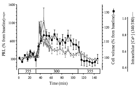
PRL cells respond rapidly to changes in extracellular osmolality (mOsmolal, indicated in the bar above the time axis). After hyposmotic stimulation, an increase in cell volume precedes a rise in [Ca2+]i, which in turn precedes PRL release. Cell volume and PRL release were measured from the same preparation of cells [54]. The trace for [Ca2+]i was recorded from cells treated identically and aligned to match the temporal scale of the other two parameters. Intracellular Ca2+ concentration was monitored by fluorescence imaging with the Ca2+-sensitive dye, fura-2, whose excitation wavelengths are 340 and 380 nm, for fura-2 bound to Ca2+ and free fura-2, respectively [54].
From: Stimulus–Secretion Coupling in the Osmoreceptive Prolactin Cell of the Tilapia
NCBI Bookshelf. A service of the National Library of Medicine, National Institutes of Health.