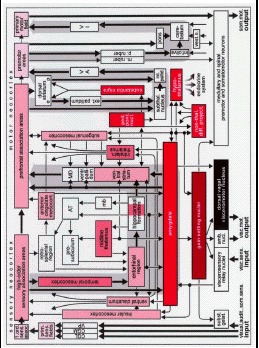
Schematic diagram of the limbic, somatomotor, and visceromotor systems. The components of the autonomic and limbic loops are interconnected so that the resulting comprehensive
schema combines both loops from (). The most important centers of the striatal and cerebellar loops have been added to the scheme to make the motor system more complete. Furthermore, additional details are supplied both in the form of select brain stem centers and of a few limbic circuit relay nuclei. The three gray arrows in the background facilitate recognition of the limbic, striatal, and cerebellar loops. Even in this
more complicated schema, it becomes clear that normal functions of the limbic and autonomic circuits depend on the structural integrity of the insular, subgenual, and temporal mesocortex. Lesions in the autonomic, limbic, and motor systems: Damaged structures are marked in black (from stage 1 onwards), rust (from stage 2 onwards), red (from stage 3 onwards), light red (from stage 4 onwards), rose (from stage 5 onwards), and light pink (from stage 6 onwards). White indicates noninvolvement. Higher centers of the limbic and visceromotor systems are susceptible to the worst pathology. The severe involvement of the temporal mesocortex leads to a marked reduction of the essential data-transfer from the sensory neocortex via the temporal proneocortex, transentorhinal periallocortex, entorhinal allocortex, hippocampal formation, and amygdala to the prefrontal cortex. Abbreviations: amb. nucl. - ambiguus nucleus; ant. cingulate
mesocort. - anterior cingulate mesocortex; AT - anterior thalamic nuclei; CGL - corpus geniculatum laterale/lateral geniculate body; CGM - corpus geniculatum mediale/medial geniculate body; ext. pallidum - external pallidum; inf. oliv. - inferior olive; int. pallid. - internal pallidum; intralam. thalamus - intralaminar nuclei of the thalamus; mb - mamillary body; e - encephalin-containing cells; p - substance P-containing cells; MD - mediodorsal nuclei of the thalamus; m. ruber - magnocellular portion of the red nucleus; p. ruber - parvocellular portion of the red nucleus; nonthal. diff. project. - nonthalamic nuclei with diffuse projections; ped. pont. nucl. - tegmental pedunculopontine nucleus; prim. sens. fields - primary sensory fields; som. mot. output - somatomotor output; subst. gelat. - substantia gelatinosa; subthal. nucleus - subthalamic nucleus; VA - ventral anterior nuclei of the thalamus; ventr. pallidum - ventral pallidum; vest.
n. - vestibular nuclei; VI - ventral intermediate nucleus of the thalamus; VP - ventral posterior complex
of the thalamus; visc. mot. output - visceromotor output; visc. sens. input - viscerosensory input; visual,
audit., som. sens. input - visual, auditory, and somatosensory input; 1. ord. sens. assoc. - first order sensory
association fields.