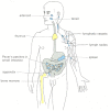
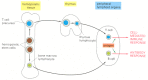
Lymphocytes are responsible for the astonishing specificity of adaptive immune responses. They occur in large numbers in the blood and lymph (the colorless fluid in the lymphatic vessels that connect the lymph nodes in the body to each other and to the bloodstream) and in lymphoid organs, such as the thymus, lymph nodes, spleen, and appendix ().
Human lymphoid organs. Lymphocytes develop in the thymus and bone marrow (yellow), which are therefore called central (or primary) lymphoid organs. The newly formed lymphocytes migrate from these primary organs to peripheral (or secondary) lymphoid organs (more...)
In this section, we discuss the general properties of lymphocytes that apply to both B cells and T cells. We shall see that each lymphocyte is committed to respond to a specific antigen and that its response during its first encounter with an antigen ensures that a more rapid and effective response occurs on subsequent encounters with the same antigen. We consider how lymphocytes avoid responding to self antigens and how they continuously recirculate between the blood and lymphoid organs, ensuring that a lymphocyte will find its specific foreign antigen no matter where the anitgen enters the body.
Lymphocytes Are Required for Adaptive Immunity
There are about 2 × 10¹² lymphocytes in the human body, making the immune system comparable in cell mass to the liver or brain. Despite their abundance, their central role in adaptive immunity was not demonstrated until the late 1950s. The crucial experiments were performed in mice and rats that were heavily irradiated to kill most of their white blood cells, including lymphocytes. This treatment makes the animals unable to mount adaptive immune responses. Then, by transferring various types of cells into the animals it was possible to determine which cells reversed the deficiency. Only lymphocytes restored the adaptive immune responses of irradiated animals, indicating that lymphocytes are required for these responses ().
A classic experiment showing that lymphocytes are required for adaptive immune responses to foreign antigens. An important requirement of all such cell-transfer experiments is that cells are transferred between animals of the same inbred strain. Members (more...)
The Innate and Adaptive Immune Systems Work Together
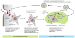
As mentioned earlier, lymphocytes usually respond to foreign antigens only if the innate immune system is first activated. As discussed in Chapter 25, the innate immune responses to an infection are rapid. They depend on pattern recognition receptors that recognize patterns of pathogen-associated molecules (immunostimulants) that are not present in the host organism, including microbial DNA, lipids, and polysaccharides, and proteins that form bacterial flagella. Some of these receptors are present on the surface of professional phagocytic cells such as macrophages and neutrophils, where they mediate the uptake of pathogens, which are then delivered to lysosomes for destruction. Others are secreted and bind to the surface of pathogens, marking them for destruction by either phagocytes or the complement system. Still others are present on the surface of various types of host cells and activate intracellular signaling pathways in response to the binding of pathogen-associated immunostimulants; this leads to the production of extracellular signal molecules that promote inflammation and help activate adaptive immune responses.
Some cells of the innate immune system directly present microbial antigens to T cells to initiate an adaptive immune response. The cells that do this most efficiently are called dendritic cells, which are present in most vertebrate tissues. They recognize and phagocytose invading microbes or their products at a site of infection and then migrate with their prey to a nearby peripheral lymphoid organ. There they act as antigen-presenting cells, which directly activate T cells to respond to the microbial antigens. Once activated, some of the T cells then migrate to the site of infection, where they help other phagocytic cells, mainly macrophages, destroy the microbes (). Other activated T cells remain in the lymphoid organ and help B cells respond to the microbial antigens. The activated B cells secrete antibodies that circulate in the body and coat the microbes, targeting them for efficient phagocytosis.
One way in which the innate immune system helps activate the adaptive immune system. Specialized phagocytic cells of the innate immune system, including macrophages (not shown) and dendritic cells ingest invading microbes or their products at the site (more...)
Thus, innate immune responses are activated mainly at sites of infection, whereas adaptive immune responses are activated in peripheral lymphoid organs. The two types of responses work together to eliminate invading pathogens.
B Lymphocytes Develop in the Bone Marrow; T Lymphocytes Develop in the Thymus
T cells and B cells derive their names from the organs in which they develop. T cells develop in the thymus, and B cells, in mammals, develop in the bone marrow in adults or the liver in fetuses.
Despite their different origins, both T and B cells develop from the same pluripotent hemopoietic stem cells, which give rise to all of the blood cells, including red blood cells, white blood cells, and platelets. These stem cells (discussed in Chapter 22) are located primarily in hemopoietic tissues—mainly the liver in fetuses and the bone marrow in adults. T cells develop in the thymus from precursor cells that migrate there from the hemopoietic tissues via the blood. In most mammals, including humans and mice, B cells develop from stem cells in the hemopoietic tissues themselves (). Because they are sites where lymphocytes develop from precursor cells, the thymus and hemopoietic tissues are referred to as central (primary) lymphoid organs (see ).
The development and activation of T and B cells. The central lymphoid organs, where lymphocytes develop from precursor cells, are labeled in yellow boxes. Lymphocytes respond to antigen in peripheral lymphoid organs, such as lymph nodes or spleen.
As we discuss later, most lymphocytes die in the central lymphoid organ soon after they develop, without ever functioning. Others, however, mature and migrate via the blood to the peripheral (secondary) lymphoid organs—mainly, the lymph nodes, spleen, and epithelium-associated lymphoid tissues in the gastrointestinal tract, respiratory tract, and skin (see ). As mentioned earlier, it is in the peripheral lymphoid organs that T cells and B cells react with foreign antigens (see ).
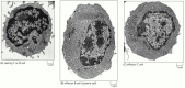
T and B cells become morphologically distinguishable from each other only after they have been activated by antigen. Nonactivated T and B cells look very similar, even in an electron microscope. Both are small, only marginally bigger than red blood cells, and contain little cytoplasm (). Both are activated by antigen to proliferate and mature into effector cells. Effector B cells secrete antibodies. In their most mature form, called plasma cells, they are filled with an extensive rough endoplasmic reticulum (). In contrast, effector T cells () contain very little endoplasmic reticulum and do not secrete antibodies.
Electron micrographs of nonactivated and activated lymphocytes. (A) A resting lymphocyte, which could be a T cell or a B cell, as these cells are difficult to distinguish morphologically until they have been activated to become effector cells. (B) An (more...)
There are two main classes of T cells—cytotoxic T cells and helper T cells. Cytotoxic T cells kill infected cells, whereas helper T cells help activate macrophages, B cells, and cytotoxic T cells. Effector helper T cells secrete a variety of signal proteins called cytokines, which act as local mediators. They also display a variety of costimulatory proteins on their surface. By means of these cytokines and membrane-bound costimulatory proteins, they can influence the behavior of the various cell types they help. Effector cytotoxic T cells kill infected target cells also by means of proteins that they either secrete or display on their surface. Thus, whereas B cells can act over long distances by secreting antibodies that are distributed by the bloodstream, T cells can migrate to distant sites, but there they act only locally on neighboring cells.
The Adaptive Immune System Works by Clonal Selection
The most remarkable feature of the adaptive immune system is that it can respond to millions of different foreign antigens in a highly specific way. B cells, for example, make antibodies that react specifically with the antigen that induced their production. How do B cells produce such a diversity of specific antibodies? The answer began to emerge in the 1950s with the formulation of the clonal selection theory. According to this theory, an animal first randomly generates a vast diversity of lymphocytes, and then those lymphocytes that can react against the foreign antigens that the animal actually encounters are specifically selected for action. As each lymphocyte develops in a central lymphoid organ, it becomes committed to react with a particular antigen before ever being exposed to the antigen. It expresses this commitment in the form of cell-surface receptor proteins that specifically fit the antigen. When a lymphocyte encounters its antigen in a peripheral lymphoid organ, the binding of the antigen to the receptors activates the lymphocyte, causing it both to proliferate and to differentiate into an effector cell. An antigen therefore selectively stimulates those cells that express complementary antigen-specific receptors and are thus already committed to respond to it. This arrangement is what makes adaptive immune responses antigen-specific.
The term “clonal” in clonal selection theory derives from the postulate that the adaptive immune system is composed of millions of different families, or clones, of lymphocytes, each consisting of T or B cells descended from a common ancestor. Each ancestral cell was already committed to make one particular antigen-specific receptor protein, and so all cells in a clone have the same antigen specificity (). According to the clonal selection theory, then, the immune system functions on the “ready-made” principle rather than the “made-to-order” one.
The clonal selection theory. An antigen activates only those lymphocyte clones (represented here by single cells) that are already committed to respond to it. A cell committed to respond to a particular antigen displays cell-surface receptors that specifically (more...)
There is compelling evidence to support the main tenets of the clonal selection theory. For example, when lymphocytes from an animal that has not been immunized are incubated in a test tube with a number of radioactively labeled antigens, only a very small proportion (less than 0.01%) bind each antigen, suggesting that only a few cells are committed to respond to these antigens. Moreover, when one antigen is made so highly radioactive that it kills any cell that it binds to, the remaining lymphocytes can no longer produce an immune response to that particular antigen, even though they can still respond normally to other antigens. Thus, the committed lymphocytes must have receptors on their surface that specifically bind that antigen. Although most experiments of this kind have involved B cells and antibody responses, other experiments indicate that T cells, like B cells, operate by clonal selection.
How can the adaptive immune system produce lymphocytes that collectively display such an enormous diversity of receptors, including ones that recognize synthetic molecules that never occur in nature? We shall see later that the antigen-specific receptors on both T and B cells are encoded by genes that are assembled from a series of gene segments by a unique form of genetic recombination that occurs early in a lymphocyte's development, before it has encountered antigen. This assembly process generates the enormous diversity of receptors and lymphocytes, thereby enabling the immune system to respond to an almost unlimited diversity of antigens.
Most Antigens Activate Many Different Lymphocyte Clones
Most large molecules, including virtually all proteins and many polysaccharides, can serve as antigens. Those parts of an antigen that combine with the antigen-binding site on either an antibody molecule or a lymphocyte receptor are called antigenic determinants (or epitopes). Most antigens have a variety of antigenic determinants that can stimulate the production of antibodies, specific T cell responses, or both. Some determinants of an antigen produce a greater response than others, so that the reaction to them may dominate the overall response. Such determinants are said to be immunodominant.
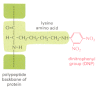
The diversity of lymphocytes is such that even a single antigenic determinant is likely to activate many clones, each of which produces an antigen-binding site with its own characteristic affinity for the determinant. Even a relatively simple structure, like the dinitrophenyl (DNP) group in , can be “looked at” in many ways. When it is coupled to a protein, as shown in the figure, it usually stimulates the production of hundreds of species of anti-DNP antibodies, each made by a different B cell clone. Such responses are said to be polyclonal. When only a few clones are activated, the response is said to be oligoclonal; and when the response involves only a single B or T cell clone, it is said to be monoclonal. Monoclonal antibodies are widely used as tools in biology and medicine, but they have to be produced in a special way (see ), as the responses to most antigens are polyclonal.
The dinitrophenyl (DNP) group. Although it is too small to induce an immune response on its own, when it is coupled covalently to a lysine side chain on a protein, as illustrated, DNP stimulates the production of hundreds of different species of antibodies (more...)
Immunological Memory Is Due to Both Clonal Expansion and Lymphocyte Differentiation
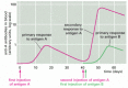
The adaptive immune system, like the nervous system, can remember prior experiences. This is why we develop lifelong immunity to many common infectious diseases after our initial exposure to the pathogen, and it is why vaccination works. The same phenomenon can be demonstrated in experimental animals. If an animal is immunized once with antigen A, an immune response (either antibody or cell-mediated) appears after several days, rises rapidly and exponentially, and then, more gradually, declines. This is the characteristic course of a primary immune response, occurring on an animal's first exposure to an antigen. If, after some weeks, months, or even years have elapsed, the animal is reinjected with antigen A, it will usually produce a secondary immune response that is very different from the primary response: the lag period is shorter, and the response is greater. These differences indicate that the animal has “remembered” its first exposure to antigen A. If the animal is given a different antigen (for example, antigen B) instead of a second injection of antigen A, the response is typical of a primary, and not a secondary, immune response. The secondary response must therefore reflect antigen-specific immunological memory for antigen A ().
Primary and secondary antibody responses. The secondary response induced by a second exposure to antigen A is faster and greater than the primary response and is specific for A, indicating that the adaptive immune system has specifically remembered encountering (more...)
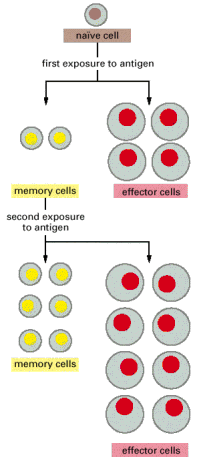
The clonal selection theory provides a useful conceptual framework for understanding the cellular basis of immunological memory. In an adult animal, the peripheral lymphoid organs contain a mixture of cells in at least three stages of maturation: naïve cells, effector cells and memory cells. When naïve cells encounter antigen for the first time, some of them are stimulated to proliferate and differentiate into effector cells, which are actively engaged in making a response (effector B cells secrete antibody, while effector T cells kill infected cells or help other cells fight the infection). Instead of becoming effector cells, some naïve cells are stimulated to multiply and differentiate into memory cells—cells that are not themselves engaged in a response but are more easily and more quickly induced to become effector cells by a later encounter with the same antigen. Memory cells, like naïve cells, give rise to either effector cells or more memory cells ().
A model for the cellular basis of immunological memory. When naïve lymphocytes are stimulated by their specific antigen, they proliferate and differentiate. Most become effector cells which function and then die, while others become long-lived (more...)
Thus, immunological memory is generated during the primary response in part because the proliferation of antigen-stimulated naïve cells creates many memory cells—a process known as clonal expansion—and in part because memory cells are able to respond more sensitively and rapidly to the same antigen than do naïve cells. And, unlike most effector cells, which die within days or weeks, memory cells can live for the lifetime of the animal, thereby providing lifelong immunological memory.
Acquired Immunological Tolerance Ensures That Self Antigens Are Not Attacked
As discussed in Chapter 25, cells of the innate immune system recognize molecules on the surface of pathogens that are not found in the host. The adaptive immune system has a far more difficult recognition task: it must be able to respond specifically to an almost unlimited number of foreign macromolecules, while avoiding responding to the large number of molecules made by the host organism itself. How does it do it? For one thing, self molecules do not induce the innate immune reactions that are required to activate adaptive immune responses. But even when an infection triggers innate reactions, self molecules still do not normally induce adaptive immune responses. Why not?
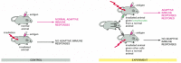
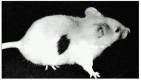
One answer is that the adaptive immune system “learns” not to respond to self antigens. Transplantation experiments provide one line of evidence for this learning process. When tissues are transplanted from one individual to another, as long as the two individuals are not identical twins, the immune system of the recipient usually recognizes the donor cells as foreign and destroys them. (For reasons we discuss later, the foreign antigens on the donor cells are so powerful that they can stimulate adaptive immune responses in the absence of infection or an adjuvant.) If, however, cells from one strain of mouse are introduced into a neonatal mouse of another strain, some of these cells survive for most of the recipient animal's life, and the recipient will now accept a graft from the original donor, even though it rejects “third-party” grafts. Apparently, nonself antigens can, in some circumstances, induce the immune system to become specifically unresponsive to them. This antigen-specific unresponsiveness to foreign antigens is known as acquired immunological tolerance ().
Immunological tolerance. The skin graft seen here, transplanted from an adult brown mouse to an adult white mouse, has survived for many weeks only because the white mouse, at the time of its birth, received an injection of cells from the brown mouse (more...)
The unresponsiveness of an animal's adaptive immune system to its own macromolecules (natural immunological tolerance) is acquired in the same way. Normal mice, for example, cannot make an immune response against one of their own protein components of the complement system called C5 (discussed in Chapter 25). Mutant mice, however, that lack the gene encoding C5 (but are otherwise genetically identical to the normal mice) can make a strong immune response to this blood protein when immunized with it. Natural immunological tolerance for a particular self molecule persists only for as long as the molecule remains present in the body. If a self molecule such as C5 is removed, an animal gains the ability to respond to it after a few weeks or months. Thus, the immune system is genetically capable of responding to self molecules but learns not to do so.
The learning process that leads to self-tolerance can involve killing the self-reactive lymphocytes (clonal deletion), functionally inactivating them (clonal anergy or inactivation), stimulating the cells to produce modified receptors that no longer recognize the self antigen (receptor editing), or the suppression of self-reactive lymphocytes by a special type of regulatory T cell. The process begins in the central lymphoid organs when newly formed self-reactive lymphocytes first encounter their self antigen. Instead of being activated by binding antigen, the immature lymphocytes are induced to either alter their receptors or die by apoptosis. Lymphocytes that could potentially respond to self antigens that are not present in the central lymphoid organs often die or are either inactivated or suppressed after they have matured and migrated to peripheral lymphoid organs.
Why does the binding of self antigen lead to tolerance rather than activation? As we discuss later, for a lymphocyte to be activated in a peripheral lymphoid organ, it must not only bind its antigen but must also receive a costimulatory signal. The latter signal is provided by a helper T cell in the case of a B lymphocyte and by an antigen-presenting cell in the case of a T lymphocyte. The production of costimulatory signals usually depends on exposure to pathogens, and so a self-reactive lymphocyte normally encounters its antigen in the absence of such signals. Without a costimulatory signal, an antigen tends to kill or inactivate a lymphocyte rather than activate it ().
Induction of immunological tolerance to self antigens in central and peripheral lymphoid organs. When a self-reactive immature lymphocyte binds its self antigen in the central lymphoid organ where the cell is produced, it may be induced to alter the receptor (more...)
Tolerance to self antigens sometimes breaks down, causing T or B cells (or both) to react against the organism's own tissue antigens. Myasthenia gravis is an example of such an autoimmune disease. Affected individuals make antibodies against the acetylcholine receptors on their own skeletal muscle cells. These antibodies interfere with the normal functioning of the receptors so that the patients become weak and may die because they cannot breathe. The mechanisms responsible for the breakdown of tolerance to self antigens in autoimmune diseases are unknown. It is thought, however, that activation of the innate immune system by infection may help trigger certain anti-self responses in genetically susceptible individuals.
Lymphocytes Continuously Circulate Through Peripheral Lymphoid Organs
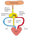
Pathogens generally enter the body through an epithelial surface, usually through the skin, gut, or respiratory tract. How do the microbial antigens travel from these entry points to a peripheral lymphoid organ, such as a lymph node or the spleen, where lymphocytes are activated (see )? The route and destination depend on the site of entry. Antigens that enter through the skin or respiratory tract are carried via the lymph to local lymph nodes; those that enter through the gut end up in gut-associated peripheral lymphoid organs such as Peyer's patches; and those that enter the blood are filtered out in the spleen. In most cases, dendritic cells carry the antigen from the site of infection to the peripheral lymphoid organ, where they become antigen-presenting cells (see ), specialized for activating T cells (as we discuss later).
But the lymphocytes that can recognize a particular microbial antigen in a peripheral lymph organ are only a tiny fraction of the total lymphocyte population. How do these rare cells find an antigen-presenting cell displaying their antigen? The answer is that they continuously circulate between the lymph and blood until they encounter their antigen. In a lymph node, for example, lymphocytes continually leave the bloodstream by squeezing out between specialized endothelial cells lining small veins called postcapillary venules. After percolating through the node, they accumulate in small lymphatic vessels that leave the node and connect with other lymphatic vessels that pass through other lymph nodes downstream (see ). Passing into larger and larger vessels, the lymphocytes eventually enter the main lymphatic vessel (the thoracic duct), which carries them back into the blood (). This continuous recirculation between the blood and lymph ends only if a lymphocyte encounters its specific antigen (and a costimulatory signal) on the surface of an antigen-presenting cell in a peripheral lymphoid organ. Now the lymphocyte is retained in the peripheral lymphoid organ, where it proliferates and differentiates into effector cells. Some of the effector T cells then leave the organ via the lymph and migrate through the blood to the site of infection (see ).
The path followed by lymphocytes as they continuously circulate between the lymph and blood. The circulation through a lymph node is shown here. Microbial antigens are carried into the lymph node by dendritic cells, which enter via afferent lymphatic (more...)
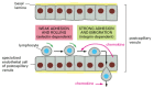
Lymphocyte recirculation depends on specific interactions between the lymphocyte cell surface and the surface of the specialized endothelial cells lining the postcapillary venules in the peripheral lymphoid organs. Many cell types in the blood come into contact with these endothelial cells, but only lymphocytes adhere and then migrate out of the bloodstream. The lymphocytes initially adhere to the endothelial cells via homing receptors that bind to specific ligands (often called counterreceptors) on the endothelial cell surface. Lymphocyte migration into lymph nodes, for example, depends on a homing receptor protein called L-selectin, a member of the selectin family of cell-surface lectins discussed in Chapter 19. This protein binds to specific sugar groups on a counterreceptor that is expressed exclusively on the surface of the specialized endothelial cells in lymph nodes, causing the lymphocytes to adhere weakly to the endothelial cells and to roll slowly along their surface. The rolling continues until another, much stronger adhesion system is called into play by chemo-attractant proteins (called chemokines; see below) secreted by endothelial cells. This strong adhesion is mediated by members of the integrin family of cell adhesion molecules (discussed in Chapter 19), which become activated on the lymphocyte surface. Now the lymphocytes stop rolling and crawl out of the blood vessel into the lymph node ().
Migration of a lymphocyte out of the bloodstream into a lymph node. A circulating lymphocyte adheres weakly to the surface of the specialized endothelial cells lining a postcapillary venule in a lymph node. This initial adhesion is mediated by L-selectin (more...)
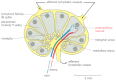
Chemokines are small, secreted, positively charged proteins that have a central role in guiding the migrations of various types of white blood cells. They are all structurally related and bind to the surface of endothelial cells, and to negatively charged proteoglycans of the extracellular matrix in organs. By binding to G-protein-linked receptors (discussed in Chapter 15) on the surface of specific blood cells, chemokines attract these cells from the bloodstream into an organ, guide them to specific locations within the organ, and then help stop migration. (The AIDS virus (HIV) also binds to chemokine receptors, which allows the virus to infect white blood cells.) T and B cells initially enter the same region of a lymph node but are then attracted by different chemokines to separate regions of the node—T cells to the paracortex and B cells to lymphoid follicles (). Unless they encounter their antigen, both types of cells soon leave the lymph node via lymphatic vessels. If they encounter their antigen, however, they remain in the node, proliferate, and differentiate into either effector cells or memory cells. Most of the effector cells leave the node, expressing different chemokine receptors that help guide them to their new destinations—T cells to sites of infection and B cells to the bone marrow.
A simplified drawing of a human lymph node. B cells are primarily clustered in structures called lymphoid follicles, whereas T cells are found mainly in the paracortex. Both types of lymphocytes are attracted by chemokines to enter the lymph node from (more...)
Summary
Innate immune responses are triggered at sites of infection by microbe-specific molecules associated with invading pathogens. In addition to fighting infection directly, these responses help activate adaptive immune responses in peripheral lymphoid organs. Unlike innate immune responses, adaptive responses provide specific and long-lasting protection against the particular pathogen that induced them.
The adaptive immune system is composed of millions of lymphocyte clones, with the cells in each clone sharing a unique cell-surface receptor that enables them to bind a particular antigen. The binding of antigen to these receptors, however, is usually not sufficient to stimulate a lymphocyte to proliferate and differentiate into an effector cell that can help eliminate the pathogen. Costimulatory signals provided by another specialized cell in a peripheral lymphoid organ are also required. Helper T cells provide such signals for B cells, while antigen-presenting dendritic cells usually provide them for T cells. Effector B cells secrete antibodies, which can act over long distances to help eliminate extracellular pathogens and their toxins. Effector T cells, by contrast, act locally at sites of infection to either kill infected host cells or help other cells to eliminate pathogens. As part of the adaptive immune response, some lymphocytes proliferate and differentiate into memory cells, which are able to respond faster and more efficiently the next time the same pathogen invades. Lymphocytes that would react against self molecules are either induced to alter their receptors, induced to kill themselves, inactivated, or suppressed, so that the adaptive immune system normally reacts only against foreign antigens. Both B and T cells circulate continuously between the blood and lymph. Only if they encounter their specific foreign antigen in a peripheral lymphoid organ do they stop migrating, proliferate, and differentiate into effector cells or memory cells.