From: Chapter 14, Otitis Media
Copyright © 2002, ASM Press.
NCBI Bookshelf. A service of the National Library of Medicine, National Institutes of Health.
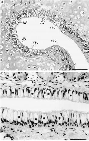
Cross-sections in mid-Eustachian tube 14 days after intranasal inoculation. (a) Note many vacuolated epithelial cells (vac) and those demonstrating late stages of intranuclear inclusions typical of AV. (b) Note marked goblet cell (*) hyperplasia. Bars = 50 μm. (Reprinted from The Journal of Infectious Diseases [13] with permission of the publisher.)
From: Chapter 14, Otitis Media
NCBI Bookshelf. A service of the National Library of Medicine, National Institutes of Health.