By agreement with the publisher, this book is accessible by the search feature, but cannot be browsed.
NCBI Bookshelf. A service of the National Library of Medicine, National Institutes of Health.
Kufe DW, Pollock RE, Weichselbaum RR, et al., editors. Holland-Frei Cancer Medicine. 6th edition. Hamilton (ON): BC Decker; 2003.

Holland-Frei Cancer Medicine. 6th edition.
Show detailsFungal infections began to emerge as a significant problem among cancer patients once effective antibacterial agents became available and immunocompromised patients were surviving for prolonged periods. Initially, Candida spp accounted for the vast majority of fungal infections, but in recent years, other organisms, especially Aspergillus spp, have been responsible for the continuing increased frequency. Most fungal infections occur in patients with hematologic neoplasms.90 At the present time, at least 40% to 50% of fatal infections are caused by fungi. Fungal infections occur in approximately 10% of patients with lymphoma but remain an infrequent complication in patients with metastatic carcinoma. A disturbing observation in recent years is the increasing frequency of systemic fungal infections in patients undergoing initial remission induction chemotherapy for acute leukemia and lymphoma. In the past, these infections tended to occur predominantly in patients with far-advanced neoplasms who were no longer responding to chemotherapy.
Fungal infections occurring in cancer patients can be divided into two major categories: the pathogenic fungi (Cryptococcus neoformans, Histoplasma capsulatum, and Coccidioides immitis) and the opportunistic fungi (Candida spp, Aspergillus spp, and other fungi). The former organisms cause infections in the general population but are more likely to cause disseminated infection in the cancer patient. Acute infection during cancer therapy often represents reactivation of latent infection. Patients with lymphoma are most susceptible to these infections. Opportunistic fungi usually cause only superficial infection in immunocompetent hosts but are the most common cause of systemic fungal infection in patients with impaired host defense mechanisms.
Fungemia is uncommon even in patients with disseminated infection. Most cases of fungemia are caused by Candida spp. In the past, the majority of infections were caused by Candida albicans, but in recent years Candida tropicalis, Candida glabrata, Candida parapsilosis, and other species have emerged as significant pathogens.91 Aspergillus spp and Mucorales are rarely cultured from the blood even in patients with widely disseminated infection. Fungi that are cultured from blood specimens of patients with disseminated infection more often include Candida neoformans, H. capsulatum, Fusarium spp, and Trichosporon beigelii. The extensive use of intravascular catheters has resulted in an increased frequency of fungemia, especially caused by Candida spp. Because 70% to 80% of these patients have evidence of deep-seated infection, all of these patients should be given antifungal therapy. The duration of fungemia during therapy is prolonged if intravascular catheters are not removed, hence, it is advisable to remove them, whenever possible. If the infection is caused by C. parapsilosis, therapy is rarely effective without removal of the catheter.92 Local infections caused by Aspergillus spp and Rhizopus spp may occur at catheter sites and progress to pulmonary or disseminated disease.
The setting in which fungal infection develops is complex, and multiple factors are responsible for the rapid increase in these infections. Frequently, this has been attributed to the widespread use of broad-spectrum antibiotics. Bacteria that inhibit the growth of Candida spp are suppressed by antibiotic therapy permitting the overgrowth of Candida spp Antibiotics probably do not directly predispose patients to fungal infections; they facilitate fungal overgrowth in the oropharynx and gastrointestinal tract. At high concentrations, Candida spp cross the intact gastrointestinal mucosa and enter the circulation by a process known as persorption.
Adrenal corticosteroids can interfere with macrophage function, and the macrophage is one of the primary defenses against fungal invasion. Prolonged and severe neutropenia also predisposes to fungal infection. Neutrophils ingest and kill Candida spp in vitro and serve as a major defense against systemic infection. In an animal model of aspergillosis, it was demonstrated that the macrophage represents the primary defense mechanism against spores and that the neutrophil represents the primary defense mechanism against mycelia.93 The administration of adrenal corticosteroids interferes with macrophage function, allowing ingested spores to germinate. The administration of nitrogen mustard (and many other cancer chemotherapeutic compounds) causes neutropenia, which facilitates establishment of infection by activated mycelia. The role of lymphocytes and lymphokines is complex and has not been fully elucidated.
Fungal infections often become established at sites of previous infection or necrosis. For example, more than 70% of cases of pulmonary aspergillosis occur in association with a previous or concomitant bacterial pneumonia. Candida spp often infect gastrointestinal ulcerations caused by antitumor agents, or herpes or bacterial infection. The availability of effective antitumor agents and supportive care measures has prolonged the survival of many patients with severely compromised host defense mechanisms, permitting the development of fungal infections in patients who previously would have died of bacterial infections.
Candida Species
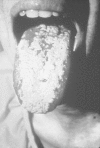
Most superficial fungal infections of the oropharynx and gastrointestinal tract are caused by C. albicans. Oropharyngeal candidiasis occurs in approximately 25% to 30% of cancer patients undergoing chemotherapy, and is even more frequent in patients with acute leukemia. This infection is especially prevalent in patients receiving antitumor agents or radiotherapy that causes mucosal damage, patients receiving adrenal corticosteroids, and patients already colonized with Candida spp at the onset of chemotherapy. Some patients with solid tumors develop mild, asymptomatic infection following chemotherapy that is not recognized and resolves without therapy. Also, patients with oropharyngeal candidiasis may have associated esophageal candidiasis that is asymptomatic.94 The lesions appear as white or grayish white plaques surrounded by an erythematous halo (Figure 160-4). The plaques are friable with a freely bleeding base. These lesions may be difficult to differentiate from the necrotic mucosa caused by antitumor agents, such as methotrexate. Candida plaques may occur in association with herpes simplex infection. Topical agents, such as nystatin, are often of minimum benefit, and not infrequently, the lesions progress. Orally absorbable antifungal agents, such as fluconazole and itraconazole solution, are generally effective. Occasional patients may require a short course of parenteral amphotericin B, which produces dramatic improvement in most cases after the initial dose.

Figure 160-4
Severe oropharyngeal candidiasis in a patient with acquired immunodeficiency syndrome. (Four-color version of figure on CD-RM)
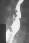
Candida esophagitis is no longer a rare infection in patients with hematologic neoplasms. The most common symptom is dysphagia, often accompanied by severe retrosternal pain, nausea, and vomiting, and occasionally, gastrointestinal bleeding. Rarely, this infection can lead to perforation.95 The pain during swallowing may become so severe that the patient is unable to eat. It may be present with or without oral candidiasis. A characteristic cobblestone or moth-eaten appearance of the esophageal mucosa is present on barium-contrast radiographic studies (Figure 160-5). However, these abnormalities may be caused by other organisms, such as herpes simplex and cytomegalovirus. Furthermore, in 25% of cases, the radiographic studies are negative. The diagnosis is established by esophagoscopy, which reveals characteristic ulcerations with pseudomembrane formation. This procedure permits biopsy for definitive diagnosis. Appropriate therapy consists of an orally absorbable antifungal agent, but some patients may have difficulty swallowing these medications. Parenteral amphotericin B is dramatically effective within 24 to 48 h, and approximately 80% of patients respond after a 5-day course of therapy. Fluconazole is an equally effective and less-toxic parenteral preparation. Because Candida spp are the most common cause of infectious esophagitis, it is often appropriate to initiate antifungal therapy empirically and perform esophagoscopy if the patient fails to respond after several days.

Figure 160-5
The characteristic “moth-eaten” appearance of the esophageal mucosa demonstrated on barium-swallow examination in a patient with esophageal candidiasis.
Superficial Candida infections of other areas of the gastrointestinal tract have been identified in approximately 3% of cancer patients. They are found at autopsy examination in 10% of patients with lymphoma, in 15% of patients with acute leukemia, and in only 2% of patients with carcinoma. Candidiasis may involve any portion of the gastrointestinal tract, and multiple sites are often involved.96 Usually, infection below the esophagus is asymptomatic and only diagnosed at autopsy examination.
Systemic Candida infections may be localized to a single organ, but usually multiple organs are involved.97 Occasional cases of chronic laryngitis caused by Candida spp have been recognized. Pulmonary infection is usually a manifestation of disseminated disease, but occasional primary infections of the lung occur because of aspiration of contaminated oropharyngeal secretions. It is difficult to establish the diagnosis of primary Candida pneumonia because there are no characteristic findings.98 Isolation of Candida spp from the sputum does not establish the diagnosis because it may represent contamination from the oropharynx. In one study of 25 patients who died in an intensive care unit, Candida spp was isolated from a percutaneous needle biopsy or bronchoalveolar lavage specimen of 10 and 9 patients, respectively, but histopathologic confirmation was found in only 2 at autopsy examination.99 A substantial proportion of patients with prolonged urinary catheterization develop candiduria which often represents only colonization. Patients with surgery or abnormalities of the genitourinary tract, neutropenia or diabetes mellitus should always be treated with systemic therapy because they are at risk of serious Candida infection. Candida peritonitis may occur following intestinal perforation or in association with peritoneal catheters.
Disseminated candidiasis is found at autopsy examination in 20% to 30% of patients with acute leukemia, in 2% of patients with lymphoma, and in less than 1% of patients with metastatic cancer. Approximately 55% of episodes are caused by C. albicans, 20% by Candida tropicalis, 15% by C. glabrata and the remainder primarily by C. parapsilosis and Candida krusei. Two patterns of organ involvement have been described. In one, occurring predominantly in patients with acute leukemia, the most frequent organs infected include the gastrointestinal tract, liver, spleen, and lungs. This pattern of involvement suggests the gastrointestinal tract as the primary site of origin. The second pattern of distribution involves predominantly the heart, kidneys, and lung, which is the pattern of distribution in animals following direct intravenous injection.97
There are no characteristic physical signs and symptoms of disseminated candidiasis. Some patients present with the acute onset of tachycardia, tachypnea, and hypotension suggestive of endotoxin shock. Often, the only indications of this infection are a gradual worsening of the patient's clinical condition, associated with fever unresponsive to antibiotic therapy. Some patients have ocular infection causing blurred vision, pain, scotomas, or loss of visual acuity. Ocular lesions include white fluffy retinal exudates with vitreous haze or hemorrhage, hypopyon, or iritis, but these lesions are rarely found in neutropenic patients. Approximately 10% of patients, especially those with severe neutropenia, develop characteristic erythematous macronodular skin lesions. These skin lesions may be single or multiple, localized or diffuse, often resembling a drug rash. Candida organisms can be identified in the subcutaneous tissue of skin biopsies of these lesions and can be cultured from the tissue in 50% of cases. In occasional patients, these skin lesions are associated with a myositis in which the patient has exquisitely tender muscles.
Central nervous system involvement occurs in up to 50% of patients with disseminated candidiasis. It may be manifested as cerebritis, cerebral abscesses, or meningitis. Often the only symptom is mental obtundation. A stiff neck may be detected in some patients with meningitis, but often this sign is absent. Examination of the cerebrospinal fluid usually reveals nonspecific abnormalities such as an elevated protein and occasional mononuclear cells. Candida organisms are seldom visualized or cultured from the cerebrospinal fluid.
A chronic form of Candida infection known as hepatosplenic candidiasis—or, more appropriately, chronic disseminated candidiasis—has been described during the past 30 years.100 This infection typically occurs in patients with acute leukemia undergoing chemotherapy while they are experiencing prolonged severe neutropenia. They develop fever that fails to respond to broad-spectrum antibacterial antibiotics, as well as antifungal agents. After achieving remission of their leukemia with neutrophil recovery, they remain febrile and debilitated with substantial weight loss. Symptoms, including right upper quadrant or shoulder pain, may now appear. Hepatosplenomegaly may be detected. Characteristically, the alkaline phosphatase concentration becomes highly elevated, and other liver function tests may also become abnormal. Ultrasonography, magnetic resonance imaging (MRI), or computed tomography (CT) of the liver and spleen reveals multiple lesions (Figure 160-6). Other organs may also be infected. Only 50% to 60% of these patients respond to amphotericin B plus flucytosine, whereas approximately 80% respond to fluconazole or lipid formulations of amphotericin B.101 Usually several weeks of therapy are required before the patient improves. This type of Candida infection has virtually disappeared from institutions where fluconazole is used prophylactically for marrow transplant recipients and acute leukemia patients.

Figure 160-6
Multiple “punched-out” lesions in the liver, spleen, and kidneys in a patient with chronic disseminated candidiasis.
Because the frequency of infections caused by Candida spp other than C. albicans has increased substantially in recent years, unique features of some of these infections have been recognized. At some institutions C. tropicalis has surpassed C. albicans as the most common cause of disseminated infection.102 It appears to be more virulent because only 3% to 15% of neutropenic patients colonized by C. albicans subsequently develop disseminated infection, as compared to 40% to 80% of those colonized by C. tropicalis. Skin lesions and myositis are associated more often with C. tropicalis infection. C. parapsilosis is less virulent than C. albicans, and infection is nearly always associated with intravascular catheters and parenteral nutrition. Only 10% of patients with intravascular catheters who develop C. parapsilosis fungemia respond to antifungal therapy without removal of the catheter.92
Colonization and infection caused by C. krusei and C. glabrata is associated with the use of fluconazole prophylaxis.103 Unlike most other Candida spp, C. krusei has been isolated from many foods and beverages, but infrequently from normal humans. It is inherently resistant to fluconazole and variably susceptible to itraconazole. It may be more easily acquired from nosocomial and environmental sources in patients receiving azole prophylaxis. C. glabrata is a common contaminant of the skin and urine. It has variable susceptibility to fluconazole, and itraconazole but many infections have been treated successfully with high doses of fluconazole. C. glabrata is often a cause of candidemia among patients already receiving fluconazole or amphotericin B as therapy or prophylaxis.104
The diagnosis of disseminated candidiasis is often not established before death. Only 50% of patients have abnormal chest radiographs at the onset of infection involving the lungs. Candida spp are isolated from blood specimens of about 70% of patients with disseminated candidiasis using the most sensitive cultural techniques such as lysis-centrifugation. A variety of noncultural methods, including monoclonal antibodies to detect mannoproteins, enzyme-linked immuno- sorbent assay (ELISA) technique to detect enolase, and PCR analyses have been developed, but none are reliable.105 The isolation of C. albicans from throat or urine specimens is of little diagnostic importance, whereas the isolation of C. tropicalis indicates the presence of infection in approximately 60% of neutropenic patients.
Amphotericin B (AMB) has been effective for the treatment of disseminated candidiasis in patients with adequate neutrophil counts, but it has undesirable acute toxicities including fever, chills, and headaches. Chronic administration often results in nephrotoxicity which is of concern in cancer patients who often are receiving other nephrotoxic agents. The administration of AMB in lipid formulations reduces the frequency of nephrotoxicity and allows for the administration of much higher daily doses, but it is not clear that these higher doses result in enhanced efficacy. Combining AMB with 5-fluorocytosine may increase therapeutic efficacy, especially if the infection is caused by C. tropicalis. In vitro and animal studies indicate that these agents interact synergistically against Candida spp. The myelosuppressive toxicity of 5-fluorocytosine can be problematic in patients whose marrow function has been compromised already by chemotherapy. Fluconazole is as effective as AMB and is much less toxic.106,107 C. krusei is inherently resistant to fluconazole and about 15% of isolates of C. glabrata are also resistant. Other agents available for therapy of Candida infections include itraconazole, voriconazole and caspofungin.
The therapy of candidemia and disseminated candidiasis in neutropenic patients is often unsuccessful. AMB, although considered to be a fungicidal agent, is rarely effective unless neutrophil recovery occurs. Fluconazole is as effective as AMB, but also is of limited efficacy in patients with persistent neutropenia. The efficacy of other antifungal agents in these patients has not been determined.
Aspergillus Species
Aspergillosis is an increasing problem in neutropenic patients and patients receiving chronic adrenal corticosteroid therapy. Recent studies report a frequency of 20% to 50% among patients with acute leukemia. This infection is also common in bone marrow transplant recipients during the posttransplant period when they are neutropenic and later if they develop graft-versus-host disease. The most common pathogen is Aspergillus fumigatus. Other human pathogens include Aspergillus terreus, Aspergillus flavus, Aspergillus niger, Aspergillus glaucus, and Aspergillus nidulans. Infection is usually acquired by inhalation of spores, which are deposited in the paranasal sinuses or lungs.108 Outbreaks of aspergillosis have occurred on leukemic and bone marrow transplantation units, usually associated with construction within or adjacent to the hospital. Studies show that the concentration of Aspergillus spores is much higher at construction sites within the hospital than at other hospital sites. Disturbance of dust above false ceilings represents a significant risk to susceptible patients, and they should be protected from such exposure.
More than 70% of infections involve the lungs, and approximately 35% of patients with pulmonary aspergillosis have hematogenous dissemination to other organs. Pulmonary infection may be manifested as necrotizing bronchopneumonia, hemorrhagic pulmonary infarction, solitary or miliary lung abscesses, lobar pneumonia, or bronchitis. A few patients will develop exsanguinating pulmonary hemorrhage early in the course of their infection. The classic clinical presentation of pulmonary aspergillosis is the sudden onset of pleuritic chest pain with fever, hemoptysis, and a pleural friction rub suggestive of pulmonary embolus and infarction. Unfortunately, this classic syndrome occurs in less than 30% of patients. Often, the only evidence of infection is prolonged fever with pulmonary infiltrates that fail to respond to antibacterial therapy. Occasional patients present only with fever and a normal physical and chest radiographic examination.
The earliest abnormality on chest radiographic examination is the appearance of single or multiple rounded dense areas (Figure 160-7). Patients with symptoms of pulmonary infarction may develop typical wedge-shaped lesions on radiographic examination. As the infection progresses, one of several patterns may emerge, including single or multiple abscesses with cavitation, lobar pneumonia, or patchy or diffuse pulmonary infiltrates located unilaterally or bilaterally. Patients whose infection is controlled often develop cavities with or without fungus balls. High-resolution CT scanning of the lung can be helpful in the early diagnosis of aspergillosis. Nodular lesions may be detected in the lungs of patients with normal chest radiographs. Characteristic findings in early stage disease are multiple nodules with a halo of surrounding ground-glass attenuation which represents hemorrhage surrounding a region of pulmonary infarction.109 As healing occurs, the infarcted tissue becomes necrotic and retracts from the viable tissue leaving an air crescent.

Figure 160-7
Multiple rounded pulmonary infiltrates compatible with invasive pulmonary aspergillosis.
Pathologically, pulmonary infection consists of nodular zones of consolidation surrounded by hemorrhage, abscess formation, or typical wedge-shaped infarcts. On microscopic examination, Aspergillus spp appear as uniform septate hyphae, about 4 microns in diameter with dichotomous branching. These organisms have a propensity for invading blood vessels, causing thrombosis and infarction.
Aspergillus sino-orbital infection is being diagnosed with increasing frequency in patients with acute leukemia and in marrow transplant recipients, accounting for at least 15% of cases of aspergillosis (Figure 160-8). Signs and symptoms include fever, retro-orbital pain, headache, circumorbital erythema, nasal obstruction, and necrotic encrustation of the nasal septum, palate, or external nares. Infections may erode through the base of the skull and invade the brain or cause destruction of the paranasal and facial structures and the eye. About half the infections disseminate to other organs. The fungus can often be isolated from nasal cultures. It can be visualized histopathologically and often cultured from biopsy specimens.

Figure 160-8
Pansinusitis caused by Aspergillus spp in an allogeneic bone marrow transplant recipient with persistent fever.
Overall, approximately 35% of infections are widely disseminated. Organs involved include the lung, brain, gastrointestinal tract, liver, kidney, and thyroid.110 Aspergillus infection of the liver may cause multiple abscesses or vascular thrombosis and infarction, occasionally resulting in Budd-Chiari syndrome. The organism is seldom isolated from blood culture specimens of patients with disseminated disease.
Although central nervous system aspergillosis sometimes results from local extension of sinus infection, more often, it follows hematologic dissemination. Multiple lesions are usually present, with substantial vascular invasion leading to cerebral thrombosis, infarction, and abscess formation. These patients are lethargic and have focal neurologic signs indicative of the area of brain involvement. The cerebrospinal fluid is normal in most instances, although leukocytosis and increased protein concentrations may be found.
A localized form of aspergillosis has been described in association with intravascular catheters. Aspergillus spores may be deposited from the air at the time of insertion or may be impregnated in materials used for catheter dressings. These infections are potentially serious because they can disseminate. Skin lesions, manifested as sharply defined black eschars, occur in about 5% of patients with disseminated infection. Aspergillus stomatitis occurs in neutropenic patients.
Antemortem diagnosis of aspergillosis is difficult because the organism is cultured from clinical specimens of less than 30% of infected patients, hence, many infections are diagnosed only at autopsy examination. Although Aspergillus spp can be cultured from normal, uninfected subjects and is a potential laboratory contaminant, if it is cultured from respiratory secretions of susceptible patients, there is a high probability of infection. Hyphal elements can be identified in infected sinus tissue biopsies, but the organism is cultured from only 75% of these cases. PCR and glucomannan detection by sandwich ELISA, are promising noncultural methods for identifying infected patients.111 Serial glucomannan assays may be useful for assessing response to therapy.
AMB is effective for the treatment of aspergillosis, provided the patient's underlying deficiency in host defense mechanisms has been corrected. Patients with persistent deficiencies in host defense mechanisms, especially neutropenia, seldom respond to therapy. Recovery often requires prolonged therapy with AMB, which usually causes dose-limiting nephrotoxicity, hence, a lipid formulation is preferable to AMB desoxycholate. Because substantially higher doses of AMB can be administered by the lipid formulations they may be more effective, although the evidence is only suggestive.
Itraconazole is an azole compound that has activity against Aspergillus spp.112 In the past, its use was limited because only an oral capsule was available and oral absorption was variable. Currently, an oral solution and an intravenous solution are available that provide more reliable serum concentrations. It appears to be as effective as amphotericin B but no comparative trial has been conducted.
Two new agents are available for treatment of aspergillosis.113 Caspofungin inhibits the synthesis of β-(1,3)-glucan, a major cell wall component. Its response rate in patients failing to respond to other therapies is approximately 50% and it is well-tolerated. Voriconazole is a new azole that was more effective than AMB in an initial randomized trial. It is also better tolerated than AMB, although some patients develop transient phototoxicity.
A major problem is the management of the patient who has recovered from pulmonary aspergillosis and who has a persistent cavity with or without a fungus ball. Acute pulmonary hemorrhage can occur, leading to death. Furthermore, the potential for reactivation of infection interferes with subsequent cancer chemotherapy. Surgical excision of the residual cavity should be given consideration, but that may not always be technically possible due to the presence of multiple cavities or the location of the lesion. Antifungal therapy should be administered during periods of neutropenia associated with chemotherapy in patients with residual lesions to prevent reactivation. The appropriate duration of such therapy is unknown, but should be continued for a minimum of two courses.
Cryptococcus Species
Cryptococcosis is primarily a disease of patients with impaired cellular immunity, hence patients with lymphoma, Hodgkin disease, and other lymphatic malignancies are at risk from this infection. Occasionally, infection occurs in patients with solid tumors. Unlike candidiasis and aspergillosis, cryptococcosis is acquired prior to hospitalization. The organism is ubiquitous in animals and soil specimens. Infection begins in the lungs, where in healthy humans, it may remain asymptomatic and resolve without therapy. Approximately 40% of infections in cancer patients involve the lungs.114 A variety of abnormalities may be found on chest radiographs, including miliary, nodular or cavitary lesions. On pathologic examination, the typical lesions have a gelatinous appearance. The lesions consist of aggregations of encapsulated budding yeast cells with minimal inflammatory response. Granulomas may be present in some cases. Primary cryptococcal pneumonia may follow a fulminant course, leading to the death of the patient within1 to 2 weeks after onset of symptoms.
The most common form of cryptococcal infection is meningoencephalitis, accounting for approximately 50% of infections in cancer patients.114 It is a consequence of hematogenous dissemination from the lung where usually the infection is no longer apparent. Infection may be acute, subacute, or chronic, and may arise suddenly or insidiously. Symptoms of central nervous system infection include headache, vertigo, nausea, and vomiting; physical findings consist of fever, meningitis, stupor, signs of increased intracranial pressure, and focal neurologic defects. Leukocytosis (predominantly with lymphocytes), hypoglycorrhachia, and elevated protein concentrations are usually found in the cerebrospinal fluid. Yeast cells can be visualized in the cerebrospinal fluid of nearly 60% of patients. The organisms may be confused with mononuclear cells, but they can be differentiated by an India ink preparation that defines the capsule of the yeast cells. Although Cryptococcus neoformans usually is cultured easily from the cerebrospinal fluid of patients with meningitis, occasional cancer patients have normal cerebrospinal fluid, and the organism cannot be isolated from repeated culture specimens. The serologic test for detection of cryptococcal antigen in cerebrospinal fluid and blood is useful for rapid diagnosis of this infection, especially in cases where the organism cannot be visualized.
Disseminated infection may involve multiple organs with the skin and bone being frequent sites of involvement. Skin lesions may be acneiform, nodular, pustular, plaque-like or ulcerated and are present in approximately 10% of patients with disseminated infection. Occasional patients develop a cryptococcal panniculitis. The yeast may be isolated from blood, urine, or sputum culture specimens of patients with disseminated infection.
Initial therapy of meningitis should consist of AMB plus 5-fluorocytosine for about 2 weeks. 5-Fluorocytosine can cause myelosuppression, especially when combined with AMB, because it is excreted in the urine and AMB causes renal impairment. Subsequently, therapy can be continued with fluconazole for at least 10 weeks. Fluconazole can also be used as primary therapy for subacute and chronic infections of other sites.
Other Opportunistic Fungi
A variety of fungi of low pathogenicity have been recognized as cause of significant infection in occasional cancer patients. These organisms include Trichosporon spp, Blastoschizomyces capitatus, Fusarium spp, Pseudallescheria boydii, Scedosporium spp, Geotrichum candidum, and Malassezia furfur. In addition, members of the order Mucorales cause infection in occasional cancer patients. Most of these infections occur sporadically, although there have been clusters of Fusarium infections in recent years at some institutions. The majority of infected patients have hematologic neoplasms, especially acute leukemia.
Mucormycosis is infection caused by fungi of the order Mucorales. Rhizopus spp cause the majority of these infections. Mucormycosis was first described in patients with diabetic ketoacidosis, in whom infection characteristically involves the sinuses, orbit, and brain. Pneumonia is the most common form of infection in cancer patients, although some patients develop sinusitis or cutaneous infection. Infection may become disseminated, involving the heart, kidney, gastrointestinal tract, liver, and spleen. The clinical presentation of mucormycosis is similar to that of aspergillosis.
Microscopically, these organisms appear as broad nonseptate branching hyphae. Like Aspergillus spp, Mucorales invade blood vessels, causing thrombosis and infarction. Most cases of mucormycosis in cancer patients are diagnosed at autopsy; consequently, therapeutic measures have not been evaluated extensively. AMB is only marginally active against these fungi. Aggressive surgical debridement is an important component of the therapy of sino-orbital infection and should be considered in patients with residual lesions after pulmonary infection. Neutropenic patients uniformly fail to respond to therapy unless neutrphil recovery occurs.
Trichosporon spp cutaneum can cause disseminated infection, primarily in patients with hematologic neoplasms but also in patients undergoing chemotherapy for metastatic carcinoma. The majority of patients have been severely neutropenic.115 A few patients, especially those with adequate neutrophil counts, develop localized pneumonia without dissemination. There are no characteristic signs and symptoms suggestive of Trichosporon infection. A variety of skin lesions have been described, and skin lesions occur in approximately 30% of infected patients. Portals of entry include the gastrointestinal tract, respiratory tract, and intravenous catheter sites. Trichosporon infection is often associated with other concurrent opportunistic infections. Although these organisms may be susceptible to AMB, the azole compounds appear to be more effective therapeutic agents.
Fusarium spp have emerged as significant pathogens in neutropenic patients during the past decade.116,117 These organisms produce a potent toxin that, when ingested, causes aplastic anemia. Localized infections of the lung, sinuses, and skin occur, but most patients have disseminated infection. Cutaneous and subcutaneous skin lesions are frequent in disseminated infection. Like Aspergillus spp, these organisms invade blood vessels, causing thrombosis and infarction. Usually Fusarium spp can be isolated readily from blood culture or tissue specimens. It may be difficult to distinguish Fusarium from some other fungi on histopathologic examination. Recovery from this infection depends on resolution of neutropenia, and currently available antifungal agents are at best only marginally active against these fungi.
Empiric Therapy
The high frequency of fungal infections in neutropenic patients at autopsy examination and the inadequacy of diagnostic procedures have led to empiric administration of antifungal agents in patients suspected of having this infection. Empiric therapy should be considered in patients with neutrophil counts of less than 100/mm3 for greater than 1 week who develop fever that fails to respond to 4 to 7 days of broad-spectrum antibacterial therapy.118,119 Other supportive indications are indwelling catheters, chronic adrenal corticosteroid therapy, the presence of unexplained pulmonary infiltrates, and deteriorating renal or hepatic function. AMB has been used most often as empiric therapy, but other alternatives are effective, including lipid formulations of AMB, fluconazole, itraconazole, and voriconazole. Fluconazole is less attractive than other agents for empiric therapy because it is not active against aspergillosis. Responding patients probably should remain on antifungal therapy for at least 1 or 2 weeks. Empiric antifungal therapy is not indicated for a majority of patients who have only transient or modest degrees of neutropenia.
- Fungal Infections - Holland-Frei Cancer MedicineFungal Infections - Holland-Frei Cancer Medicine
Your browsing activity is empty.
Activity recording is turned off.
See more...